Introduction
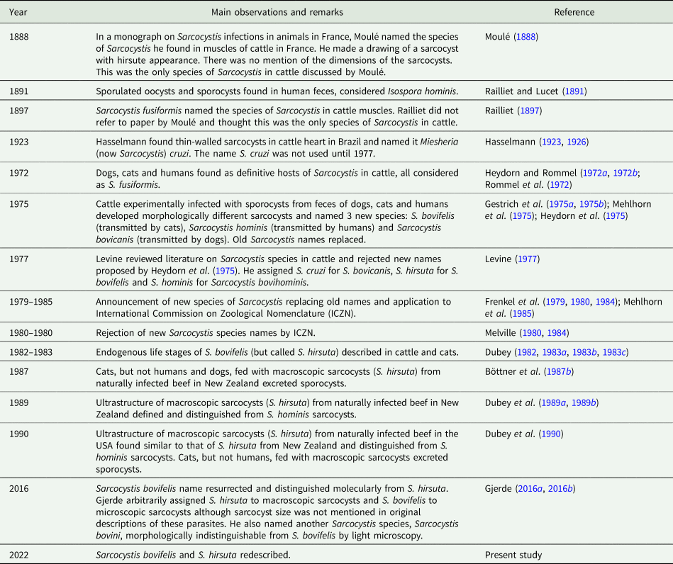
Infections by Sarcocystis in cattle are ubiquitous. Sarcocysts were first observed in cattle musculature more than 2 centuries ago (Levine, Reference Levine1977). The taxonomy of Sarcocystis species in cattle remains debated, including at least 8 named species, Sarcocystis cruzi, Sarcocystis heydorni, Sarcocystis bovini, Sarcocystis hirsuta, Sarcocystis rommeli, Sarcocystis sinensis, Sarcocystis hominis and Sarcocystis bovifelis (Dubey et al., Reference Dubey, Calero-Bernal, Rosenthal, Speer and Fayer2016; Gjerde, Reference Gjerde2016a, Reference Gjerde2016b; Hu et al., Reference Hu, Huang, Wen, Esch, Liang and Li2017). Among these, a complete description of the life cycle is known only for S. cruzi, which cycles between cattle and canids.
Before the discovery of the obligatory 2-host life cycle of Sarcocystis species in 1972, only 1 species of Sarcocystis (Sarcocystis fusiformis) was recognized in cattle. Then, researchers believed it parasitized both cattle (Bos taurus) and water buffalo (Bubalus bubalis). Currently, the name S. fusiformis is restricted to the macroscopic sarcocyst-forming species in water buffalo; no species of Sarcocystis sharing its morphology occurs in cattle (Dubey et al., Reference Dubey, Fayer, Rosenthal, Calero-Bernal and Uggla2014, Reference Dubey, Hilali, van Wilpe, Verma, Calero-Bernal and Abdel-Wahab2015).
Until cats were established as definitive hosts for Toxoplasma gondii in 1970 (reviewed in Dubey and Beattie, Reference Dubey and Beattie1988), no cyst-forming coccidia (in the genera Sarcocystis, Toxoplasma or Besnoitia) were completely understood. I have been actively involved in these discoveries for the past 5 decades. Soon after the discovery of the coccidian oocysts in cats in 1970, an intensive search began to find definitive hosts for Sarcocystis. In 1972 the 2-host life cycle of Sarcocystis was discovered when cats, dogs and humans were found as definitive hosts for various species of Sarcocystis in livestock (Heydorn and Rommel, Reference Heydorn and Rommel1972a, Reference Heydorn and Rommel1972b; Rommel and Heydorn, Reference Rommel and Heydorn1972; Rommel et al., Reference Rommel, Heydorn and Gruber1972).
Subsequent investigations, in Germany, revealed the existence of 3 species of Sarcocystis infecting cattle: 1 transmitted exclusively by canids, 1 exclusively by cats and the third exclusively by humans. Based on these findings, Heydorn et al. (Reference Heydorn, Gestrich, Mehlhorn and Rommel1975) proposed new names for species of Sarcocystis in cattle; Sarcocystis bovicanis for the species transmitted via dogs, S. bovifelis for the species transmitted by cats and Sarcocystis bovihominis for the species transmitted by humans.
An intense debate followed the proposal to rename these species. Scientists from Germany and their collaborators (see Table 1) sought a rational system, combining information from the intermediate and definitive hosts (e.g. S. bovicanis). They suggested new names for Sarcocystis species of livestock, and even suggested replacing the type species of the genus, Sarcocystis miescheriana with Sarcocystis muris (Frenkel et al., Reference Frenkel, Heydorn, Mehlhorn and Rommel1979; Mehlhorn et al., Reference Mehlhorn, Heydorn, Frenkel and Göbel1985).
Summary of Sarcocystis bovifelis and Sarcocystis hirsuta

However, Levine (Reference Levine1977) upheld the principle of nomenclatural priority. This principle seeks stability in scientific communication by retaining names for described species. He therefore diligently searched the literature and documented, chronologically, each named species of Sarcocystis (see Table 1). Levine (Reference Levine1977) concluded that the previously used names for Sarcocystis species must be retained, even though the descriptions of species were incomplete; he assigned S. cruzi for S. bovicanis, S. hirsuta for S. bovifelis and S. hominis for S. bovihominis. The German researchers applied to the Secretary of International Code of Zoological Nomenclature (ICZN) requesting permission to adopt their proposed naming system. However, ICZN rejected their application (Table 1).
Recently, Gjerde (Reference Gjerde2016a, Reference Gjerde2016b) reviewed the literature on Sarcocystis species in cattle and concluded that there were 2 more species of Sarcocystis in cattle, transmissible via cats, and resurrected the name S. bovifelis and assigned the name S. hirsuta to another species in cattle transmitted via cats. He molecularly distinguished S. hirsuta from S. bovifelis and another new species with unknown life cycle as S. bovini. Here, I redescribe S. bovifelis and S. hirsuta.
History of S. bovifelis
Table 1 summarizes chronologically information on the biology of S. bovifelis and S. hirsuta. To clarify ambiguities the original German studies are described in the following section.
Feeding of naturally infected bovine muscle to cats
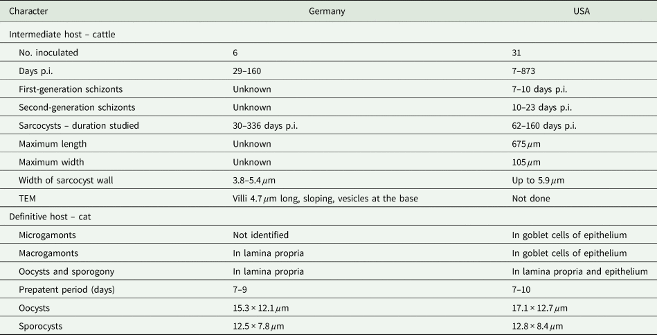
In their pioneering experiment 1, initiated in 1971, muscles from cattle oesophagi (number of cattle not stated) naturally infected with Sarcocystis were homogenized and fed to 8 cats (aged 3–12 months) over a period of 4 days (Heydorn and Rommel, Reference Heydorn and Rommel1972a). Cats excreted sporocysts after a prepatent period of 7–9 days; sporocysts were 10.8–13.9 × 6.9–9.3 μm (Heydorn and Rommel, Reference Heydorn and Rommel1972a).
In experiment 2, performed in 1972, development of Sarcocystis in cats was studied (Heydorn and Rommel, Reference Heydorn and Rommel1972b). For this, pieces of fresh beef (tissue not stated) from naturally infected cattle (how many cattle, not stated) were fed to 2-month-old cats. Inoculated cats were euthanized at 4, 6 and 8 h post-inoculation (p.i.) and on days 1, 2, 3, 4 and 5 p.i. No gametogonic stages were detected at 4 and 6 h p.i. At 8 h p.i., bradyzoites had entered intestinal mucosa, and young macrogamonts were found in the lamina propria of the small intestine. Macrogamonts were 6.1 × 4.8 μm on day 1, 10.2 × 8.3 μm on day 2 and 12.5 × 8.1 μm on day 3. On day 4, oocysts were recognized. On day 5, sporogony was initiated. Microgamonts were not identified (Heydorn and Rommel, Reference Heydorn and Rommel1972b).
Experiment 3 was performed in Berlin by Gestrich et al. (Reference Gestrich, Heydorn and Baysu1975a). Naming of the species S. bovifelis by Heydorn et al. (Reference Heydorn, Gestrich, Mehlhorn and Rommel1975) was primarily based on this experiment. This paper does not have an English summary and there are misinterpretations. Therefore, details are provided here.
Six calves aged 7–8-week-old were orally inoculated with 500 000 (2 calves) or 2 000 000 (4 calves) sporocysts; the calves were necropsied on days 29, 30, 48, 98, 160 and 194 p.i. The sporocysts, less than 6-month-old, had been obtained from feces of 8 cats that were fed with naturally infected cattle oesophagi muscles (presumably from experiment 1 of Heydorn and Rommel, Reference Heydorn and Rommel1972a, but not stated in the paper). One calf died on day 29 p.i. due to causes unrelated to Sarcocystis; no parasitic stages were detected (Gestrich et al., Reference Gestrich, Heydorn and Baysu1975a). The other 5 calves remained asymptomatic. No parasitic stages were detected at days 29, 30 or 42 p.i. Sarcocysts were observed in calves necropsied on days 98 and 160 p.i. but nothing is stated for the calf necropsied on day 198 p.i. Tissues from the calf necropsied on day 160 p.i. were fed to 3 cats, 1 dog, 4 rhesus monkeys and 1 human volunteer (Gestrich et al., Reference Gestrich, Heydorn and Baysu1975a; Heydorn et al., Reference Heydorn, Gestrich and Janitschke1976; Heydorn, Reference Heydorn1977). The cats excreted sporocysts and the sporocysts were 10.8–13.9 × 6.9–9.3 μm. Neither the dog, monkeys nor the human volunteer excreted sporocysts.
Morphologically, sarcocyst dimensions were not stated, except that sarcocyst wall was 3.8–5.4 μm thick in the calf necropsied on day 98 p.i. (Gestrich et al., Reference Gestrich, Heydorn and Baysu1975a). Sarcocysts from calves necropsied on days 98 and 160 p.i. were studied by transmission electron microscopy (TEM) (Gestrich et al., Reference Gestrich, Mehlhorn and Heydorn1975b). The villar protrusions (vp) on the sarcocyst wall were 3.8–5.4 μm long and 1.5 μm wide. The vp were sloping and contained vesicles at the base. The vp were circular at the base and spindle shaped at the tip. The vp had 200–300 microtubules (fibrils) running parallel to each other from the tip of the vp into the interior of the sarcocyst wall (Gestrich et al., Reference Gestrich, Mehlhorn and Heydorn1975b). The microtubules had a regular periodicity and the internal diameter was 15–18 nm. Metrocytes were 12–14 × 5–7 μm and bradyzoites were 13–17 μm long and 2.5–3.0 μm wide.
Experiment 4 was performed at the University of Munich, Germany by another group of researchers (Boch et al., Reference Boch, Laupheimer and Erber1978). A 30-day-old calf was gavaged with 300 000 S. bovifelis sporocysts (the source of sporocysts was not stated). Several muscle biopsies were taken from day 40 to 90 p.i., every 4–7 days, and from day 90 to 150 p.i., every 10th day; no mention is made concerning the size of biopsies or method of examination. Illustrations provided are from unfixed muscle squashes. Immature sarcocysts, with barely perceptible walls, were first found on day 50 p.i. On day 61 p.i., the sarcocyst wall had vp that increased in length and width; vp on day 110 p.i. were 6 × 0.7 μm and were 7.2 × 1.0 μm on day 140 p.i. The sarcocysts from day 50 p.i. were about 250 × 15 μm in size and contained only metrocytes. On day 90 p.i., the sarcocysts were about 1000 × 30 μm in size and contained bradyzoites. No sarcocysts were detected in trypsin-digested tongue, oesophagus, diaphragm and thigh muscles of the calf when it was necropsied on day 160 p.i. The identity of Sarcocystis species investigated is uncertain.
In 1980s, I studied the full life cycle of a Sarcocystis species transmissible from cats to cattle in Bozeman, Montana, USA; I had designated this species as S. hirsuta (Dubey, Reference Dubey1982, Reference Dubey1983b, Reference Dubey1983c), following the view prevailing at that time (Levine, Reference Levine1977) but now considered S. bovifelis. All life cycle work with this parasite was conducted using 1 isolate (designated the Bozeman isolate, discussed later) of Sarcocystis. For the life-cycle studies with the Bozeman isolate, I used calves and cats raised in captivity (Dubey, Reference Dubey1982, Reference Dubey1983a) to strictly monitor their diets.
As the nomenclature of bovine Sarcocystis remains controversial (Gjerde, Reference Gjerde2016a, Reference Gjerde2016b; Hu et al., Reference Hu, Huang, Wen, Esch, Liang and Li2017), I recently reviewed earlier experiments (including Dubey, Reference Dubey1982 and additional experiments a few years later). Fortunately, I was able to find records and histological slides from the studies performed in 1980s. Unfortunately, paraffin blocks from those studies do not remain. Considering the efforts, expense and the ethical concerns constraining experimental studies in cats, I here seek to summarize and correct errors in past representations of life-cycle descriptions, and deposit reference material (in the Smithsonian Museum) for the benefit of future researchers.
The objective of the present paper is to redescribe S. bovifelis and S. hirsuta, and not to discuss validity of the other named Sarcocystis species in cattle.
Materials and methods
Origin of the Bozeman isolate of S. bovifelis
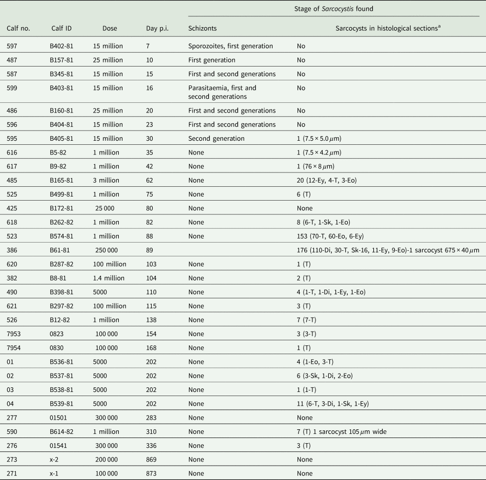
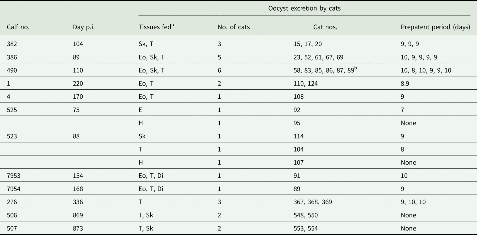
While studying congenital transmission of T. gondii, a 700 kg cow (no. 2 in Dubey, Reference Dubey1983b) was inoculated intra-ruminally with T. gondii oocysts at 130 days of pregnancy. This Holstein cow was born and bred at the Montana State University Agricultural Experiment Station. The cow was necropsied on 22 August 1980 on the day of parturition. Gluteal muscle (~250 g) of the cow was fed to a cat (no. 29) over a period of 7 days. The cat excreted T. gondii oocysts as well as Sarcocystis sporocysts. The cat was euthanized on 9 October 1980, 40 days after its last ingesting beef. Its small intestinal mucosa and submucosa were scraped from musculature, homogenized, centrifuged and suspended in antibiotic saline. Sporocysts from the intestinal scrapings from this cat were fed to a 7-day-old calf (calf no. 382) on the day of the euthanasia of the cat (Table 2). This was done to exclude any Toxoplasma oocysts from the inoculum infecting the calf (in the intestine, Toxoplasma oocysts are unsporulated and non-infectious, whereas Sarcocystis oocysts in the intestine are fully sporulated). Forty-one years later, I have recently re-examined histological sections of this cow, finding no sarcocysts in sections of its oesophagus, tongue, diaphragm or skeletal muscle. Calf no. 382 was euthanized on day 104 p.i. and its muscles were fed to 3 cats and sporocysts from these cats were used to infect other calves (Table 3). Subsequently, 2 other calves (calf nos. 386 and 425) were also fed sporocysts from cat no. 29 and subsequently cycled between calves and cats (Table 3).
Sarcocystosis in calves orally inoculated with S. bovifelis sporocysts from cats

a Di, diaphragm; Eo, esophagus; Ey, eye; Sk, skeletal muscle; T, tongue.
Sporocyst excretion in feces of cats fed with muscles of calves inoculated with S. bovifelis

a Eo, esophagus; H, heart; Sk, skeletal muscles; T, tongue.
b Histological sections from this cat used for description of sporogony.
In general, materials and methods resembled those previously described (Dubey, Reference Dubey1982, Reference Dubey1983b). Thirty-one calves were inoculated orally with 5000 to 100 million sporocysts, obtained from experimentally infected cats (Table 2). The calves were necropsied on days 7–873 p.i. (Table 2). For the determination of parasitaemia, 7 mL of blood was drawn from each inoculated calf between days 7 and 62 p.i., into vacuum tubes containing ethylenediaminetetraacetic acid twice weekly and on the day of necropsy. Smears of the entire buffy coat were air dried, fixed with methanol, stained with Giemsa's stain and examined at 1000× magnification (Dubey, Reference Dubey1982).
The calves were euthanized and necropsied immediately. Portions of various tissue were fixed in 10% buffered formalin: bone marrow, brain, spinal cord, eyes, pituitary, salivary and adrenal glands, thymus, lungs, heart, diaphragm, spleen, kidneys, liver, gallbladder, urinary bladder, omentum, rumen, reticulum, omasum, abomasum, small and large intestines, oesophagus, skeletal muscle, lymph nodes (superficial cervical, mandibular, retropharyngeal, mediastinal, hepatic, gastric, mesenteric, subiliac), cerebrum, cerebellum, pons, medulla and tongue. In 2 calves necropsied on days 7 and 10 p.i., sections of every 0.5 m of intestine were also examined. Paraffin-embedded sections were cut at 3–5 μm. Selected tissues (especially tongue and oesophagus) were also fixed in Bouin's fluid (BF) or Helly's fixative (HF). Selected tissues were embedded in glycol methacrylate and sectioned at 3 μm. Sections were stained with haematoxylin and eosin (HE), Heidenhain's iron haematoxylin (IH), periodic acid-Schiff's haematoxylin (PASH) or Giemsa's stain. Gluteal muscle and diaphragm were sectioned longitudinally.
The number of sarcocysts shown in Table 2 in each muscular tissue is based on an approximately 2 × 1 cm of gluteal muscle, 3 × 1 cm of tongue, 4 × 0.5 cm of diaphragm, half cross-section of oesophagus and 3 × 0.5 cm of ocular muscle.
For the study of gametogony, a 2-day-old kitten was inoculated orally by a stomach tube with 100 million bradyzoites freed from intramuscular sarcocysts by pepsin digestion (Dubey, Reference Dubey1983a). The calf had been fed with 100 million sporocysts of the Bozeman isolate, 103 days previously. The kitten was administered with bradyzoites in 2 doses at 2 h apart and necropsied 4 h later. The small intestine was cut into 20 segments and from each segment 2–4 small (2–5 mm wide) segments were fixed in a mixture of formaldehyde and glutaraldehyde. Tissues were embedded in glycol methacrylate, and 3 μm sections from each of 5 segments were examined after staining with HE, IH, PASH or Giemsa's stain (1982). All sections were examined at 1000× magnification.
For the study of sporogony, sections of the small intestine of a cat fed with muscles from calf no. 490 were examined microscopically after staining with HE. This calf had been inoculated with S. bovifelis sporocysts and euthanized on day 110 p.i. (Table 2). The cat was fed ad lib muscles over a period of 1 week and euthanized 12 days from the first ingestion of infected beef; the cat started excreting sporocysts 9 days later. The measurements of sporogonic stages are from intestinal homogenate of the cats and not from feces.
For TEM examination of merozoites, a paraffin block of myocardium of calf no. 599 on day 16 p.i. was deparaffinized, fixed in osmium tetroxide and processed using TEM. Ultrathin sections were examined using a JEOL electron microscope (JEOL, Inc., Peabody, MA, USA).
Recently, for the present study, I photographed life-cycle stages and measured them using a digital DP73 camera (Olympus Optical Ltd.) fitted on an Olympus AX 70 microscope (Olympus Optical Ltd., Tokyo, Japan).
Results
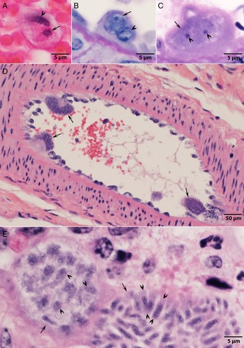
Information concerning the life-cycle stages of S. bovifelis (Bozeman isolate) is summarized in Tables 2–5 and briefly stated here. Two generations of schizonts were detected. First-generation schizonts were found on days 7–23 p.i. in arteries associated with mesenteric lymph nodes and intestines (Fig. 1). Schizonts matured between days 7 and 10 p.i. Intracellular sporozoites were observed in arterial blood (Fig. 1A) and in endothelial cells of the arteries; infected endothelial cells were bulging towards the arterial lumen (Fig. 1B–D). First-generation schizonts were 37.2 × 22.3 μm and contained more than 100 merozoites, which measured 5.1 × 1.2 μm (Fig. 1E).
Development of first-generation schizonts of Sarcocystis bovifelis in mesenteric arteries of calves. (A) A sporozoite (arrow) in a leucocyte in mid-lumen of a mesenteric artery of calf no. 597, day 7 p.i. The leucocyte nucleus (arrowhead) is out of focus. BF, HE. (B) Cross-section of an artery in mesentery of calf no. 597, day 7 p.i. Note an early schizont (arrow). The infected endothelial cell is protruding in the lumen. The endothelial cell nucleus (arrowhead) is slightly out of focus. BF, PASH. (C) A schizont (arrow) protruding in the lumen. Note nuclei (arrowheads) of the schizont. BF, IH. (D) Maturing schizonts protruding (arrows) in lumen of artery in mesentery of calf no. 487, day 10 p.i. HE. (E) Immature schizont (arrow on the left) with vesicular nuclei (arrowheads) and an adjacent mature schizont (arrow on the right) with few longitudinally cut merozoites (opposing arrowheads). BF, IH.

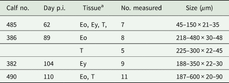
Measurements of live unstained S. bovifelis sarcocysts from fresh muscle squashes

a Eo, esophagus; Ey, eye; T, tongue.
Comparison of S. bovifelis studies of Heydorn et al. in Germany with that of the Bozeman isolate in the USA

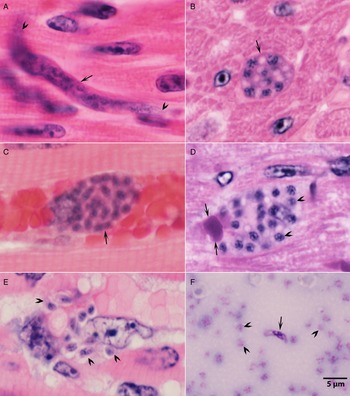
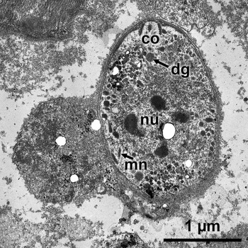
Second-generation schizonts were found in capillaries of striated muscles and heart on days 15–23 p.i. (Fig. 2A–E). Second-generation schizonts were 13.9 × 6.5 μm and contained up to 35 merozoites that measured 4.0 × 1.5 μm. Second-generation individual merozoites were observed within myocytes and in tissue macrophages in muscles on days 16–23 p.i. One of these merozoites in the myocardium of calf no. 599, day 16 p.i. is shown in Fig. 3.
Second-generation schizonts and a free merozoite in calf no. 599, day 16 p.i. Scale bar applies to all panels. (A) Immature narrow serpentine multinucleated immature schizont (arrow) in myocardium; arrowheads point to its ends. BF, HE. (B) Small immature schizont with 8 nuclei in the myocardium. BF, HE. (C) Multinucleated schizont (arrow) in an ocular capillary. HE. (D) Immature schizont with 22 nuclei (arrowheads); compare the size of the parasite nuclei with the size of an erythrocyte (double arrows of the capillary). HE. (E) Ruptured schizont with spilled merozoites (arrows). BF, HE. (F) An extracellular merozoite (arrow) in smear of buffy coat of blood from a jugular vein. Arrowheads point to thrombocytes. Methanol, Giemsa's stain.

Transmission electron micrograph of an intracellular S. bovifelis merozoite in the myocardium of calf no. 599, day 16 p.i. Note a conoid (co), micronemes (mn), dense granules (dg), a nucleus (nu) and absence of rhoptries.

Parasitaemia was transient and of low degree. Merozoites measuring 5.4 × 1.5 μm were found free in the peripheral blood of calf no. 599, day 16 (incorrectly stated as day 11 in Dubey, Reference Dubey1982) p.i. (Table 1) (Fig. 1F). The size of merozoites and the timing indicate that parasitaemia was due to first-generation merozoites. The occurrence of second-generation meronts only in muscles is unusual when compared to occurrences of the other species of Sarcocystis in domestic animals (Dubey et al., Reference Dubey, Calero-Bernal, Rosenthal, Speer and Fayer2016). The occurrence of merozoites in macrophages in muscles suggests that the second-generation merozoites are transported locally from capillaries to myofibres; this might explain why second-generation merozoites were not found in peripheral blood.
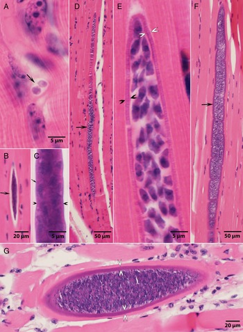
Sarcocysts formed between days 25 and 75 p.i. in striated muscles, but not in the heart. At days 30 and 35 p.i., a single sarcocyst from each calf was cut in cross-section. The sarcocyst at day 30 p.i. was 5 × 3 μm and contained 2 metrocytes (Fig. 4A). The identification of early sarcocysts was facilitated because of the presence of a parasitophorous vacuole, which is absent in schizogonic stages of Sarcocystis (Dubey et al., Reference Dubey, Calero-Bernal, Rosenthal, Speer and Fayer2016). At day 42 p.i., a single sarcocyst was observed; it was 76 × 8 μm with 35 metrocytes in 1 plane of section contained within a very thin sarcocyst wall (Fig. 4B and C). At day 62 p.i., several immature sarcocysts were observed (Table 2). One of these sarcocysts was cut longitudinally, measured 544 × 17 μm long and contained only metrocytes within a 3 μm thick sarcocyst wall (Fig. 4D and E). Bradyzoites developed between days 62 and 75 p.i. Live sarcocysts measured from 4 calves, on days 62–110 p.i., were up to 600 μm long (incorrectly stated as 800 μm long in Dubey, Reference Dubey1982, because of my miscalculation of the multiplication factor) (Table 3). Maximum thickness of the Sarcocystis wall was 5.5 μm (Fig. 5A and B). Only 2 sarcocysts were cut longitudinally; the longest sarcocyst was 675 μm (Fig. 4F). Maximum width of a mature sarcocyst was 105 μm. Metrocytes were globular to elongated (Fig. 4E). Bradyzoites in sections were 13 × 3 μm. In calves examined at 103 days p.i. with sporocysts, only a few sarcocysts were observed (Table 2). Only a few degenerating sarcocysts were observed, and they were not associated with eosinophilic myositis.
Sarcocysts of S. bovifelis. (A) Sarcocyst in gluteal muscle of calf no. 595, 30 days p.i. Arrow points to 2 metrocytes in a parasitophorous vacuole. BF, HE. (B, C) Longitudinal section of immature sarcocyst in gluteal muscle of calf no. 617, day 42 p.i. Note thin sarcocyst wall (opposing arrowheads). HF, HE. (D, E) Longitudinally cut immature sarcocyst (arrow) in tongue of calf no. 485, day 62 p.i. HF, HE. Note thickness (opposing arrowheads) of the sarcocyst wall. (F) Longitudinally cut immature sarcocyst (arrow) in gluteal muscle of calf no. 523, day 88 p.i. This is the longest sarcocyst observed in sections. BF. HE (G) Section of a mature sarcocyst in oesophagus of calf no. 523, 88 days p.i. Note thickness of the sarcocyst wall (opposing arrowheads). HF, HE.

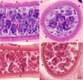
Comparison of mature sarcocysts of S. bovifelis from the study in the USA and in Germany. HE. Opposing arrows point to thickness of the sarcocyst wall. Note metrocytes (me) and bradyzoites (br). Scale bar applies to all panels. Longitudinal (A) and cross-section (B) of sarcocysts in calf no. 386, 89 days p.i. from the present study in the USA. Longitudinal (C) and cross-section (D) of sarcocysts in calf no. 12, day 98 p.i. from the study in Germany (Gestrich et al., Reference Gestrich, Heydorn and Baysu1975a).

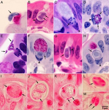
Gamonts were observed in parasitophorous vacuoles of goblet cells in the epithelium of the small intestine of the newborn kitten (Fig. 6). The host cell nucleus was indented, misoriented, but not hypertrophied (Fig. 6B–D). Numerous free bradyzoites were observed in contents of stomach, small intestine and colon (Fig. 6A). All stages of microgametogony, from the single-nucleated stage to fully developed gametes were observed (Fig. 6B–D). Microgamonts were 6.9 × 6.0 μm and contained up to 12 nuclei or microgametes around a prominent residual body. Microgametes were 3.5 × 0.5 μm (Fig. 6D and E). Macrogamonts were elongated (Fig. 6G) to circular (Fig. 6H) and measured 6.0 × 5.0 μm.
Sexual stages of S. bovifelis in sections of small intestine of cats. Scale bar applies to all panels. (A)–(H) are sections from a kitten orally inoculated with free bradyzoites; the epithelial brush border is oriented up towards the intestinal lumen. The host cell nucleus (arrowheads) is misoriented and indented. (I)–(M) are from the same HE-stained section of small intestine of a cat fed with S. bovifelis-infected meat. (A) Bradyzoite (arrow) in lumen, adjacent to a goblet cell. PASH. (B) A binucleate microgamont. Note vesicular nuclei (arrows). HE. (C) Immature microgamont in a goblet cell with 6 nuclei (arrow). (D) Mature microgamont with peripherally arranged microgametes (arrows) and a large residual body (rb). (E) A rupturing microgamont with 2 gametes (arrow) in lumen around a residual body (rb). Giemsa. (F) A macrogamont (arrow) in a goblet cell. (G) An elongated macrogamont (arrow) in a goblet cell. HE. (H) A near mature macrogamont (arrow) in a goblet cell. PASH. (I) An immature oocyst with 2 polar nuclei (arrows). (J) An oocyst with 2 sporocysts (arrows). HE. (K) An oocyst with 2 sporocysts. Arrows point to microgamete-like structures. (L) A sporulated sporocyst (arrow) with 4 sporozoites. (M) A sporocyst with 2 sporozoites (arrow) in focus.

Sarcocysts were infectious to cats at days 75 s p.i. Cats excreted sporocysts on days 7–10 after ingesting infected meat (Table 3). The 2 cats fed with heart of calves euthanized on days 75 and 88 p.i. did not excrete sporocysts, whereas those fed with other muscles from the same calf did (Table 3). Neither of 2 cats fed with muscles of cattle euthanized on 869 and 873 days p.i. excreted sporocysts. The absence of sporocysts in these cats was verified by digesting small intestinal scrapings in chlorox and by microscopic examination of the centrifuged homogenate.
Histologically, oocysts were observed in the lamina propria and in the villar epithelial tips of the small intestine of cats. Unsporulated oocysts with 2 unsporulated sporocysts, and fully sporulated sporocysts were observed in the lamina propria (Fig. 6I–M). In intestinal scrapings of cats, sporulated oocysts were 17.1 × 12.7 μm (16–18 × 11–14). They contained sporocysts measuring 12.8 × 8.4 μm (11–14 × 7–9), each with 4 elongate sporozoites. No Stieda body was present. The sporocystic residuum varied from being compact to being a few scattered granules. Living sporozoites were 8.2 × 1.9 μm (7.5–9 × 1.5–2.0), with anterior pointed ends. In sections of sporozoites, the nucleus was subterminal and there were several PAS-positive granules.
Sarcocystis bovifelis was only mildly pathogenic to calves (Dubey, Reference Dubey1983c).
Examination of specimens from the description of S. bovifelis (Heydorn et al., 1975)
Mehlhorn et al. (Reference Mehlhorn, Heydorn, Frenkel and Göbel1985) had submitted histological sections of sarcocysts of their newly named Sarcocystis species to several individuals/institutions, including Beltsville, Maryland. Among these was an HE-stained cross-section of oesophagus of a calf, day 98 after oral inoculation with 500 000 S. bovifelis sporocysts (calf no. 12 of Gestrich et al., Reference Gestrich, Heydorn and Baysu1975a). Numerous sarcocysts were present. Most sarcocysts were cut in cross-section. However, 2 sarcocysts were longitudinally cut; the longest sarcocyst was 570 × 42 μm and it contained bradyzoites. The maximum thickness of sarcocyst wall in any sarcocyst was 5.5 μm (Fig. 5C and D). These sarcocysts were morphologically like sarcocysts of the Bozeman isolate (Fig. 5A and B). Also included in this collection is a section of skeletal muscle of calf labelled Sarcocystis bovifelis on day 62 p.i.; however, I could not locate any sarcocyst in this section. Possibly, there was an error in the paper by Gestrich et al. (Reference Gestrich, Heydorn and Baysu1975a); they stated biopsy examination of calf muscle inoculated with sporocysts derived from feces of a human and dogs, but not cats.
A comparison of data obtained with the Bozeman isolate and the studies of Heydorn et al. (Reference Heydorn, Gestrich, Mehlhorn and Rommel1975) (Table 5) indicates a similar parasite was involved in both studies. However, it is uncertain if more than 1 species of Sarcocystis was present in the German study because the inoculum was initially derived from muscles of several cattle.
Taxonomic summary of Sarcocystis bovifelis Heydorn, Gestrich, Mehlhorn and Rommel, 1975; amend, present study
Diagnosis
Intermediate host: Cattle (Bos spp.) Sarcocysts in skeletal muscle but not in heart. First- and second-generation schizonts in blood vessels. Sarcocysts mature between 62 and 75 days p.i. Sarcocysts microscopic, <1 mm long and <110 μm wide. Sarcocyst wall <6 μm thick. The vp contain vesicles at the base and taper distally, designated as a new type 10g following the classification of Dubey et al. (Reference Dubey, Calero-Bernal, Rosenthal, Speer and Fayer2016).
Definitive host: Felids. Gamonts in epithelium, primarily in goblet cells of the small intestine. Sporogony in the lamina propria of the small intestine. Prepatent period 7–10 days. Sporulated sporocysts ~12–13 × 8–9 μm.
Specimens deposited
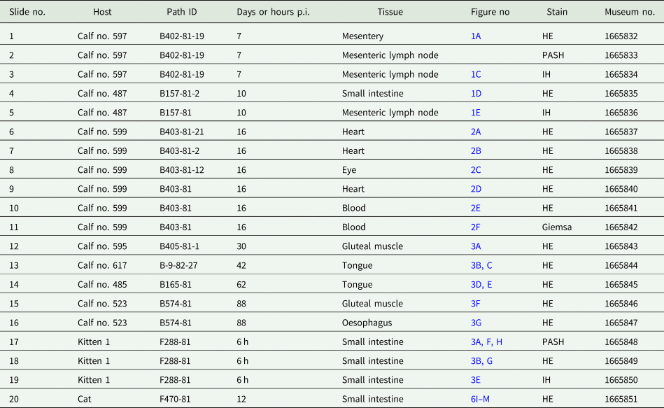
Bozeman isolate: The specimens were deposited in the United States National Parasite Collection in the Division of Invertebrate Zoology and National Museum of Natural History, Smithsonian Institution, Museum Support Center, MRC 534, 4210 Silver Hill Road, Suitland, Maryland 20746, USA, under numbers USNM nos. 1665832-166851 (Table 6).
Details of S. bovifelis (Bozeman isolate) specimens deposited in the Smithsonian Museum

Section from the study by Heydorn and associates: An HE-stained section of oesophagus from calf no. 12 of Gestrich et al. (Reference Gestrich, Heydorn and Baysu1975a) has also been deposited in the USNM no. 166852.
Molecular characteristic: DNA was not extracted from experimentally infected cattle or cats either the Bozeman or the German study and there are no archived samples available. However, Gjerde (Reference Gjerde2016b) molecularly characterized 45 S. bovifelis sarcocysts from naturally infected beef from Argentina and Germany using cox1, 18S rRNA, ITS1 and 28S rRNA genes. These sarcocysts were 1–3 mm long and up to 0.2 mm wide (Gjerde, Reference Gjerde2016a, Reference Gjerde2016b) and much bigger in size than those observed in experimentally infected cattle.
Redescription of S. hirsuta Moulé, 1888
The following account is based on the report of Dubey et al. (Reference Dubey, Udtujan, Cannon and Lindsay1990). In addition to the data reported in that paper, additional details are provided here. In 1988, I was contacted by Dr Udtujan of USDA's Food and Safety Inspection Service laboratory in Alameda, California concerning condemnation of cattle due to grossly visible sarcocysts at a meat packing plant in San Leandro, California, USA (Dubey et al., Reference Dubey, Udtujan, Cannon and Lindsay1990). Dr Udtujan reported that carcases of 18 cows (whole carcases or in parts) were condemned because of grossly visible sarcocysts (see Graphical abstract). The cows were 5–7-year-old Holstein and originated from Oregon, Nevada or California. The records do not indicate whether all condemnations occurred on a particular day or were identified over a period of weeks.
The index case was a cow condemned in 1988; the cow had been purchased from a livestock auction yard; no other information about the cow is available. Skeletal muscles from the cow were shipped cold (not frozen) by air and received at my laboratory in Beltsville on 1 September 1988. After photographing sarcocysts in situ, individual sarcocysts were dissected, separated easily from muscles. In total, 49 sarcocysts were isolated from muscles, 4 sarcocysts were ingested by a human volunteer (Dr Lindsay, coauthor of the report, who was aware of the potential zoonotic risks); no sporocysts were detected in his feces examined up to day 30 p.i. Isolated sarcocysts were fed to 2 littermate 17-day-old kittens (15 sarcocysts to kitten no. 543, and 30 sarcocysts to littermate kitten no. 544) that were nursed by their queen (cat no. 529). Additionally, cat no. 529 and a male cat no. 256 were fed ad lib 500 g of beef from the cow over a period of 3 days. All 4 cats were euthanized on day 12 after ingesting sarcocysts, and their intestinal scrapings were digested in chlorox as described (Dubey et al., Reference Dubey, Calero-Bernal, Rosenthal, Speer and Fayer2016); no sporocysts were found in the kitten fed with 15 sarcocysts. A few sporocysts were detected in the intestinal digest of the kitten fed with 30 sarcocysts; however, sporocysts from this kitten were not measured. Ten sporocysts from the intestine of the male cat were 15 × 10 μm. The sporocysts were measured at 400× magnification so it is only an estimate of size; this issue was not critical at the time of investigation. Lacking any control of their exposure history, we cannot know whether the 500 g of beef fed to these 2 adult cats was infected solely with S. hirsuta. No sarcocysts were observed in HE-stained histological sections of several pieces of this cow's muscles which lacked visible sarcocysts. Therefore, the size of sporocysts of S. hirsuta requires confirmation.
The sarcocysts in the cow were 2–7 mm long and approximately 0.5 mm wide. Three sarcocysts were fixed in glutaraldehyde and examined by TEM at the Armed Forces of Institute of Pathology, Washington, DC. The following description is based on these 3 sarcocysts.
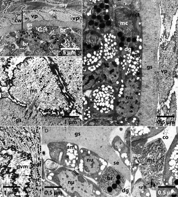
All 3 sarcocysts were structurally similar. The sarcocyst wall was not well demarcated from the myocytes (Fig. 7A); these images are new and were not published earlier. The total thickness of the sarcocyst wall (measured from the base of the vp to the area juxtaposed with myocytes) varied from 3.0 to 6.8 μm long within the same sarcocyst, depending on the folding of villi. The sarcocyst wall consisted of an outermost parasitophorous vacuolar membrane (pvm) that was lined by a 45 nm thick electron-dense layer that was folded into electron-dense blebs that were up to 160 nm long and up to 65 nm wide (Fig. 7B and C). The pvm was folded into vp. The vp had a narrow stalk attached to the ground substance (gs) layer (Fig. 7B). These vp were up to 8 μm long, constricted at the base, expanded laterally at the mid-region and tapered distally. However, not all vp were expanded laterally, and thus appeared upright. The villar core contained microfilamentous tubular structures, most of which were arranged haphazardly. The microfilaments had electron-dense granules (Fig. 7B and C). A relatively electron-lucent gs layer was present immediately beneath the vp (Fig. 7D and E). The gs immediately below the pvm contained few granules, with indistinct boundaries. These granules were absent or infrequent in the gs extending into the interior of the sarcocyst. The gs was approximately 2.2 μm thick, except at the origination of septa that was of variable thickness (Fig. 7D).
Transmission electron microscopic views of sarcocysts of Sarcocystis hirsuta in a naturally infected cow from the USA. am, amylopectin granules, br, bradyzoites, co, conoid, dg, dense granules, edg, electron-dense granules, edl, electron-dense layer, gs, ground substance layer, me, metrocytes, mc, micronemes, mp, micropore, mt, microtubules, mct, mitochondrion, my, myocyte, nu, nucleus, pvm, parasitophorous vacuolar membrane, rh, rhoptries, cw, sarcocyst wall, se, septa, vp, villar protrusions. (A) Low magnification of sarcocyst showing vp butted against my. Also note folded vp, relatively smooth gs and numerous br. (B, C) Sections of pedunculated vp, attached to gs by a narrow stalk (double arrows). The pvm is highly folded, is lined by edl and has blebs (triple arrows). The vp contains numerous criss-crossing mt and edg (arrowheads). (D) Sections of me (arrows) below gs and se. Note numerous am, and a large nucleus. Also note section of br with many micronemes and dg. (E) Longitudinal section of a br (double arrowheads) butted against the smooth gs. Note numerous mc, a group of dg, an elongated (mct), numerous am and a terminal nu. (F) Anterior (conoidal) end of a br. Note a co, numerous mc, a micropore (triple arrows) and 2 rh.

Only a few metrocytes were observed; they were observed in the gs, had relatively low electron density, compared with bradyzoites (Fig. 7D). They were oblong in shape and contained lipid droplets, amylopectin granules, no micronemes or rhoptries but had a large nucleus (Fig. 7D). They were difficult to measure but appeared 5–7 μm long in sections. Numerous bradyzoites were present and they appeared structurally intact. Bradyzoites were arranged in packets. It was rare to find bradyzoites cut longitudinally (Fig. 7E). The bradyzoites were like bradyzoites of other Sarcocystis species (Dubey et al., Reference Dubey, Calero-Bernal, Rosenthal, Speer and Fayer2016), and contained a conoid, hundreds of micronemes, a micropore but only 2 rhoptries (Fig. 6F). They were approximately 12 × 3 μm (Fig. 7E).
In general, structure of the sarcocyst wall from these samples from the USA was like 11 macroscopic sarcocysts (2–7 mm long) from New Zealand cattle that were sent to me for ultrastructural studies (Dubey et al., Reference Dubey, Speer and Fayer1989a, Reference Dubey, Speer and Charleston1989b). Böttner et al. (Reference Böttner, Charleston and Hopcroft1987a, Reference Böttner, Charleston, Pomroy and Rommel1987b) also made similar observations but their material was infected with other Sarcocystis species with thick sarcocyst wall, therefore not compared with observations of samples from the USA.
Concerning transmission experiments, Böttner et al. (Reference Böttner, Charleston and Hopcroft1987a, Reference Böttner, Charleston, Pomroy and Rommel1987b) fed isolated 554 (134 to the first, 230 to the second and 190 to the third) sarcocysts to 3 cats and 1 dog. Only 1 cat excreted few sporocysts, 11–17 days p.i.; sporocyst size was not stated. No sporocysts were observed in feces of a human volunteer who ingested 80 isolated sarcocysts, 200 g of raw beef and an unknown number of macroscopic sarcocysts from another 800 g of raw beef (Böttner et al. (Reference Böttner, Charleston and Hopcroft1987a, Reference Böttner, Charleston, Pomroy and Rommel1987b).
These transmission trials, in New Zealand and in the USA, indicate that the infectivity of macroscopic S. hirsuta sarcocysts to cats is very low and there is no evidence of the zoonotic nature of S. hirsuta.
Taxonomic summary of Sarcocystis hirsuta Moulé, 1888; amend, present study
Diagnosis
Sarcocysts: macroscopic, up to 7 mm long and up to 1 mm wide, appearing thick-walled by light microscopy. Ultrastructurally, the sarcocyst wall with type 28 of Dubey et al. (Reference Dubey, Calero-Bernal, Rosenthal, Speer and Fayer2016). Vp are up to 8 μm long, constricted at the base, expanded laterally in the mid-region and tapered distally. Some of the villar tips are folded to form 2–4 conical projections. The villar core contains microfilaments and electron-dense granules. The gs layer ~2 μm thick, relatively homogenous. Bradyzoites ~12 × 3 μm.
Intermediate hosts: cattle (Bos spp.).
Definitive hosts: cats (Felis catus).
Specimens deposited: There are no archived specimens (histologic sections, paraffin blocks or frozen tissue).
However, Gjerde (Reference Gjerde2016b) molecularly characterized 56 S. hirsuta sarcocysts from naturally infected beef from Argentina, New Zealand, Brazil and Germany using cox1, 18S rRNA, ITS1 and 28S rRNA genes. These sarcocysts were 3–8 mm long and up to 0.5 mm wide (Gjerde, Reference Gjerde2016b).
Discussion and remarks
In the present paper, I have reviewed morphology of life-cycle stages of S. bovifelis and S. hirsuta. All evidence indicates that S. bovifelis sarcocysts are microscopic. In experimentally infected calves observed for more than a year, sarcocysts remained <1 mm; macroscopic sarcocysts were never found in hundreds of calves used in Sarcocystis research in our laboratory (Dubey et al., Reference Dubey, Calero-Bernal, Rosenthal, Speer and Fayer2016). Boch et al. (Reference Boch, Laupheimer and Erber1978) stated that in a calf biopsied at 90 days p.i., with sporocysts from cat feces, the maximum sarcocyst size was 1000 × 30 μm. They also stated that cyst walls were up to 7.2 μm. However, the source of inoculum used to infect the calf is uncertain. With respect to thickness of the sarcocyst wall, it is important to measure structures in stained histological sections from fresh samples fixed in formalin or other fixatives. Freezing can distort morphology of the parasites. Use of wet mounts can also add to variation, because the villar characteristics can vary with the amount of pressure applied to squeeze samples; osmolarity of the medium (saline vs water) is also important. In histological sections, the S. bovifelis sarcocyst wall was never more than 6 μm thick.
The discussion in the present paper is confined to experimental infections of cattle with S. bovifelis. Gjerde (Reference Gjerde2016a, Reference Gjerde2016b) has reviewed molecular characterization in detail and proposed a new species, S. bovini, with sarcocysts measuring 1–3 mm × 0.1–0.2 mm in naturally infected cattle, morphologically indistinguishable from sarcocysts of S. bovifelis. However, based on cox1 Gjerde (Reference Gjerde2016b) distinguished S. bovifelis from S. hirsuta and S. bovini. Further studies are needed to describe life-cycle stages of Sarcocystis species in cattle with thick sarcocyst walls (S. bovini and S. rommeli).
After the ingestion of bradyzoites in sarcocysts by the definitive host, bradyzoites directly transform into gamonts without any asexual multiplication. Therefore, it is difficult to find early stages of transformation. To overcome some of these obstacles, a 2-day-old kitten was fed with 100 million free bradyzoites; these were concentrated from infected musculature by pepsin digestion and by reducing the volume to <0.5 mL. This ensured no spontaneous infection. Within 6 h, bradyzoites had converted to gamonts. Unlike other species of Sarcocystis studied (see Dubey et al., Reference Dubey, Calero-Bernal, Rosenthal, Speer and Fayer2016), S. bovifelis gamonts were predominantly in goblet cells. Staining with PASH facilitated identification of goblet cells, because the granules in goblet cells stain bright red. Heydorn and Rommel (Reference Heydorn and Rommel1972b) did not identify microgamonts. Additionally, they found macrogamonts only in the lamina propria and not in the epithelium.
The mechanism by which infected epithelial cells drop down to the lamina propria to complete the sexual development is unknown. The fertilization of macrogamonts by microgamonts must be very fleeting because fertilization has been observed by TEM in S. cruzi only twice among all coccidian parasites studied (see Fig. 1.67 in Dubey et al., Reference Dubey, Calero-Bernal, Rosenthal, Speer and Fayer2016). In the present study, microgamete-like structures were observed twice around oocysts of S. bovifelis (Fig. 5K); significance of these findings is unknown because the oocyst wall had already formed.
To study the development of S. bovifelis in the present study, calves were infected with as many as 100 million sporocysts. To minimize extraneous infections, newborn calves were bottle fed, kept indoors and euthanized on days 7–62 p.i. No thin-walled sarcocysts, resembling S. cruzi, were detected in these calves. Other calves, infected for more than 2 months and kept outdoors, did acquire a few S. cruzi-like sarcocysts, primarily in the heart and tongue. Sarcocystis bovifelis sarcocysts were not found in the heart. This site predilection is biologically and historically important. It explains why my 1969 attempts (with my supervisor Dr J. K. Frenkel) to transmit from cattle hearts, heavily infected with Sarcocystis, to cats were unsuccessful because we chose the wrong tissue to feed the cats (unpublished). It is now clear that the Sarcocystis species transmitted by cats do not parasitize the myocardium.
The number of S. bovifelis sarcocysts found after 3 months p.i. was inconsistent; only a few sarcocysts were detected despite examining multiple sections. Based on these findings, most sarcocysts were present in the tongue and oesophagus. Surprisingly, only 1 sarcocyst was observed in calf no. 620, despite feeding 100 million sporocysts. Overall, these findings suggest that the number of S. bovifelis sarcocysts decreases over time.
I hope this paper, and the reference material I have deposited, conclusively clarify distinctions between 2 species of Sarcocystis with thick sarcocyst walls that occur in cattle: S. bovifelis and S. hirsuta.
Data availability
none
Acknowledgements
I am indebted to Fernando Murata and Joe Mowery for help with illustrations. I thank Oliver Kwok for assistance with bibliography. I thank Dr Christian Bauer, emeritus Professor of Parasitology, Institute of Parasitology, Justus Liebig University Giessen, Germany for careful translation of the paper by Gestrich et al. (Reference Gestrich, Heydorn and Baysu1975a).
Author contributions
I (J. P. D.) conceived, wrote and revised the text.
Financial support
This research received no specific grant from any funding agency, commercial or not-for-profit sectors.
Conflict of interest
The author declares there are no conflicts of interests.