Implications
Newcastle disease (ND) is a communicable disease and destroys physiological function of birds. Fat supplies energy to the animal for growth and production. This study attempts to clarify the changes in chicken fat metabolism in the early stages of ND. There will be some benefit for research on fat metabolism or related pathologies.
Introduction
Newcastle disease (ND) is a fatal disease in poultry caused by the Newcastle disease virus (NDV), an enveloped RNA-containing virus of the paramyxovirus group. NDV matures at the host cell membrane surface by a budding process and has proved to be seriously harmful to the poultry breed industry (Alexander, Reference Alexander1995). Currently, ND in poultry remains a major problem because no effective treatment has been developed. In addition, some variations of NDV have been found to be more potent than others. Therefore, it is necessary that more research be conducted that focuses on ND.
Like other infectious illnesses induced by a bacteria or virus, infection with the NDV disrupts the normal functions of physiological systems and destroys the balance of metabolism (Butler, Reference Butler1975; Sijtsma et al., Reference Sijtsma, West, Rombout and Zijpp1989). Some researchers have shown that birds infected with the NDV represent a model for biochemical responses resulting from infection (Squibb et al., Reference Squibb, Beisel and Bostian1971; Oliveira et al., Reference Oliveira, Figueiredo-Lima, Faria Filho, Marques and Moraes2009; Sun et al., Reference Sun, Li, She, Wang, Han, Li, Ding and Yue2009). Experimental infection trials could answer many questions about the epidemiology of NDV, such as the pathogenicity and infectivity (Beard and Brugh, Reference Beard and Brugh1975; Carrasco et al., Reference Carrasco, Seki, Raso, Paulillo and Pinto2008).
Normal fat metabolism is an important basis for a balance of energy balance in the body. Disruption of this balance therefore threatens the healthy growth and production. It is known that infection with the NDV will break the balance of fat metabolism, but details of these changes are unclear, especially at the early stages of the infection (<72 h). The aim of this study is to clarify the effects of NDV infection in black-bone chicken fat metabolism.
Material and methods
Experimental design
A total of 100 male black-bone chickens (Gallus gallus) were housed in a clean room with constant lighting. A commercial diet and water was offered ad libitum. At 5 weeks of age, 60 healthy chickens were randomly divided into control group and infection group. The infection group was housed in an isolation welling after intraocular infection with 108 EID50 of the NDV. After all were injected, six chickens were killed at 0, 12, 24, 48 and 72 h, respectively. After each kill time, liver and abdominal fat pads were collected for RNA and protein extraction. The tissue samples were snap-frozen in liquid nitrogen as soon as they were obtained and stored at −80°C until analysis.
Blood lipids and enzyme activity
Birds were bled by cardiac puncture at 0, 12, 24, 48 and 72 h after infection before abattage. Some of the blood was used to match the hemagglutination inhibition (HI) antibodies, and some was collected to obtain plasma by centrifugation at 1500 × g for 15 min. The concentration of triglyceride (TG), total cholesterol (TC), high-density lipoprotein cholesterol (HDL-C) and low-density lipoprotein cholesterol (LDL-C) in the serum samples were analyzed with an automatic biochemical analyzer (CL7200 Shimadzu, Kyoto, Japan) using colorimetric methods. Finally, the activity of lipoprotein lipase (LPL), hepatic lipase and lipase in the serum was analyzed using respective kits that could be used in chicken (A054 and A067, Jiancheng, Nanjing, China).
HI assay for ND antibody titer
Microbeta HI tests were used to determine the HI antibodies in the serum, performing the method as some authors described (Yeo et al., Reference Yeo, Nagy and Krell2003; Kong et al., Reference Kong, Hu, Yin, Wu, Rui, Wang and Yang2006). Blood samples (2 ml per chicken) were collected and placed at 37°C for 2 h and then centrifuged at 1500 × g for 15 min for serum collection. The serum was serially diluted twofold in sterile phosphate-buffered saline (pH 7.2) on a 96-well plate. Dilutions ranged from 1 : 2 to 1 : 2048. 4 HA units of NDV were added into each well, except the last well, which served as a control. The mixture was incubated at 37°C for 30 min to prepare for the HI assay. The highest dilution of serum that causes complete inhibition was considered the end point. Titers of HI antibody were calculated from the geometric mean, which was expressed as a reciprocal log2 value.
RNA extraction and real-time PCR
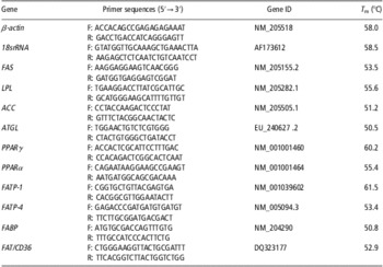
Total RNA was extracted from frozen abdominal fat pad and liver samples using the RNAiso Plus Kit (TaKaRa Biotech, Dalian, China) according to the manufacturer's instruction. Reverse transcription was performed for the first-strand cDNA synthesis by using the Primescript RT reagent kit (TaKaRa, Dalian, China). Real-time PCR was performed with an iQ5 system (BioRad, USA) using a 20-μl reaction mixture containing 12.5 μl of SYBR Premix Ex Taq™ II (Tiangen, Beijing, China), 1 μl of forward primer, 1 μl of reverse primer, 2 μl of template cDNA and 8.5 μl of ddH2O. Cycling conditions consisted of an initial, single cycle for 30 s at 95°C followed by 40 cycles of 5 s at 95°C and 35 s at T m (Table 1). The mRNA levels were calculated relative to that of β-actin and 18srRNA, and the normalized values were expressed as ratios, with the value at 0 h defined as 1.0.
PCR parameter of primers

FAS = fatty acid synthase; LPL = lipoprotein lipase; ACC = acetyl-CoA carboxylase; ATGL = adipose triglyceride lipase; PPAR = peroxisome proliferator-activated receptor; FATP = fatty acid transport protein; FABP = fatty acid-binding protein; FAT = fatty acid translocase.
Western blot analysis
The adipose tissue was homogenized and lysed with lysis buffer for 30 min at 4°C. The buffer contained 50 mM Tris (pH7.4), 150 mM NaCl, 1% NP-40 and 0.1% SDS (sodium dodecyl sulfate). After centrifugation at 12 000 × g for 10 min at 4°C, supernatants were removed and protein concentrations were determined using the bicinchoninic acid method with bovine serum albumin as the standard. Total protein extracts were separated by 12% SDS-polyacrylamide gel electrophoresis and transferred to polyvinylidene membrane (60 V for 4 h). The membranes were blocked with 5% non-fat milk in TBST (Tris-buffered saline containing 0.1% tween 20) at room temperature for 2 h, and probed overnight with anti-PPARγ (peroxisome proliferator-activated receptor gamma; 1 : 250, Santa Cruz Biotechnologies, Santa Cruz, CA, USA), anti-ATGL (adipose triglyceride lipase; 1 : 400, Santa Cruz Biotechnologies, Santa Cruz, CA, USA), anti-FAS (fatty acid synthase; 1 : 300, Santa Cruz Biotechnologies, Santa Cruz, CA, USA) and anti-β-actin (1 : 500, Santa Cruz Biotechnologies, Santa Cruz, CA, USA) primary antibodies at 4°C. After washing with TBST, the membranes were probed with horseradish peroxidase labeled secondary antibody (1 : 4000, Santa Cruz Biotechnologies, Santa Cruz, CA, USA) at room temperature for 1.5 h. Blots were visualized with chemiluminescence reagent (Millipore, Billerica, MA, USA) and an imaging system (BioRad, Hercules, CA, USA).
Statistical analyses
SPSS 13.0 statistics software package was used for one-way ANOVA and t-test. Data were presented as means ± s.e.m. Differences were considered statistically significantly at P < 0.05 or P < 0.01.
Results
Clinical changes
Some clinical symptoms post NDV infections were observed including febricity, drowsiness, coprorrhea, plumery tangle and decreased appetite. Figure 1 shows that the chickens’ cloacal temperatures rapidly increase and feed consumption gradually decrease after NDV infection.
The effect of Newcastle disease virus infection on cloacal temperature (a) and average feed intake (b) of chickens. n = 6, **P < 0.01, *P < 0.05 v. 0 h.

Changes of anti-NDV antibody titer in serum
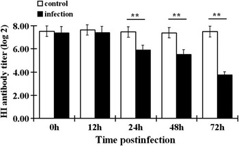
The changes of HI antibody titer in the serum are showed in Figure 2. At 0, 12 and 24 h, serum HI antibody titer levels did not conspicuously differ (P > 0.05) between the NDV-infected and control groups. However, HI antibody titers were markedly decreased at 48 and 72 h in the infected group (P < 0.05), whereas the control groups’ levels remained unchanged.
The effect of Newcastle disease virus (NDV) infection on the Hemagglutination inhibition antibody titers in serum. n = 6, **P < 0.01, *P < 0.05 v. 0 h.

Blood lipids and enzyme activity
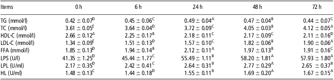
Table 2 shows decreasing plasma HDL-C concentrations over time, while the concentrations of TC and LDL-C increased. There was a decrease followed an increase in concentrations of TG and free fatty acid (FFA). In addition, NDV infections markedly increased the activity of LPL, hepatic lipase and lipase at every point in time (P < 0.01).
Effects of the NDV on the blood lipids and enzymatic activity of black-bone chicken

NDV = Newcastle disease virus; TG = triglyceride; TC = total cholesterol; HDL-C = high-density lipoprotein cholesterol; LDL = low-density lipoprotein cholesterol (LDL-C); FFA = free fatty acid; LPS = lipopolysaccharide; LPL = lipoprotein lipase; HL = hepatic lipase.
Data are expressed as the mean ± s.e.m.; n = 6.
In the same row, means with the different letters are different (P < 0.05%), means with same letters are not significantly different (P > 0.05%).
Gene expression and protein contents associated with fat metabolism
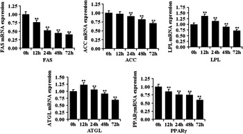
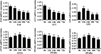
To clarify the changes in fat metabolism, we examined the mRNA expression of genes involved in fat metabolism in both adipose tissue and liver tissue. Over time, NDV infection markedly decreased mRNA expression of FAS (P < 0.01), acetyl-CoA carboxylase (ACC; P < 0.01) in both adipose tissue (Figure 3) and liver tissue (Figure 4). There was a rise in the mRNA expression of LPL (P < 0.01), peroxisome proliferator-activated receptor alpha (PPARα) (P < 0.01) and ATGL (P < 0.01) right after infection, but these expressions decreased with time. It was obvious that the initiation of changes was predominant after 24 h of NDV infection. These results suggested that the infection affected the balance between synthesis and oxidation of fatty acids (FAs), potentially down-regulating FAS and more FFAs were transported to various organs for oxidation.
The effect of the Newcastle disease virus infection on the expression of genes involved in lipid metabolism in the adipose tissue: fatty acid synthase (FAS), acetyl-CoA carboxyl's (ACC), lipoprotein lipase (LPL), adipose triglyceride lipase (ATGL) and peroxisome proliferators – activated receptor γ (PPARγ). The expression levels of mRNA were measured by RT-PCR analysis and normalized to β-actin and 18srRNA. Data are expressed as the mean ± s.e.m. n = 6, *P < 0.05, **P < 0.01 v. 0 h.

The effect of the Newcastle disease virus infection on the expression of genes involved in lipid metabolism in the liver, fatty acid synthase (FAS), acetyl-CoA carboxyl's (ACC), lipoprotein lipase (LPL), adipose triglyceride lipase (ATGL) and peroxisome proliferators – activated receptor α, γ (PPARα and PPARγ). The expression levels of mRNA were measured by RT-PCR analysis and normalized to β-actin and 18srRNA. Data are expressed as the mean ± s.e.m. n = 6, *P < 0.05, **P < 0.01 v. 0 h.

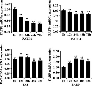
In recent years, fatty acid transporters (FATPs), fatty acid translocase (FAT) and fatty acid-binding protein (FABP) have been implicated by a variety of methods as functionally linked to FA transport. In our study, NDV infection markedly decreased mRNA expression for FATP1 (P < 0.01) and FATP4 (P < 0.01) but increased mRNA expression for FABP (P < 0.01) at all time points. At 12 and 24 h after NDV infection, the expression of FAT mRNA increased until 48 h. From 48 to 72 h, expression levels began to decrease (Figure 5).
The effect of the Newcastle disease virus infection on the expression of genes involved fatty acids transport: fatty acid transporter 1, 4 (FATP1 and FATP4), fatty acid translocase (FAT) and fatty acid bind protein (FABP). The expression levels of mRNA were measured by RT-PCR analysis and normalized to β-actin expression. Data are expressed as the mean ± s.e.m. n = 6, *P < 0.05, **P < 0.01 v. 0 h.

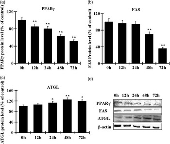
The protein levels of PPARγ, FAS and ATGL in adipose tissue were examined by Western blotting analysis and the results showed that the NDV infection caused a change in protein levels. The infection significantly decreased the protein levels of PPARγ after 12 h, decreased FAS level after 48 h and increased levels of ATGL from 24 to 72 h (Figure 6).
The effect of Newcastle disease virus infection on protein levels of peroxisome proliferators – activated receptor γ (PPARγ), fatty acid synthase (FAS) and adipose triglyceride lipase (ATGL). The protein level was measured by Western Blotting analysis and normalized to β-actin level. Quantification of PPARγ (a), FAS (b) and ATGL (c) levels through analysis the optical density of the blots (d) by using a Quantity One software. n = 3, *P < 0.05, **P < 0.01 v. 0 h.

On the basis of mRNA expression and protein levels, we concluded that synthesis of fat decreased and that the hydrolysis of lipids is strengthened during the early stage of the NDV infection. As time increases, the metabolism of fat was fully inhibited by the virus.
Discussion
ND is considered an acute and infectious disease, with plenty of previous research indicating effects of NDV on animal physiology (Brown et al., Reference Brown, King and Seal1999). This study attempted to specifically pinpoint changes in the fat metabolism of chickens infected with the NDV. Increase in the cloacal temperature and reduction in HI antibody titers in the serum showed that the artificial infection of NDV was effective. It has been shown from several animal experiments that the lipopolysaccharide (LPS) infection markedly and quickly changed the physiology of chicken. The NDV has not infected chickens as quickly as the LPS infection (which takes only several hours), but the effects of the virus were more prolonged and maladies long lasting (Riaz et al., Reference Riaz, Kenneth and Arthur1998).
Change in the concentration of plasma lipids is an important indicator of lipid metabolism. Different lipoproteins are involved in various kinds of lipid transport in different organs. FFA in the circulation appears to be correlated to stearolysis and indicates a change in lipid metabolism. We found that concentrations of HDL-C decreased, whereas concentrations of LDL-C and TC both had been increased. The concentrations of TG and FFA were elevated most at the beginning of the infection. These changes may be due to the following reasons: (1) more energy was needed to combat fever in chickens infected with NDV to maintain consumption; (2) diarrhea and decrease in feed consumption resulted in a decrease in the energy. Therefore, more fat in the body decomposed into FFAs for other organs to use. The detection of activity from several enzymes related to lipolysis supports this hypothesis. On the other hand, NDV infection did not change the chickens’ feed intake until 48 h. We concluded that the early changes of fat metabolism was just caused by the infection and not related to energy intake.
FAS and ACC are important enzymes in the lipogenesis process and fat accumulation to catalyze the synthesis of FAs (Smith et al., Reference Smith, Witkowski and Joshi2003; Tong, Reference Tong2005; Maier et al., Reference Maier, Leibundgut and Ban2008). LPL hydrolyzes lipids in lipoproteins, such as chylomicrons and very low-density lipoproteins, into FFAs and monoacylglycerols (Mead et al., Reference Mead, Irvine and Ramji2003). ATGL is a crucial lipase that catalyzes TG hydrolysis and has a key role in lipid droplet degradation (Raben and Baldassare, Reference Raben and Baldassare2005; Smirnova et al., Reference Smirnova, Goldberg, Makarova, Lin, Brown and Jackson2006). The PPARs are a group of nuclear receptor proteins that function as transcription factors regulating the expression of genes involved in energy homeostasis (Everent et al., Reference Everent, Galli and Grabb2000). PPARα is an important member of this protein family, and activated PPARα may facilitate stearolysis (Yoon, Reference Yoon2009). In contrast with PPARα, PPARγ is mainly responsible for adipocyte differentiation and lipid synthesis (Konig et al., Reference Konig, Koch, Spielmann, Christian, Frank, Gabriele and Klaus2009). The results showed that mRNA expression of FAS, ACC and PPARγ was decreased after NDV infection, but mRNA expression of LPL, ATGL and PPARα increased post infection (mainly at the beginning). These changes suggested that FA de novo synthesis was inhibited, whereas stearolysis was strengthened. Except for this, there was a tendency for all mRNA expression to decrease as the time went on. Moreover, the changes in protein levels of PPARγ, FAS and ATGL were basically consistent with the changes in mRNA expression. It has been found that changes in feed intake had significant effects on genes expression in the animal for a long time (Richards et al., Reference Richards, Poch, Coon, Rosebrough, Ashwell and McMurtry2003; Sun et al., Reference Sun, Richards, Rosebrough, Ashwell, McMurtry and Coon2006). However, NDV infection has more quick and severe changes on gene expression in our study.
Evidence from numerous sources has shown that the intestine, liver, heart, adipose tissue and other organs possess a saturable and completable FA transport system, which is controlled by several proteins FATP, FABP and FAT (Stahl et al., Reference Stahl, Gimeno, Tartaglia and Lodish2001; Pohl et al., Reference Pohl, Ring, Hermaann and Stremmel2004; Stahl, Reference Stahl2004). It was taken into consideration that FFA rapidly diffused through the phospholipids bilayer in high concentrations. However, the protein-mediated FA transport system produces a marked effect at low substrate concentrations (Chabowski et al., Reference Chabowski, Górsk and Bonen2006; Thompson et al., Reference Thompson, Lobo and Bernlohr2010). The specific role of each protein within this system is still unclear. It has been reported that in hamster LPS infection decreased expression of FAT in the liver and increased expression of FATP and FAT in heart and adipose tissue (Riaz et al., Reference Riaz, Kenneth and Arthur1998). In this study, NDV infection decreased expression of FATP1 and FATP4 and increased expression of FAT and FABP. This result indicted that these proteins played different roles in FFA transport.
In conclusion, the NDV infection destroys the dynamic balance of fat metabolism in chickens. At the start of infection, lipogenesis is inhibited, whereas lipolysis is strengthened. Eventually, fat metabolism becomes fully depressed.
Acknowledgments
This work was supported by a grant from The National Nature Science Foundation of China (30871785) and the Program for New Century Excellent Talents in Universities, Chinese Ministry of Education (NCET-06-0865).