Introduction
Paranasal sinus carcinomas represent 3–6 per cent of head and neck tumours, and have an incidence of approximately one in 100 000.Reference Dulguerov, Jacobsen, Allal, Lehmann and Calcaterra1, Reference Osguthorpe2 In our institution, they account for 3 per cent of malignant head and neck tumours.
In the majority of published series, carcinomas of the parasinusal sinus most frequently occur in the maxillary sinus.Reference Dulguerov, Jacobsen, Allal, Lehmann and Calcaterra1, Reference Myers, Nussenbaum, Bradford, Teknos, Esclamado and Wolf3–Reference Grau, Jakobsen, Harbo, Svane-Knudsen, Wedervang and Larsen7 In our centre, however, more than half are located in the ethmoid sinus.Reference Gras Cabrerizo, Orus Dotu, Montserrat Gili, Fabra Llopis, Leon Vintro and De Juan Beltran8
Paranasal sinus carcinoma tumours may be of various histological types, including squamous cell carcinoma, adenocarcinoma, cystic adenoid carcinoma and undifferentiated carcinoma. Adenocarcinomas very rarely occur outside the ethmoid sinus, although squamous cell carcinomas also occur frequently at this site.
No consensus has yet been reached concerning the optimal management of Paranasal sinus carcinoma tumours. This is partly due to the small number of patients in series published to date, the great variation in histological type, the lack of specificity in the recent tumour-node-metastasis (TNM) classification of malignant tumours, and the fact that the results of ethmoid carcinoma treatment are often reported jointly with those of carcinomas originating in the maxillary sinus or nasal fossa.Reference Sobin and Wittekind9, Reference Sobin and Wittekind10
The major treatment modality for ethmoid carcinoma is surgery with post-operative radiotherapy. Survival rates have improved in recent years, compared with those for maxillary sinus carcinoma, mainly as the result of new surgical techniques (such as craniofacial resection), improvements in radiotherapy treatment and early diagnosis.
This study aimed to present the results of our experience with the management of patients with carcinomas of the ethmoidal sinus complex.
Materials and methods
Patient and tumour characteristics
The data used in this study were obtained retrospectively from a database that prospectively collected information on clinical status, tumour extension, histological type, treatment and follow up for all patients with ethmoid sinus carcinoma.Reference León, Orús and Quer11
From 1984 to 2005, 52 patients with paranasal sinus carcinoma were diagnosed and treated in our centre. Thirty-four of these carcinomas (65 per cent) were located in the ethmoidal sinus complex, seventeen (33 per cent) in the maxillary sinus and one in the sphenoid sinus (2 per cent). This study includes only the 34 patients with ethmoid sinus carcinoma.
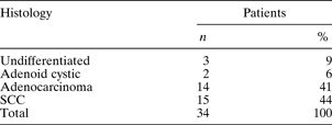
These 34 patients comprised 26 men (76 per cent) and eight women (24 per cent), with a median age of 64 years (range 27–86 years). Squamous cell carcinoma was involved in 44 per cent of patients (15/34), adenocarcinoma in 41 per cent (14/34), undifferentiated carcinoma in 9 per cent (3/34) and adenoid cystic carcinoma in 6 per cent (2/34) (see Table I).
Patient distribution by histological tumour type
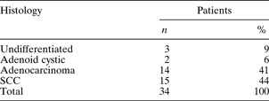
SCC = squamous cell carcinoma
A history of occupational wood dust exposure was recorded in eight patients (24 per cent), seven of whom had adenocarcinoma and one undifferentiated carcinoma.
Staging was assigned according to the sixth UICC-TNM edition.Reference Sobin and Wittekind10 Eleven patients were classified as T2, six as T3, six as T4a and 11 as T4b. Two patients (6 per cent) already had nodal metastases at the time of diagnosis.
All patients underwent pre-operative computed tomography (CT) and magnetic resonance imaging (MRI) to assess the tumour extent. The anterior skull base (cribriform plate and/or ethmoid roof) was involved in 17 patients (50 per cent). The tumour invaded the dura and/or the brain in eight patients (24 per cent). The anterior orbital contents were invaded in seven patients (21 per cent).
Treatment
Treatment decisions were made by a head and neck oncology panel comprising an otorhinolaryngologist, an oncologist and a radiotherapist, with the collaboration of a radiologist and/or pathologist in some cases.
Eight patients (24 per cent) received palliative treatment (seven classified as T4b and one as T4a).
The remaining patients (26/34) received treatment with radical intent.
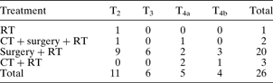
In these patients, treatment consisted of surgery in combination with radiotherapy in 85 per cent (22/26), chemotherapy and radiotherapy in 11 per cent (3/26) and radiotherapy alone in 4 per cent (1/26). All surgery consisted of a transfacial (n = 19) or endonasal (n = 3) approach, combined in some cases with a neurosurgical approach. This combined procedure had been performed in our department since 1993 in cases in which the tumour involved the cribriform plate, ethmoid roof or dura mater, as visualised on CT and MRI. The neurosurgical approach consisted of a bifrontal craniotomy via a bicoronal incision. Five patients required craniofacial resection. One patient required orbital exenteration for advanced disease. Two patients were treated with neck dissection for clinical nodes. In the absence of clinical and radiological evidence of cervical nodes, no prophylactic neck treatment was performed.
Table II shows the patient distribution according to tumour type and radical treatment received.
Patient distribution by tumour category and treatment*
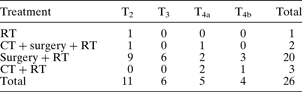
Data represent patient numbers.
* With radical intent. T = tumour; RT = radiotherapy; CT = chemotherapy
All patients were treated with external-beam radiotherapy using linear accelerators. Radiation was delivered using conventional fractionation with a continuous course of once-daily, five days per week treatment for all patients. The initial treatment volume was designed to encompass the primary tumour site with 1–2 cm margins, depending on the pathological findings and the proximity of critical structures. The median dose to the primary tumour was 60 Gy (range 50–64 Gy). The median dose to the primary tumour for patients undergoing post-operative and definitive radiotherapy was 54 and 64 Gy, respectively.
The most common chemotherapy induction regimen used was cisplatin and 5-fluorouracil.
Statistical analysis and follow up
Adjusted survival was analysed using the Kaplan-Meier method, and curves were compared using the Mantel-Haenszel test (log rank). Endpoints were calculated from the date of diagnosis until the event of interest, this being death for observed survival and local, regional or distant failure for disease-free survival. The minimum follow up was two years.
Results
Locoregional control
Nine of the 26 patients who underwent radical treatment (34 per cent) suffered a recurrence: four patients with squamous cell carcinoma, three with adenocarcinoma and two with undifferentiated carcinoma. Recurrence was local in eight patients and locoregional in one. The locoregional control rate was thus 66 per cent (17/26). As two of these nine patients were successfully rescued with surgical treatment, the final locoregional control rate was 73 per cent (19/26).
Local control rates were significantly worse in patients with dural or brain involvement.
In patients with tumour invasion of the anterior skull base but not the dura, the three-year local control rate was 55 per cent (five of nine). In those in whom the tumour affected the dura and/or the brain, the three-year local control rate was 12 per cent (one of eight).
Survival
The actuarial observed survival at five years for all ethmoid sinus carcinoma patients was 44 per cent (95 per cent confidence intervals (CI) 41–47 per cent). In the 26 patients who received radical treatment, observed survival at five years was 57 per cent (95 per cent CI 53–61 per cent). The eight patients treated with palliative measures had a median survival of 0.97 years.
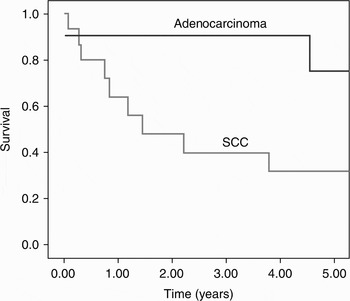
Analysis of the total population showed that patients with adenocarcinoma (14/34) had better five-year survival rates than those with squamous cell carcinoma (15/34): 72 versus 34 per cent, respectively (Figure 1). Similar results were observed in the patients undergoing treatment with radical intent, in whom the survival percentage was 78 per cent in those with adenocarcinoma, compared with 47 per cent in those with squamous cell carcinoma. Two patients with undifferentiated carcinoma and one with adenoid cystic carcinoma died within three years of diagnosis.
Kaplan-Meier representation of patient survival by histological tumour type. SCC = squamous cell carcinoma

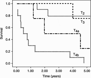
Survival rates at three and five years were 80 and 78 per cent, respectively, for T2 patients, 75 per cent for both rates for T3 patients, 50 and 26 per cent, respectively, for T4a patients, and 20 and 0 per cent, respectively, for T4b patients (Figure 2).
Kaplan-Meier representation of patient survival by tumour (T) category.

Major complications
After craniofacial surgery, there was one case of frontal osteomyelitis. There were no cases of blindness secondary to surgery or radiotherapy treatment, and there was no post-operative mortality.
Discussion
Ethmoid sinus carcinomas often follow a silent course and do not generally produce symptoms until they acquire large dimensions. Early symptoms such as rhinorrhoea, nasal obstruction or hyposmia simulate benign pathologies and are often mistakenly diagnosed and treated as an infectious or inflammatory condition, thereby delaying correct diagnosis. In our study, 68 per cent of patients were classified as T3 or T4a–b at diagnosis.
The main objective of the management of ethmoid sinus tumours is to obtain good local control of the disease, performing en bloc resection with healthy margins under frozen section control. This is often difficult due to the anatomical complexity of the region and the proximity of the skull base and orbital structures. For these reasons, most institutions employ combined treatments, such as surgery followed by radiotherapy. Surgery plus radiotherapy was used in 85 per cent (22/26) of our patients treated with radical intent; chemotherapy plus radiotherapy was used in 11 per cent (3/26), and radiotherapy alone was used in 4 per cent (1/26). Tumours restricted to the nasoethmoidal complex were treated with endonasal endoscopic sinus surgery. A transfacial approach was used when a tumour extended to the maxillary sinus, floor of the orbit, lachrymal sac or pterygoid plate. If the tumour involved the cribriform plate or the ethmoid roof, an external approach was combined with cranial resection. All patients received post-operative radiotherapy. Locoregional control was 66 per cent with this management.
A few institutions, however, reserve surgery for salvage of persistent or progressive disease, and treat these tumours with high-dose radiotherapy. They report similar results but also a high percentage of visual damage (i.e. blindness or impaired vision) in at least one eye.Reference Waldron, O'Sullivan, Warde, Gullane, Lui and Payne12, Reference Logue and Slevin13 Another disadvantage of this treatment combination is that ethmoid tumours frequently involve the skull base, where radioresistance is high.
The majority of institutions, nevertheless, consider that optimal curative treatment for ethmoidal sinus carcinoma should include surgery.
In patients with ethmoid sinus carcinoma, a poor prognosis is strongly correlated with advanced T category, lymph node involvement, absence of radiation therapy and involvement of the anterior skull base.Reference Bhattacharyya14, Reference Alvarez, Suarez, Rodrigo, Nunez and Caminero15
In our study, as expected, the higher the T category, the worse the prognosis. The five-year survival of patients in stages T2 and T3 (78 and 75 per cent, respectively) was clearly better than that of those in advanced stages (i.e. T4a and T4b; 26 and 0 per cent, respectively); these differences were statistically significant.
The latest edition of the sixth UICC-TNM classification of malignant tumours created the two advanced tumour subgroups T4a and T4b based on their resectability.Reference Sobin and Wittekind10 Although these subgroups may be interpreted differently depending on the surgeon and the centre, we consider them to be valid, as they distinguish between a patient group with a poor prognosis (i.e. T4a) and a group with a dismal prognosis (i.e. T4b). In our series, five-year survival was 26 per cent for patients with T4a tumours, compared with 0 per cent for those with T4b tumours. This clearly shows the difference in prognosis and management in these two subgroups.
Ethmoid sinus carcinoma frequently affects the anterior skull base. Of our patients, half had involvement of the cribriform plate or ethmoid roof, and 24 per cent had involvement of the dura or intracranial brain, according to CT, MRI or surgical findings.
Knegt et al. reported invasion of the anterior skull base in 40 per cent of patients with ethmoid sinus adenocarcinoma; Nuñez et al. reported the same in 38 per cent.Reference Knegt, Ah-See, vd Velden and Kerrebijn16, Reference Nuñez, Suarez, Alvarez, Losa, Barthe and Fresno17 Tumours which do not involve the cribriform plate or ethmoid roof can often be treated with a transfacial or endonasal approach alone, while lesions that do involve these sites require complementary treatment, fundamentally a craniofacial or an endoneurosurgical approach. The sixth edition of the UICC-TNM classification of malignant tumours categorised such lesions as T4b; in other words, they have a high risk of uncontrolled disease compared with T4a lesions, which have a reasonable chance of disease control.Reference Sobin and Wittekind10–Reference O'Sullivan and Shah18 However, several authors have reported a significantly worse prognosis when tumour extends beyond the dura to the brain, compared with tumour affecting the dura alone.Reference Patel, Singh, Polluri, Bridger, Cantu and Cheesman19
Based on these findings, therefore, tumours with dural involvement could perhaps be considered as T4a.
In our study, we observed that local control rates were significantly worse in patients with dural or brain involvement (12 per cent), compared with patients with tumour invading the anterior skull base without dura invasion (55 per cent). However, we were unable to investigate the differences between patients with and without dural or brain invasion in our series because of the small number of patients.
The histological type of ethmoid sinus carcinoma also affected survival. In the group treated with radical intent, patients with adenocarcinoma had better survival rates (78 per cent) than those with squamous cell carcinoma (47 per cent). Other authors have observed similar results. Cantu et al. reviewed 91 patients with malignant ethmoid sinus tumours and found that patients with adenocarcinoma had a better prognosis than patients with other histological types.Reference Cantu, Solero, Mariani, Salvatori, Mattavelli and Pizzi20 Uchida et al. reported poorer outcomes in patients with ethmoid squamous cell carcinoma or undifferentiated carcinoma, compared with patients with adenocarcinoma or adenoid cystic carcinoma.Reference Uchida, Shirato, Onimaru, Endou, Aoyama and Tsuchiya21
However, the influence of histological type is difficult to interpret, because of the small number of cases and the large number of influencing conditions. It is possible that our better results were due to the fact that, in early stage tumours (T2 to T3), the most frequent type was adenocarcinoma. As all patients classified as T4b and treated with radical intent had squamous cell carcinoma, adenocarcinoma had a more favourable tumour category distribution.
Regional metastasis to cervical nodes is less common in tumours of the paranasal region than in other head and neck carcinomas. The incidence of lymph node involvement at the time of ethmoid carcinoma diagnosis is very low. Previous studies have reported rates of clinical lymph node metastasis of 0–10 per cent.Reference Waldron, O'Sullivan, Warde, Gullane, Lui and Payne12, Reference Tiwari, Hardillo, Tobi, Mehta, Karim and Snow22–Reference Claus, Boterberg, Ost, Huys, Vermeersch and Braems24 In cases of paranasal tumour, there is no consensus on the treatment of the N0 neck. In our review, two patients (6 per cent) had lymph node invasion at the time of diagnosis (i.e. N+), while two further patients who were classified as N0 in the group treated with radical intent later developed lymph node metastasis (7 per cent). Because nodal failure in the absence of neck surgery or irradiation is infrequent, we do not recommend prophylactic neck treatment in N0 patients.
• In patients with ethmoid sinus carcinoma, combined surgery and post-operative radiotherapy provides good local control of disease
• In the present series, patients with adenocarcinoma had better survival than those with squamous cell carcinoma, after therapy with radical intent
• The cribriform plate or the ethmoid roof were involved in half the patients. Dural invasion was associated with poor local control
• Prophylactic neck treatment is not recommended in ethmoid sinus carcinoma patients unless there is cervical node involvement
In a meta-analysis published in 2001, Dulguerov et al. found specific actuarial five-year survival rates of 51 per cent in patients with ethmoid sinus carcinoma.Reference Dulguerov, Jacobsen, Allal, Lehmann and Calcaterra1 Cantu et al. reported a five-year survival rate of 47 per cent in 91 patients, while Salvan et al. reported a rate of 39 per cent in 31 patients.Reference Cantu, Solero, Mariani, Salvatori, Mattavelli and Pizzi20, Reference Salvan, Julieron, Marandas, Janot, Leridant and Domenge25 Waldron et al. reported observed five-year survival rates of 58 per cent in 29 patients with carcinoma of the ethmoid sinus treated with radiotherapy and salvage surgery.Reference Waldron, O'Sullivan, Warde, Gullane, Lui and Payne12 Bhattacharyya analysed 180 patients with ethmoid sinus cancer and found a five-year survival rate of 40 per cent.Reference Bhattacharyya14 We found similar results, observing a five-year survival rate of 44 per cent for the total group of ethmoid sinus carcinoma patients diagnosed in our centre, and 57 per cent in the 26 patients receiving radical therapy.
Stoll et al. and Jiang et al. reported better five-year survival results, of 80 per cent in a series of 76 adenocarcinomas and 63 per cent in 34 ethmoid sinus carcinomas, respectively.Reference Stoll, Bebear, Truilhe, Darrouzet and David23, Reference Jiang, Morrison, Garden, Geara, Callender and Goepfert26 These positive results could perhaps be explained by the fact that the majority of tumours were T3; no patients were classified as T4 in Jiang and colleagues' study.
Conclusions
In patients with ethmoid sinus carcinoma, a combination of surgery and post-operative radiotherapy enables good local control of the disease. In the present series, patients with adenocarcinoma had better survival rates than those with squamous cell carcinoma, after radical therapy. The cribriform plate or the ethmoid roof was involved in half the patients. Dura mater invasion was associated with a poor local control rate.
In view of these findings in patients with ethmoid sinus carcinoma, we do not recommend prophylactic neck treatment unless there is cervical node involvement.