Introduction
Amid the novel coronavirus (SARS-CoV-2) pandemic wreaking havoc on healthcare facilities worldwide, all hospitals are on high alert for the disease. All patients are mandatorily screened, and most are being tested. This is essential for the efficient delivery of patient care and the safety of healthcare workers. Nevertheless, an unprecedented situation like the present often makes patients have tunnel vision whereby anyone with symptoms similar to SARS-CoV-2 is presumptively assumed to have the diagnosis.
Clinical presentation
A 42-year-old gentleman was referred from the primary care physician with complaints of dyspnoea. He was seen in the fever clinic from where nasopharyngeal and oropharyngeal swabs were collected, and the patient was admitted in isolation for monitoring and supportive care. When his swabs were repeatedly negative, it indicated a possibly different aetiology. On proper history taking, he had progressively worsening dyspnoea and low back pain. He had no comorbidities and no significant past medical history. He was an ex-smoker. Clinical examination revealed a decreased percussion note and diminished breath sounds in the left hemithorax. There was diffuse bony tenderness in the lumbar spine. Complete blood count, coagulation screen, liver and renal chemistries were normal. A cardiac evaluation was within normal limits with good ventricular function on echocardiography.
Investigations
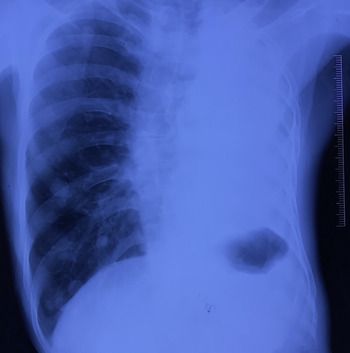
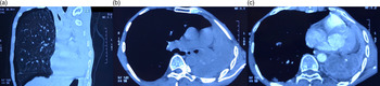
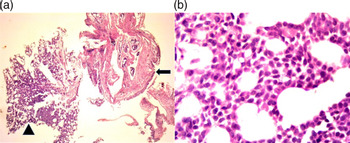
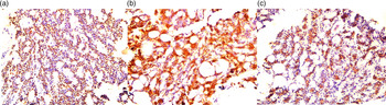
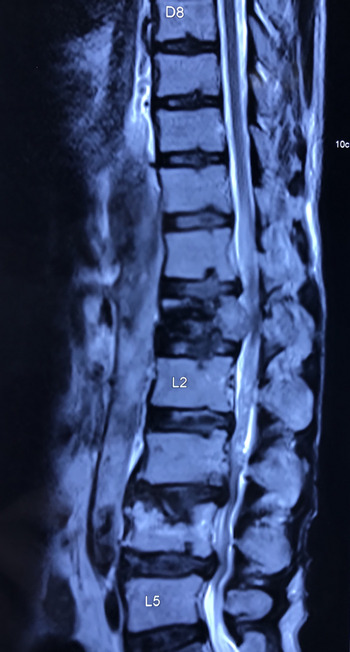
A chest radiograph (Figure 1) showed a ‘bronchial cut-off sign’, with a collapsed left lung and ipsilateral mediastinal shift. Contrast-enhanced CT (CECT) scan of the thorax (Figure 2a–c) revealed a left hilar mass lesion showing foci of calcification, with a resultant complete collapse of the left lung and ipsilateral tracheal and mediastinal shift. There was compensatory hyperinflation of the contralateral lung, and evidence of contralateral lung metastases on chest X-ray and CECT. Bronchoscopy revealed a polypoidal growth distal to the carina completely obscuring the left main bronchus. Biopsy of the lesion composed partly of a tissue lined by respiratory epithelium with tumour tissue disposed of in a cribriform pattern, composed of monotonous basaloid cells (Figure 3a and b). CK7 and CD117 were positive in luminal cells on immunohistochemistry (IHC), with p63 positive in abluminal cells (Figure 4a–c). Magnetic resonance imaging (MRI) of the spine showed a collapsed L1 vertebra and a partially collapsed L4 vertebra (Figure 5). The histopathology and IHC findings in conjunction with the imaging pointed to a diagnosis of adenoid cystic carcinoma (ACC) of the left lung with metastases to the contralateral lung and lumbar vertebrae. While the gentleman and his caregivers were relieved that he did not have the novel coronavirus, his actual prognosis was far worse.
Chest radiograph showing a collapsed left lung and ipsilateral mediastinal shift, the ‘bronchial cut-off sign’. There are soft-tissue densities suggestive of metastatic lesions in the right mid-lower zone.

CECT scan of the thorax reveals a left hilar mass lesion showing foci of calcification, with a resultant complete collapse of the left lung and ipsilateral tracheal and mediastinal shift (b and c). There is compensatory hyperinflation of the contralateral lung with nodular lesions suggestive of metastases (a).

(a) Section shows a fragmented biopsy composed partly of a tissue lined by respiratory epithelium (arrow). Tumour tissue is seen beneath the epithelium (arrowhead). (H&E 40×). (b) Section shows tumour tissue disposed of in a cribriform pattern composed of monotonous basaloid cells. (H&E 400×)

p63 positive in abluminal cells (a). CK7 positive in luminal cells (b). CD117 positive in luminal cells (c). (200×).

MRI of the spine shows a collapsed L1 vertebra and a partially collapsed L4 vertebra.

Treatment
The patient received radiotherapy to the spine to alleviate pain and was put on palliative chemotherapy.
Discussion
ACC is a rare malignancy of exocrine glands. The minor salivary glands are its most common location. They may also arise in other sites of the head and neck region, aerodigestive tract, breast, lungs, prostate and the female genitourinary tract. It usually occurs in the fifth or sixth decade of life and has a slight female preponderance.Reference Dillon, Chakraborty, Moskaluk, Joshi and Thomas1 Primary ACC of the lung constitutes only 0.1–0.2 % of all lung cancers. In the respiratory tract, they tend to occur centrally, in the trachea or central bronchi. They extensively invade inside and beyond the bronchial wall.Reference Qing, Zhou, Liu, Li, Deng and Ma2 Lung ACC manifests as a slow-growing mass, and clinical features are due to the mass lesion itself. Paraesthesia or pain occurs due to perineural invasion.Reference Spiro and Huvos3 ACCs have a low propensity for lymph nodal spread. Metastatic disease may occur in 40% of cases, with the liver, breast, bone or the brain the usual sites for distant spread.Reference Dillon, Chakraborty, Moskaluk, Joshi and Thomas1 Histopathology reveals tumour cells with angulated hyperchromatic nuclei, with minimal eosinophilic or clear cytoplasm. The original term ‘cylindroma’ is a reflection of cylindrical secretory cells with a hyaline stroma. Three growth patterns seen on histopathology are cribriform, tubular and solid.Reference Dillon, Chakraborty, Moskaluk, Joshi and Thomas1,Reference Azumi and Battifora4 IHC aids the histopathological diagnosis.Reference Qing, Zhou, Liu, Li, Deng and Ma2 Primary management is surgical. Definitive radiotherapy can be considered if surgery is not feasible. Post-operative radiotherapy may decrease local recurrences.Reference Spiro and Huvos3,Reference Balamucki, Amdur and Werning5 Metastatic disease shows low response rates to chemotherapy. There is no standard chemotherapy regimen, but, cisplatin has the strongest rationale.Reference Cerda, Sun and Vignot6 Targeted therapy specific to ACC requires further investigation, and drugs inhibiting the epidermal growth factor receptor (EGFR) pathway may have a role.Reference Mendes, Barroso and Campainha7
Funding
The authors have no funding to declare.
Conflicts of interest
The authors have no conflicts of interest to declare.
Patient consent
Patient consent has been obtained from the patient himself.