Case report
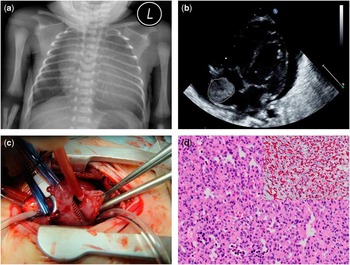
A couple of twins were delivered by caesarean section at the 34th week of gestation, because one of the foetuses was found to have massive pericardial effusion and a tumour occupying inside the right atrium by foetal echocardiography. Their 35-year-old mother had a smooth pregnancy course, and results of all other prenatal examinations, including karyotyping, were normal. The male infant with cardiac tumour weighed 1875 g, and Apgar scores were 9 and 10 at 1 and 5 minutes, respectively. On physical examination, he had subcostal retraction during inspiration, indicating that respiratory distress existed. Chest X-ray showed significant cardiomegaly (Fig 1a). Neonatal echocardiography disclosed massive pericardial effusion with right atrium diastolic collapse and one echogenic mass in the right atrium with widely based adhesion at the free wall (Fig 1b). The foramen ovale and ductus arteriosus were patent. Cardiac biventricular function was conserved.
( a ) Chest X-ray showed significant cardiomegaly. ( b ) Neonatal echocardiography revealed one echogenic mass inside the right atrium with abundant pericardial effusion. ( c ) The tumour was exposed after cardiopulmonary bypass establishment and right atriotomy. ( d ) Histopathological examination showed packed small capillary vessels (×200), and positive CD-34 immunohistochemical stain (Inset, ×200).

Although the infant did not display progression of cardiopulmonary distress, surgery was carried out on the next day after birth to determine the nature of the cardiac mass and to drain the massive pericardial effusion. Through a median sternotomy, the heart was exposed and 50 ml straw-coloured pericardial effusion was drained. A mass was seen from the outside right atrium near the inferior vena cava. After the ductus arteriosus ligated and cardiopulmonary bypass established, the right atrium was exposed and the tumour was totally removed together with the adjacent atrium free wall excised, under heart beating status (Fig 1c). The right atrium was reconstructed by equine pericardial patch and the foramen ovale was closed directly. The patient had an uneventful postoperative course. Histopathologically, the tumour was composed of closely packed, small capillaries with little luminal canalisation. Immunohistochemical staining revealed that the endothelial cells were positive for the CD-34 marker (Fig 1d). No evidence of malignancy was detected. Diagnosis of capillary haemangioma was established.
Discussion
Cardiac tumour is rare in infants with a prevalence about 0.05–0.14%, of which only 1–2% comprises vascular tumours.Reference Issacs 1 , Reference Mackie, Kozakewich, Geva, Perez-Atayde and Mulliken 2 Following the successful surgical treatment of infantile cardiac haemangioendothelioma by Ilbawi et al,Reference Ilbawi, DeLeon, Riggs and Hernandez 3 only 37 cases of haemangioma, haemangioendothelioma, or angiosarcoma in the heart of infants have been reported. Supplementary Table S1 summarises the patient characteristics, clinical presentation, surgical procedure, and outcome of these cases.
Echocardiography, either prenatal or postnatal, usually establishes the diagnosis of the cardiac tumours accurately.Reference Issacs 1 The typical echographic feature of a cardiac vascular tumour is mixed echogenicity with echogenic and hypoechogenic parts. Blood flow signal is absent by colour flow mapping in most patients, as the abnormal vascular structures are microscopic; nevertheless, the main feeding artery could be demonstrated occasionally.Reference Mackie, Kozakewich, Geva, Perez-Atayde and Mulliken 2 Unlike the more common rhombdomyoma or fibroma, cardiac vascular tumour is secretory in nature, and pericardial effusion is frequently presented.Reference Issacs 1 , Reference Sharma, Hirata and Mosca 4 In the reported 37 cases, 27 of them (73.0%) presented with pericardial effusion. Cardiac tumour or pericardial effusion was detected by foetal echocardiography in 18 patients in this review (48.6%), of which 14 (77.8%) were found to have pericardial effusion prenatally; in utero pericardiocentesis was performed on one patient at 20th week of gestation owing to severe hydrops.Reference Sharma, Hirata and Mosca 4 Congenital thyroid dysfunction, diaphragmatic hernia/eventration, and infection are other common causes of foetal pericardial effusion in the absence of hydrops.Reference Cartagena, Levin, Issenberg and Goldman 5 The presence of cardiac mass together with pericardial effusion should raise the suspicion of the diagnosis of vascular tumour. Teratoma is the other tumour frequently related to pericardial effusion and most of them originate from the pericardium, whereas cardiac vascular tumours usually arise from the heart itself.Reference Issacs 1 , Reference Cartagena, Levin, Issenberg and Goldman 5
In this review, six infants (16.2%) with cardiac vascular tumour were asymptomatic and the diagnoses were made incidentally by prenatal or postnatal echocardiography. Cardiac tamponade and cardiopulmonary distress were the usual presentations in symptomatic patients. The tumour could occur at any place of the heart, with the majority at the right atrium (27 of 37, 73.0%). Of the patients, three (8.1%) had tumour associated with cutaneous haemangiomas, and one (2.7%) had multiple visceral vascular malformations, resulting in fatal pulmonary haemorrhage. Foetal arrhythmia or cardiopulmonary compromise had occasionally developed, which resulted in preterm delivery in four foetuses (10.8%). In the presented case, we delivered the boy twins at 34th gestation week soon after the diagnosis, even without significant foetal cardiopulmonary compromise, and performed prompt surgical intervention in his very early life, concerning that cardiac tamponade might develop and the risk of respiratory distress syndrome or other neonatal morbidities was already low at this gestational age.
Surgical resection remains the choice of therapy for infants diagnosed with cardiac vascular tumour, especially for those with symptoms. Of the reviewed cases, 28 patients (75.7%) were treated with surgical resection, and complete excision was achieved in 16 patients (43.2%). No tumour recurrence was reported after complete excision. The close relationship of the tumour with the conduction system and coronary arteries might preclude the complete excision; however, tumour regression or even complete resolution might be anticipated after incomplete excision or conservative treatment.Reference Mackie, Kozakewich, Geva, Perez-Atayde and Mulliken 2 , Reference Minsart, Ridremont and Lecocq 6 Even for those more aggressive haemangioendotheliomas and malignant angiosarcomas of the heart, scarce literature also showed satisfied surgical outcome.Reference Beaton, Kuttler and Hassan 7 , Reference Rosenkranz and Murphy 8 In those with cardiac tamponade but unresectable tumours, drainage alone with or without tumour biopsy is advised. Despite with unknown mechanism, systemic steroid therapy was applied in the management of infantile cardiac vascular tumours in several reports and carried favourable outcomes.Reference Mackie, Kozakewich, Geva, Perez-Atayde and Mulliken 2 , Reference Dresse, David and Hume 9 , Reference Chang, Young, Chuu and Lue 10 Its use, however, needs further investigation and should be reserved for those who are not candidates for surgical resection or with significant residual tumour after incomplete excision.
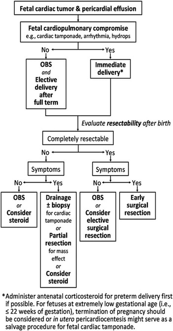
In this article, we report one small premature patient with cardiac haemangioma treated by complete excision in the neonatal period and give a review of infantile cardiac vascular tumour. Infantile cardiac vascular tumours are most commonly located in the right atrium and are frequently associated with pericardial effusion. The diagnosis can often be made by foetal echocardiography and the foetus should be delivered promptly once foetal cardiopulmonary compromise develops. Surgical removal of the tumour provides adequate result and is advised in most patients, even for those premature neonates such as the presented case. For foetuses with cardiac tumour and pericardial effusion, one algorithm regarding the decision making from prenatal diagnosis and timing of delivery to postnatal management is proposed (Fig 2).
Algorithm of decision making for foetus prenatally diagnosed with cardiac tumour and pericardial effusion. OBS=observation.

Acknowledgement
None.
Financial Support
This research received no specific grant from any funding agency, commercial, or not-for-profit sectors.
Conflicts of Interest
None.
Supplementary material
To view supplementary material for this article, please visit http://dx.doi.org/10.1017/S1047951114002121