I. INTRODUCTION
Thermoelectric materials can produce electrical power via the Seebeck effect and provide cooling capability via the Peltier effect without using coolants. Therefore, they can be considered as energy and environmentally friendly materials. The figure of merit (Z) of thermoelectric materials is determined by three transport parameters and can be expressed as Z = σS 2/κ, where σ, S, and κ are the electrical conductivity, thermopower, and thermal conductivity, respectively. Bismuth alloys show the highest dimensionless figure of merit ZT (where T is the absolute temperature) around room temperature and are one of the most commercialized thermoelectric materials. The crystal lattice of (Bi,Sb)2(Te,Se)3 has the symmetry of  . The layers of atoms A(Bi,Sb) and B(Te,Se) are arranged in the order of -B(1)-A-B(2)-A-B(1)- along the c axis.
Turning the currently best thermoelectric materials into nanoscale grain boundaries or nanostructure helps improve thermoelectric figure of merit by increasing the thermoelectric power (TEP) as a result of an enhanced density of states and by reducing thermal conductivity as a result of an increased phonon scattering.Reference Hicks and Dresselhaus1, Reference Hicks and Dresselhaus2 Recently, the theoretical concept has been fulfilled by a significant enhancement of ZT value for nanostructured bulk of Bi2-xSbxTe3.Reference Poudel, Hao, Ma, Lan, Minnich, Yu, Yan, Wang, Muto, Vashaee, Chen, Liu, Dresselhaus, Chen and Ren3, Reference Xie, Tang, Yan, Zhang and Tritt4
Several groups reported bottom-up chemical synthesis of Bi2Te3 nanocrystals at low temperatures of 140–150 °C using either organic solvent or water as reacting medium.Reference Zhao, Ji, Zhang, Zhu, Tu and Zhang5–Reference Liu, Liu, Tsao, Lu and Chang10 It is well known that the carrier concentration needs to be optimized for thermoelectric materials to achieve the best electronic transport properties due to its opposite trends with respect to electrical conductivity and thermopower. There exist complex lattice defects in Bi2-xSbxTe3. The ionized defects could change the carrier concentration and hence the electronic transport properties.Reference Schultz, McHugh and Tiller11 Furthermore, lattice defects are known to be sensitive to sintering or annealing temperatureReference Zhao, Zhang, Liu, Zhang and Li12 and play an important role to obtain good thermoelectric properties of Bi2-xSbxTe3. Although bottom-up chemical synthesis of Bi2-xSbxTe3 nanocrystals is feasible at low temperatures, it is, therefore, desirable to optimize the thermoelectric properties by finding suitable sintering conditions while consolidating Bi2-xSbxTe3 nanocrystals. In this study, the process parameter of sintering temperature is investigated to optimize the thermoelectric characteristics of pressed compacts of hydrothermally synthesized Bi0.5Sb1.5Te3 nanoplatelets. As a result, we find that the sample sintered at 340 °C has the largest power factor and its thermal conductivity is rather small having the size of 0.37 W/m·K at 295 K, which is quite small as compared to the typical value of the corresponding material. The dimensionless figure of merit ZT is 0.93 at 297 K.
II. MATERIALS AND METHODS
Nanoplatelets of Bi0.5Sb1.5Te3 were synthesized using a hydrothermal method. Te powders (6 mmol), BiCl3 (1 mmol), SbCl3 (3 mmol), ethylenediaminetetraacetic acid (8 mmol), and deionized water (70 ml) were quantitatively added into a polytetrafluoroethylene (PTFE) cylindrical container and well stirred at room temperature for 1 h. KOH (70 mmol) was then added to the above solution and stirred for 1 h, followed by adding NaBH4 (24 mmol) and stirring for another 0.5 h. The PTFE container containing the above solution was transferred to a pressure bomb and heated at 140 °C for 36 h in a box furnace. NaBH4 is required to inhibit the formation of Bi2O3. The resulting powders were washed by deionized water and then by absolute ethanol, followed by drying at 80 °C. The resulting powders were compacted into parallelepiped and then sealed in an evacuated Pyrex ampoule. The pressure of cold pressing is about 2.1 × 10⁵ N/cm2. Sintering was carried out at various temperatures of 300–380 °C for 10 h to study the effects of sintering temperature on their thermoelectric characteristics.
Powder x-ray diffraction (XRD) patterns were obtained with an XRD-6000 diffractometer (Shimadzu, Kanagawa, Japan) equipped with Fe Kα radiation. The morphology of the samples was observed with a field emission scanning electron microscope (Hitachi S-4300 FE-SEM, Hitachi, Tokyo, Japan). The thickness of nanoplatelets was determined by an atomic force microscope (AFM) using alternating current (AC) mode (PicoScan 2100, Molecular Imaging, Tempe, AZ) by oscillating the cantilever assembly at or near the cantilever’s resonant frequency using a piezoelectric crystal, for which a Si tip (force constant: 48 N/m and resonance frequency: 190 kHz, Nanosensors, Santa Clara, CA) is vibrated vertically while scanning above the film surface. Transmission electron microscope (TEM) images were taken using a JEOL JEM-2010 TEM (JEOL, Tokyo, Japan) equipped with OXFORD INCAx-sight energy dispersive x-ray (EDX, Oxford, Bucks, UK) spectrometer detector and INCAx-stream pulse processor. Electrical resistivity measurements were performed between 305 and 10 K in an Oxford closed cycle cooler cryostat using the standard four-probe techniques by reversing the current sources to cancel thermoelectric voltages. A Cernox sensor was used to monitor the ambient temperature of the sample. Temperature-dependent thermopower data were collected between 300 and 80 K using a steady technique. The thermally generated Seebeck voltage across the sample was measured using a Keithley 2182 nanovoltmeter (Keithley, Cleveland, OH). A type E differential thermocouple was used to measure the temperature difference between the hot and cold ends of the sample, which was measured using Keithley 2000 multimeter. The temperature difference was typically between 0.5 and 1 K. The TEP of the sample was obtained by subtracting the TEP of Cu Seebeck probes. Carrier concentration and mobility were determined by Hall measurements using the van der Pauw method under an applied field of 0.55 T (HMS-3000, ECOPIA, Gyeonggi-do, South Korea). Thermal conductivity measurement was carried out using transient plane source techniquesReference Gusstafsson13 with very small temperature perturbations of the sample material by the Hot Disk thermal constants analyzer (Hot Disk, Gothenburg, Sweden). The transient plane source technique makes use of a thin sensor element in the shape of a double spiral. The Hot Disk sensor acts both as a heat source for generating temperature gradient in the sample and a resistance thermometer for recording the time-dependent temperature increase. The encapsulated sensor is sandwiched between two pieces of samples. During a preset time, 200 resistance recordings are taken and from these a relation between temperature and time is established. A few parameters like the output of power to increase the temperature of the spiral, the measurement time for recording 200 point, and the size of the sensor are used to optimize the settings for the measurement. Since the vendor only provides the temperature coefficient of the resistivity (TCR) down to −80 °C for the sensor, we use TCR at room temperature as a constant in the whole investigated temperature range. Data below room temperature need further calibration for each sensor being used. However, the thermal conductivity obtained at 295 K is reliable. The uncertainty for the electrical resistivity, thermopower, and thermal conductivity is about ±3%, ±4%, and ±4%, respectively. The typical sample shape for electrical resistivity and thermopower is parallelepiped with the dimension of 12.30 × 2.30 × 0.91 mm3, whereas typical sample shape for thermal conductivity is a circular disk with the dimensions of 14 mm in diameter and 40-mm thickness.
III. RESULTS AND DISCUSSION
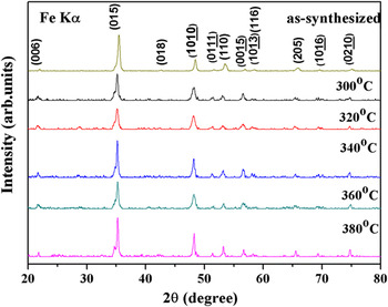
Figure 1 shows the XRD patterns of hydrothermally prepared Bi0.5Sb1.5Te3 nanoplatelets and sintered Bi0.5Sb1.5Te3 at different temperatures of 300–380 °C in an evacuated ampoule. Most of the reflection peaks can be indexed based on a rhombohedra lattice with the space group of  . The (015) and (
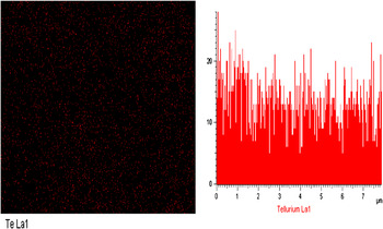
 ) reflections of the as-synthesized Bi0.5Sb1.5Te3 have a shoulder on left and right hand side, respectively. Both shoulders belong to (101) and (102) reflections of Te, respectively, indicating the presence of Te. The shoulders become more apparent as the hydrothermally prepared powders are subject to sintering. The density of our sintered pellets lies between 87 and 90% of the theoretical density of 6.878 g/cm3. EDX analyses were carried out on randomly chosen five spots of each sample. The EDX spectra reveal that the composition is not homogeneous, in particular the Te content. For sample sintered at 380 °C, there are Te-rich and Te-poor spots with a large difference of Te content, which can be seen in Fig. 2 showing the EDX mapping of Te and one of the line scans across the sample.
Powder x-ray diffraction patterns of hydrothermally prepared Bi0.5Sb1.5Te3 nanoplatelets and sintered Bi0.5Sb1.5Te3 at various temperatures of 300–380 °C in an evacuated ampoule.

The energy dispersive x-ray spectrometer mapping of tellurium and one of the line scans across the sample sintered at 380 °C.

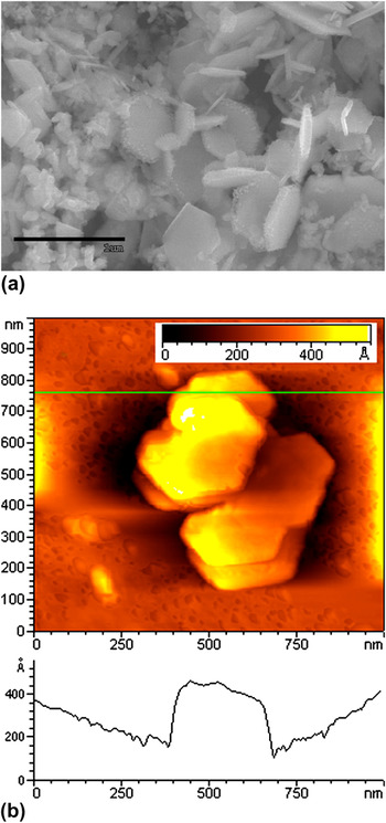
Figure 3(a) shows the scanning electron microscope (SEM) image of hydrothermally synthesized Bi0.5Sb1.5Te3 nanoplatelets having various sizes. The AFM image shown in Fig. 3(b) indicates that the nanoplatelets have a thickness of about 30–50 nm. The EDX spectra reveal that the composition of the nanoplatelets is Bi0.47Sb1.53Te2.75. The sum of atomic ratio of Bi and Sb is taken as 2 in the composition calculation.
(a) Scanning electron microscope (SEM) image of as-synthesized Bi0.5Sb1.5Te3 showing the hexagonal nanoplatelets; (b) atomic force microscope image of as-synthesized Bi0.5Sb1.5Te3 showing a thickness of about 30–50 nm. The profile shows only the lower platelet. There is another platelet on top of it.

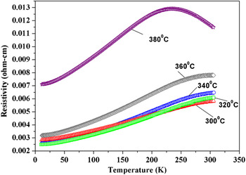
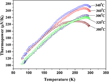
Figure 4 shows the temperature dependence of the electrical resistivity for Bi0.5Sb1.5Te3 samples sintered at different temperatures of 300–380 °C. All samples display metal-like temperature dependence between 300 and 10 K except for a change from metallic to nonmetallic temperature dependence at 235 K for the sample sintered at 380 °C. Moreover, the resistivity of all samples is lower than the one which is compacted without sintering. The resistivity of the non-sintered sample is ∼40 mΩ·cm at room temperature and increase with decreasing temperature. As shown in Fig. 4, it can be readily seen that the resistivity increases with increasing sintering temperature, and that there is a big jump of the resistivity as the sintering temperature is increased to 380 °C. Figure 5 shows the temperature dependence of the thermopower for Bi0.5Sb1.5Te3 samples sintered at different temperatures. Samples sintered at 300–340 °C show a slope in the temperature dependence of thermopower, which resembles the slope in the temperature dependence of electrical resistivity, whereas the 380 °C-sintered sample shows a flattening and downturn in thermopower above 200 K corresponding to the change in sign of the resistivity temperature dependence. For a single and nondegenerate band, the diffusion thermopower can be expressed asReference Mott and Davis14

Temperature dependence of electrical resistivity for Bi0.5 Sb1.5Te3 sintered at different temperatures of 300–380 °C in an evacuated ampoule.

Temperature dependence of thermopower for Bi0.5Sb1.5Te3 sintered at different temperatures of 300–380 °C in an evacuated ampoule.

where f, σ, E F, and k B are the Fermi distribution function, electrical conductivity, Fermi energy, and Boltzmann constant, respectively. For a system with one type of carrier, the electrical conductivity σ in Eq. (1) is proportional to the product of carrier concentration and mobility and can be expressed as

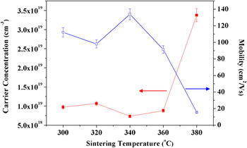
where n and μ are the carrier concentration and mobility, respectively. The mobility μ of carriers can be expressed as μ = eτ/m*, where τ is the relaxation time associated with the carrier scattering process and m* is their effective mass. Therefore, both the electrical resistivity and thermopower of a conductor depend on its carrier concentration and mean free path length between collisions. Figure 6 shows the carrier concentration and mobility for Bi0.5Sb1.5Te3 sintered at different temperatures of 300–380 °C. Although the size of carrier concentration does not show a monotonic trend, the experimental data can be discussed in two categories in terms of sintering temperature. First, samples sintered between 300 and 360 °C do not show significant variation of the carrier concentration, having values in the range (0.9 ± 0.2) × 1019 cm−3. Second, the sample sintered at 380 °C shows different behavior as for resistivity and thermopower, exhibiting a very small Hall coefficient and so a dramatically increased carrier concentration of 3.4 × 1019 cm−3, being about 4.6 times of that for the sample sintered at 340 °C. The carrier mobility estimated from Eq. (2) shows a corresponding decrease for the 380 °C-sintered sample to account for its higher resistivity. Creation of p-type carriers in (Bi,Sb)2Te3 is mainly attributed to the antistructure defects, which is generated by occupation of Te vacancy with Bi or Sb atoms.Reference Starý, Horák, Stordeur and Stölzer15, Reference Fan, Yang, Zhu, Bao, Duan, Xiao, Zhang and Xie16 Formation of antistructure defects can be described by the following equation:


Carrier concentration and mobility at room temperature for Bi0.5Sb1.5Te3 sintered at different temperatures of 300–380 °C in an evacuated ampoule.

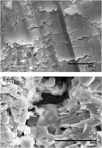
where Bi′Te and Sb′Te are the antisite defects, V Bi and V Sb are the vacancies, and h is the electron hole. As the sintering temperature increases to 380 °C, a significant increase of hole concentration could be due to more antistructure defects generated in the sample, which is caused by slight evaporation of Te atoms.Reference Hyun, Oh, Hwang and Shim17 Scattering from these defects as well as the fragmented structure of this sample [Fig. 7(b)] could cause the observed reduced mobility, as discussed below.
The cross-sectional SEM micrographs of Bi0.5Sb1.5Te3 sintered at (a) 340 °C and (b) 380 °C in an evacuated ampoule.

The thermopower of p-type (Bi,Sb)2Te3 thermoelectric materials in the extrinsic conduction region can be expressed asReference Hyun, Oh, Hwang and Shim17
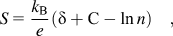
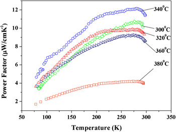
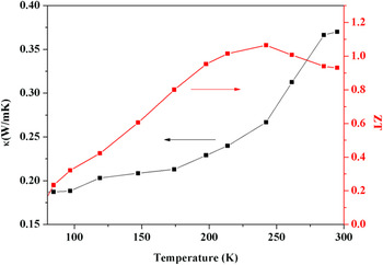
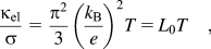
where n is the hole concentration, δ is associated with the scattering parameter, and C is a constant. According to the data shown in Figs. 5 and 6, it appears that the size of thermopower correlates well with the magnitude of the carrier concentration at room temperature. It can be readily seen that the sample having higher carrier concentration has a lower size of thermopower. According to Eq. (5), these results seem to suggest that all the samples follow a similar scattering mechanism or that the effect of the scattering parameter δ on the thermopower is negligible. It is also interesting to note that the size of the mobility shows an opposite trend to that of the carrier concentration. In general, samples having higher resistivity are expected to show larger sizes of thermopower. However, the sample sintered at 380 °C shows the highest resistivity of 11.8 mΩ·cm at 295 K but the lowest thermopower of 215 μV/K at 295 K, whereas the sample sintered at 340 °C shows the resistivity of 6.4 mΩ·cm and relatively high thermopower of 273 μV/K at 295 K. These results seem to contradict the general behavior. Even though the sample sintered at 380 °C has the highest carrier concentration of 3.4 × 1019 cm−3, its high resistivity can be ascribed to its low mobility of 15 cm2/V·s. In contrast, the sample sintered at 340 °C has the lowest carrier concentration of 7.3 × 1018 cm−3, but the highest mobility of 134 cm2/V·s among these samples. Figure 7 shows the cross-sectional SEM micrographs of Bi0.5Sb1.5Te3 sintered at 340 °C and 380 °C, respectively. Figure 7(b) clearly shows the crevice of the sample sintered at 380 °C, which might hinder the carriers from transport across the sample and hence might be responsible for its small mobility. Moreover, morphologies for left, center and right areas in Fig. 7(b) are different from one another. This morphological heterogeneity might also be responsible for its low mobility and high resistivity.Reference Liu, Wu, Hsu, Su, Wang and Shieu18Figure 8 shows the temperature dependence of the power factor for Bi0.5Sb1.5Te3 samples sintered at different temperatures of 300–380 °C in an evacuated ampoule. The sample sintered at 340 °C shows the largest power factor of 11.6 μW/cm·K2 at 295 K, whereas the sample sintered at 380 °C shows the smallest power factor of 3.9 μW/cm·K2. Since the sample sintered at 340 °C shows the largest power factor among these samples, the thermal conductivity measurement was only performed on this sample. Even though the power factor of 11.6 μW/cm·K2 for the sample sintered at 340 °C is relatively small as compared to ∼50 μW/cm·K2 for the zone-melted counterpart, its thermal conductivity is rather small with the size of 0.37 W/m·K at 295 K as shown in Fig. 9.Reference Jiang, Chen, Bai, Yao and Wang19–Reference Yang, Aizawa, Yamamoto and Ohta21 Since the thermal conductivity of a material is determined by both the electronic and lattice contributions, i.e., κ = κ el + κ ph, where κ el and κ ph represent the electronic and lattice thermal conductivity, respectively. The electronic contribution to thermal conductivity can be calculated using the Wiedemann–Franz law according to the relation between the electronic thermal conductivity κ el and electrical conductivity σ, which can be expressed asReference Kittel22
Temperature dependence of the power factor σS 2 for Bi0.5 Sb1.5Te3 sintered at different temperatures of 300–380 °C in an evacuated ampoule.

Temperature dependence of the thermal conductivity and dimensionless figure of merit for Bi0.5Sb1.5Te3 sintered at different temperatures of 300–380 °C in an evacuated ampoule. Since the vendor only provides the temperature coefficient of the resistivity (TCR) down to −80 °C for the sensor, we use TCR at room temperature as a constant in the whole investigated temperature range. The data below room temperature need further calibration for each sensor being used. However, the thermal conductivity obtained at 295 K is reliable.

where L 0 is the Lorenz number and has the size of 2.45 × 10⁻⁸ W·Ω/K2. According to Eq. (6), the calculated electronic thermal conductivity for the sample sintered at 340 °C is 0.11 W/m·K at 295 K and therefore the lattice thermal conductivity κ ph would be 0.26 W/m·K at this temperature. The low electronic thermal conductivity is associated with its low electrical conductivity as compared to typical value of 1 mΩ·cm for Bi2-xSbx xTe3 with good thermoelectric performance. The value of κ ph = 0.26 W/m·K is quite small as compared to the samples fabricated using spark plasma sintering methods with the size of 0.5–0.8 W/m·K.Reference Jiang, Chen, Bai, Yao and Wang19 HollandReference Holland23 refined a model developed by CallawayReference Callaway24 for expressing the lattice thermal conductivity:

where θ D is the Debye temperature, ν g is the velocity of phonons, τ is the relaxation time, and ω is the phonon angular frequency. An umklapp process, phonon–defect scattering, or phonon–grain boundary scattering would lead to reduction of τ. Using Matthiessen’s rule, τ can be expressed as
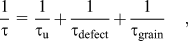
where τu, τdefect, and τgrain are the relaxation time due to the umklapp process, phonon–defect scattering, and phonon–grain boundary scattering, respectively. The magnitude of τ grain is dependent of the average grains size, as described using the expressionReference Graebner, Reiss and Seibles25:
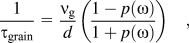
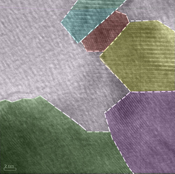
where p(ω) is the probability of specular reflection and d is the average grain size. Suppose that the phonon spectrum is not drastically altered, the significant reduction of thermal conductivity should be attributed to a strong scattering of phonons.Reference Xie, He, Kang, Tang, Zhu, Laver, Wang, Copley, Brown, Zhang and Tritt26Figure 10 shows the TEM image of the sample sintered at 340 °C. The image clearly shows nanosized grains in the microstructure of the sample. According to Eq. (9), a small average size of these nanosized grains is expected to have a small size of τ grain, which in turn one could expect a small κ ph according to Eqs. (7) and (8). Besides, since the density of compacted and sintered sample is less than 90% of its theoretical density, the porous microstructure is expected. Porosity would lead to further reduction of thermal conductivityReference Hopkins, Rakich, Olsson, El-kady and Phinney27 Therefore, the small observed κ ph could be mainly attributed to the nanosized grains and porosity of the sample. The dimensionless thermoelectric figure of merit ZT is calculated to be 0.93 at 295 K shown in Fig. 8 for the sample sintered at 340 °C.
Transmission electron microscope image showing the nanosized grains for the sample sintered at 340 °C.

IV. CONCLUSIONS
In summary, the hydrothermally synthesized nanoplatelets of Bi0.5Sb1.5Te3 are compacted and sintered in an evacuated ampoule at various temperatures of 300–380 °C for optimizing the thermoelectric characteristics. The sample sintered at 340 °C shows the largest dimensionless figure of merit ZT = 0.93 at 295 K among the samples. It has very low thermal conductivity of 0.37 W/m·K as compared to the (Bi,Sb)2Te3 alloys fabricated using other methods.
ACKNOWLEDGMENT
This work is supported by the National Science Council of Taiwan, Republic of China, Grant Nos. NSC 95-2112-M-018-006-MY3 and NSC 98-2112-M-018-005-MY3.