Introduction
Echinococcus granulosus sensu lato is the causative agent of human hydatid disease. This zoonosis is frequently encountered in countries where animal husbandry activities are prominent. Worldwide about 4 million people are infected and another 40 million run the risk of infection (Eckert et al., Reference Eckert, Conraths and Tackmann2000; McManus et al., Reference McManus, Zhang, Li and Bartley2003; Craig et al., Reference Craig, McManus, Lightowlers, Chabalgoity, Garcia, Gavidia, Gilman, Gonzalez, Lorca, Naquira, Nieto and Schantz2007). Cystic echinococcosis (CE) is transmitted in a large rural domestic cycle involving dogs as definitive hosts, livestock as intermediate hosts and humans as accidental intermediate hosts.
In Tunisia, CE causes significant socio-economic losses because of its impact on the farming economy and its high annual human surgical incidence rate (12.6/100,000 inhabitants) (Chahed et al., Reference Chahed, Bellali, Touinsi, Cherif, Ben Safta, Essoussi and Kilani2010). In Tunisia, three Echinococcus species have been observed: E. granulosus sensu stricto (G1 and G3 genotypes), E. equinus (G4 genotype) and E. canadensis (G6 genotype) (M'rad et al., Reference M'rad, Filisetti, Oudni, Mekki, Belguith, Nouri, Sayadi, Lahmar, Candolfi, Azaiez, Mezhoud and Babba2005, Reference M'rad, Oudni-M'rad, Denis, Mekki, Nouri, Sayadi, Candolfi, Azaiez, Mezhoud and Babba2010, Reference M'rad, Oudni-M'rad, Boubaker, Bouazzi, Gorcii, Nouri, Mezhoud and Babba2012; Boufana et al., Reference Boufana, Lahmar, Rebai, Ben Safta, Jebabli, Ammar, Kachti, Aouadi and Craig2014). Among these species, E. granulosus sensu stricto (G1 genotype) still remains predominant in Tunisia (M'rad et al., Reference M'rad, Filisetti, Oudni, Mekki, Belguith, Nouri, Sayadi, Lahmar, Candolfi, Azaiez, Mezhoud and Babba2005, Reference M'rad, Oudni-M'rad, Boubaker, Bouazzi, Gorcii, Nouri, Mezhoud and Babba2012).
Genotyping of Echinococcus species requires the collection of fresh cysts, dedicated spaces and specific equipment. These steps pose problems during investigations carried out in remote areas, leading to the absence or lack of data in those regions. CE may affect any human organ. With the exception of the liver and the lungs, all the other organs are considered as uncommon sites (Feki et al., Reference Feki, Ghozzi, Khiari, Ghorbel, Elarbi, Khouni and Ben Rais2008). The conventional sample collection method impedes the molecular study of cysts with unusual sites. In fact, rare cysts are generally referred to departments of anatomy–pathology for histological investigations, resulting in their exclusion from the current genotyping studies.
To overcome these difficulties, the use of formalin-fixed paraffin-embedded tissues (FF-PETs) could be a solution. FF-PETs are fixed biological materials that result from successive formalin fixation and paraffin inclusion steps. These steps preserve and maintain tissue structures in the closest state to their natural biological state. They also block their autolysis and their attack by micro-organisms. Considered as an endless source of biological material, FF-PETs have attracted the interest of many researchers during past decades and several studies have shown the possibility of DNA amplification from FF-PETs (Greer et al., Reference Greer, Lund and Manos1991; Ferrari et al., Reference Ferrari, Luvizotto, Rahal and Cardoso2007; Ortega et al., Reference Ortega, Navarro, Nicolas, Buendia, Caro, Del Rio, Martinez, Cuello, Salinas and Gallego2007; Arneson et al., Reference Arneson, Moreno, Iakovlev, Ghazani, Warren, McCready, Jurisica and Done2012). The aim of the present study was to exploit FF-PETs to identify genotypes of E. granulosus sensu lato inTunisia and to perform molecular analysis of cysts from rare sites.
Materials and methods
Histology of tissue samples
The parasitic material consisted of paraffin blocks (FF-PETs) collected from the laboratories of Pathological Anatomy and Cytology at F. Bourguiba Monastir and F. Hached Sousse teaching hospitals. All patient reports with histologically confirmed CE, and operated on between 2009 and 2010 at Monastir Hospital and between 2000 and 2001 at Sousse Hospital, were selected. Then, using patient reports on pathology, parallel blocks were selected according to the presence of parasitic elements containing DNA (protoscoleces and/or hooks and/or germinal layer). Samples with at least one of these elements were conserved for the preparation of histological sections. A total of 60 blocks of FF-PETs were harvested from 36 female and 21 male patients with an age range of 6–75 years. In addition to 31 cysts from the liver and lungs, samples from up to 16 unusual sites were processed, including the kidney (7), spleen (6), thigh (3) and one from each of the heart, pericardial region, peritoneum, submandibular region, ovary, breast, gall bladder, latero-cervical region, subcutaneous tissue, vertebral column, axillary region, pleura and the abdomen. Furthermore, four samples harvested at the Monastir Hospital included cysts from the heart, spleen, liver and kidney of a 31-year-old female who had died from multiple organ CE.
Five sections of 10 μm in thickness were cut from each paraffin block, using a microtome with a disposable blade. The sections were then placed in Eppendorf tubes. Great care was taken to avoid cross-contamination between samples and all equipment was cleaned carefully with xylene between isolates.
Molecular analysis
FF-PETs were first deparaffinized and rehydratated according to the modified protocol of Schneider et al. (Reference Schneider, Gollackner, Edel, Schmid, Wrba, Tucek, Walochnik and Auer2008). The tissue was dewaxed in 1 ml of xylene at 37°C and centrifuged for 5 min at 9300 g. Then samples were rehydrated by four decreasing ethanol washes (100%, 90%, 80% and 70%). With each ethanol wash, samples were homogenized by vortex and then centrifuged for 5 min at 9300 g. Subsequently, 70% ethanol was removed and the pellet was allowed to air-dry until ethanol evaporation was complete.
DNA extraction was then performed by phenol/chloroform method (Sambrook et al., Reference Sambrook, Fritsh and Maniatis1989). The tissues were digested overnight (Simsek et al., Reference Simsek, Kaplan and Ozercan2011) in constant agitation at 65°C (Thermomixer GeneAmp®, PCR System 9700, Applied Biosystems, Singapore) in 500 μl of lysis buffer TEX (1 m Tris–HCl, pH 8; 5 m NaCl, 0.5 m EDTA, pH 8; 10% sodium dodecyl sulphate (SDS), pH 7.2) and 20 μl proteinase K (20 mg/ml) (Fungal; Invitrogen, Carlsbad, California, USA). Genomic DNA was extracted directly from the digested products. Finally, 50 μl TE buffer was added (1 m Tris–HCl, pH 7.6; 0.5 m EDTA, pH 8) and DNA was conserved at − 20°C until use.
The mitochondrial 12S rRNA gene was amplified with Egss1 polymerase chain reaction (PCR) as described previously (Dinkel et al., Reference Dinkel, Njoroge, Zimmermann, Walz, Zeyhle, Elmahdi, Mackenstedt and Romig2004) using primers E.g.ss1 forward (5′-GTA TTT TGT AAA GTT GTT CTA-3′) and E.g.ss1 reverse (5′-CTA AAT CAC ATC ATC TTA CAA T-3′). The PCR reaction was performed in 50 μl containing 1 × PCR buffer, 2 mm MgCl2, 200 μm of each deoxynucleoside triphosphate (dNTP), 40 pmol of each primer, 3.5 μl of DNA template (not quantified) and 2.5 units Taq DNA polymerase (HotStar, Qiagen, Hilden, Germany). The PCR was performed with an initial denaturation step of 95°C for 5 min, followed by 40 cycles of denaturation (94°C for 30 s), annealing (57°C for 1 min) and extension (72°C for 40 s), and a final extension step at 72°C for 5 min.
For each reaction, a positive control (E. granulosus sensu stricto sequenced DNA from fresh protoscoleces) and a negative control (no DNA) were used. The PCR products were visualized by gel electrophoresis through a 1.5% ethidium bromide (0.25 μg/ml) stained agarose gel and finally photographed.
Data analysis
Associations between cyst occurrence and gender, cyst occurrence and age groups, and cyst site and gender were tested. Additionally, the gender distribution of patients within the age groups and the correlation between cyst site and the patients' age were verified. Differences related to age and gender were evaluated between patients with usual and unusual (excluding liver and lungs) sites. The chi-squared test, one-way analysis of variance (ANOVA) test and t-test were used to analyse all data. Tests with P < 0.05 were considered statistically significant. Statistical analysis was performed using the Statistical Package for Social Sciences Software Package (SPSS for Windows, version 17.0; Chicago, Illinois, USA).
Results
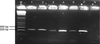
Molecular analysis by mitochondrial 12S rRNA gene amplification yielded a 254 bp fragment for all samples, corresponding to the E. granulosus sensu stricto profile. This profile was obtained whatever the cyst site and the patient's gender (fig.1).
Agarose gel electrophoresis of PCR products of E. granulosus sensu stricto to show DNA extracted from FF-PETs; Lanes: M, 1 kb DNA ladder (Invitrogen); 1–5, E. granulosus sensu stricto DNA extracted from FF-PETs (1, liver; 2, lungs; 3, peritoneum; 4, vertebral column; 5, spleen); P, positive control (E. granulosus sensu stricto sequenced DNA from fresh protoscoleces); N, negative control (no DNA).

In this study, the results showed that of the 60 positive cases examined, the highest number of cysts were localized in the liver (35%), followed by the lungs (16.6%), the kidney (11.7%), the spleen (10%), the thigh (5%) and 1.6% in the following sites with one case in each site: the heart, pericardial region, peritoneum, submandibular region, ovary, breast, gall bladder, latero-cervical region, subcutaneous tissue, vertebral column, axillary region, pleura and the abdomen. Hydatid cysts were harvested from patients in different age groups ( ≤ 10, 11–20, 21–30, 31–40, 41–50, 51–60 and ≥ 61 years). The youngest patient was 6 years old with a hydatid cyst in the latero-cervical region, while the oldest patient was 75 years old with a mammary hydatid cyst. The average age was 37.31 ± 2.641 years. The most affected age groups were those aged 11–20 years, 31–40 years and over 60 years (16.7% for each group), representing 50.1% of all cases. The highest proportions of cysts were found in 65% of female patients, compared with 35% of male patients, with a statistically significant difference (P= 0.02). The age group has no statistically significant effect on the occurrence of cysts (P= 0.934). In the present study, the sex-ratio (F/M) was in equilibrium only in the age group 51–60 years. The results indicated that females were more affected than males (0.75 ≤ sex-ratio F/M ≤ 5) in all age groups except for patients aged between 41 and 50 years, where the sex ratio F/M was slightly biased towards males (0.75:1). No association was identified with the patients' gender distribution within all age groups as all P values were not statistically significant ( ≤ 10 years, P= 0.329; 11–20 years, P= 0.722; 21–30 years, P= 0.527; 31–40 years, P= 0.284; 41–50 years, P= 0.198; 51–60 years, P= 0.348 and ≥ 61 years, P= 0.722). In the chi-squared test, no statistically significant association was found between cyst site and gender (P= 0.405). Cyst site and patient's age were not statistically correlated as the ANOVA test showed no significant results (P= 0.119). A comparison of patients with usual and unusual cyst sites demonstrated that the differences in age (P= 0.718) and/or gender (P= 0.664) between the both groups were not statistically significant.
Discussion
The present molecular analysis showed that all investigated human cysts were due to an infection with E. granulosus sensu stricto. Based on previous studies, E. granulosus sensu stricto (G1 genotype) is the most common species found in Tunisia (M'rad et al., Reference M'rad, Filisetti, Oudni, Mekki, Belguith, Nouri, Sayadi, Lahmar, Candolfi, Azaiez, Mezhoud and Babba2005, Reference M'rad, Oudni-M'rad, Boubaker, Bouazzi, Gorcii, Nouri, Mezhoud and Babba2012; Lahmar et al., Reference Lahmar, Boufana, Jebabli, Craig, Ayari, Basti, Dhibi and Torgerson2014) and is responsible for the majority of human CE cases (Alvarez Rojas et al., Reference Alvarez Rojas, Romig and Lightowlers2014). In this study, only the presence of E. granulosus sensu stricto was noted regardless of the cyst site (usual or unusual). This finding confirmed the adaptation of E. granulosus sensu stricto species to various human organs and its high infectivity in humans.
In the present study, the liver (35%) and lungs (16.6%) were the most common organs involved in human CE, as described previously in many parts of the world (Mushtaque et al., Reference Mushtaque, Mir, Malik, Arif, Khanday and Dar2012; Noomene et al., Reference Noomene, Ben Maamer, Bouhafa, Haoues, Oueslati and Cherif2013). The high prevalence in the liver is explained by this organ's role as the first filter of E. granulosus embryos migrating from the intestine via the portal stream. In our series, the other sites (excluding the liver and the lungs) represent 48.4% of cases. The exact incidence of these unusual sites is difficult to ascertain because only case reports are described in the literature (Wani et al., Reference Wani, Wani, Malik, Parray, Wani and Dar2012). In the present study, the most common rare sites of infection are the kidney, the spleen and the thigh, respectively. In many previous reports, the kidney was cited as the third most common site of hydatid cysts after the liver and lungs (Geramizadeh, Reference Geramizadeh2013). Our study yielded a rate of 11.7% for renal CE, which is much higher than the occurrence rates reported in the literature (2–3%) (Gogus et al., Reference Gogus, Safak, Baltaci and Turkolmez2003). The spleen was the second most common unusual site (10%) in our series, which is close to earlier estimates (0.9–8%) (Polat et al., Reference Polat, Kantarci, Alper, Suma, Koruyucu and Okur2003). CE is the only parasitic cyst of the spleen and it is twice as common as the non-parasitic variety (Wani et al., Reference Wani, Wani, Malik, Parray, Wani and Dar2012). Hydatid cysts of the kidney and the spleen affected all age groups, as is the case in other countries (Eris et al., Reference Eris, Akbulut, Yildiz, Abuoglu, Odabasi, Ozkan, Atalay and Gunay2013; Rexiati et al., Reference Rexiati, Mutalifu, Azhati, Wang, Yang, Sheyhedin and Wang2014). Hydatid cyst of the muscle is rare even in endemic countries (Moretti et al., Reference Moretti, Panella, Moretti, Garofalo and Notarnicola2010). In nearly all previous reports, muscle CE was estimated to range from 1 to 5% (Madhar et al., Reference Madhar, Aitsoultana, Chafik, Elhaoury, Saidi and Fikry2013), which is in accordance with our results where thigh cysts were observed in 5% of the studied cases. In general, voluntary muscles are rarely affected in human hosts because musculoskeletal tissues are considered unfavourable for parasite survival due to the presence of lactic acid and muscular contractions that prevent the fixation of larvae to the muscular tissue (Abi Saad et al., Reference Abi Saad, Musallam, Korban, Reslan and Mneimne2009).
CE can occur regardless of age or gender. Thus, our study showed that cyst site was not correlated with the patients' age or gender and that no significant differences were found between patients with usual and unusual cyst sites with respect to the same demographic parameters.
In this study, females were more commonly affected than males (0.75 ≤ sex-ratio F/M ≤ 5). As most of the patients originated from rural areas (data not shown), this difference is due to the female role in home activities and the fact that, in rural areas, women are usually housewives and often care for dogs and cattle, which increases the risk of infection. Besides, adult females undergo more regular medical check-ups (e.g. during pregnancy), which result in fortuitous cyst discoveries on ultrasound examinations.
The use of FF-PETs made it possible to genotype rare cyst sites because in this case FF-PETs constituted the unique DNA source available for such molecular studies. To our knowledge; this is the first publication on molecular genotyping of hydatid cysts with unusual sites.
Currently, FF-PETs represent an area of intensive research activity as their use presents several advantages. FF-PETs have been exploited for decades and represent the largest source of archival biological material available for genomic studies (Aviel-Ronen et al., Reference Aviel-Ronen, Qi Zhu, Coe, Liu, Watson, Lam and Tsao2006). They also allow simple access to archival tissue banks in hospitals. In addition, compared with frozen fresh tissues which need specific collection and equipment (Farrugia et al., Reference Farrugia, Keyser and Ludes2010), paraffin blocks are easier to store and transport to laboratories (Schneider et al., Reference Schneider, Gollackner, Edel, Schmid, Wrba, Tucek, Walochnik and Auer2008). The exploitation of FF-PETs enables the retrospective molecular genotyping of E. granulosus samples, which promotes CE-related data in Tunisia. Furthermore, FF-PETs obviate the need for fresh tissues and make molecular and genetic investigation possible even after the death of the proband if the tissues obtained after surgery or autopsy have been stored (Mies, Reference Mies1994). Thus, four cysts obtained from autopsies were easily studied, which could not have been feasible with the classic sampling method.
In conclusion, FF-PETs allowed us to genotype E. granulosus from fixed human samples and, especially, to investigate hydatid cysts with atypical sites. Hence, it is worth exploiting fixed material in such retrospective studies. In addition, the present study, apart from confirming the predominance of E. granulosus sensu stricto species in Tunisia, has shown that this species is likely to infect the majority of human organs.
Acknowledgements
The authors thank the staff of the laboratories of Pathological Anatomy and Cytology at F. Bourguiba Monastir and F. Hached Sousse teaching hospitals for providing samples and information related to this study. We are also grateful to Mrs Imen Hizem Attig for providing linguistic assistance. We thank Pr. Moncef Rassas for the final check of the English part of the manuscript.
Financial support
Financial support for this study was provided by the Tunisian Ministry of Higher Education and Scientific Research.
Conflict of interest
None.