Transcatheter occlusion of small arterial ducts, of less than 4 millimetres in diameter, and closure of larger ducts with devices, have now become accepted as attractive alternatives to surgery. Indeed, interventional closure has become the treatment of choice in most of the patients now born with patency of the arterial duct.1, 2 Despite the tremendous improvements in technique and hardware available, closing large ducts in very sick and underweight infants remains a challenge for paediatric interventionists. This is because bigger devices require larger delivery sheaths, and carry the risk of possible obstruction to aorta and left pulmonary artery.3, 4 Moreover, negotiating a large device around the curve of the right ventricular outflow tract and through the pulmonary trunk, which is very tight and at right angles in infants, is difficult or impossible, the sheath becoming kinked and thereby increasing the procedural and fluoroscopic time. This subset of patients with very large tubular ducts, typically weighing less than 5 kilograms, in left ventricular failure with associated lesions, and failing to thrive, are usually referred for surgery, which in turn carries higher morbidity and mortality. Our aim, therefore, was to investigate the feasibility and efficacy of closing such large ducts by inserting the newly available modified and angled variants of the Amplatzer occluder through a specially braided kink-resistant delivery sheath using a special pusher system.
Materials and methods
In total, 67 infants presented to us with large patent arterial duct. Of these, 55 infants (82%) underwent multiple ligation, while closure was attempted in 10 using the modified Amplatzer occluder in 6 cases, and the angled occluder in 4 cases. It is these latter 10 patients that form the material for our study. Of the patients, 5 were male and 5 female, their age ranging from 4 to 12 months, with a mean of 8.2 months, and weight ranging from 3.9 to 8 kilograms, with a mean of 5.5 kilograms. The ducts were measured at 4.1 to 8.6 millimetres in diameter, with a mean of 6.3 millimetres.
In 2 of the patients, both with severe pulmonary hypertension, the oval foramen was patent, this also closed spontaneously subsequent to closure of the arterial duct with the device. In 2 other patients, there was Rubella syndrome with bilateral cataracts. In addition, one patient had cerebral palsy, one had Down's Syndrome, and one infant had aortic valvar stenosis, with a gradient of 45 over 25 millimetres of mercury measured across the valve echocardiographically. Another infant was found during the catheterization procedure to have right hydronephrosis and hydroureter, with an unascended and hypoplastic left kidney.
Our period of follow-up is from nine month to two years. All children had a history of repeated infections of the lower respiratory tract, needing repeated hospitalization, and all were failing to thrive, exhibiting difficulties in feeding.
Modifications made in the device and the system for its delivery
The modifications were made specifically by Kurt Amplatz, of AGA, Minnesota, United States of America, in order to overcome common problems encountered while closing the large patent arterial ducts encountered in infants, such as kinking of the sheath, the device obstructing the aorta and left pulmonary artery, and the difficulties encountered in manoeuvring the large retention disc in the narrow aorta. In the first place, he prepared a special braided kink-resistant sheath for delivery, hoping to overcome the problems of kinking, together with obviating the difficulties in negotiating the device through the curve of the right ventricular outflow tract and the pulmonary trunk. Second, he created a special “pusher” catheter, similar to that used in delivering the device designed to close perimembranous ventricular septal defects. This catheter has a capsule at its end, which aligns with the screw on the device, fixing it in the correct direction. This helps in pushing the angled ductal device smoothly round the bend, as well as automatically orientating the platinum marker at its tip, avoiding the problem of manoeuvring the large retention disc in the narrow aorta. (Fig. 1). Third, rather than having 3 layers of Gore-Tex polyester material sewn at the retention disk, the pulmonary end, and in the middle of the device, only a single layer of polyester material is sewn in the middle of the modified occluder, thus reducing the bulk of the device so that it can be delivered though a smaller sheath, hopefully avoiding obstruction to left pulmonary artery or the aorta (Fig. 2). Finally, in the angled modification, so as to avoid protrusion of the large device into the aorta, the aortic retention disk is made concave towards aorta, and angled at 32 degrees to the body of the device. The cylindrical body is tapered from its aortic to pulmonary end. Due to the angled retention disk, the length of the top edge of the body is only 4 millimetres, and that of the bottom edge is 9 millimetres, permitting the angled device to fit to the shape and size of the large tubular patent arterial duct. The nitinol used to create the device is closely knit, without incorporating any polyester material, permitting even the 14/12 device to be introduced easily through a 6 or 7 French delivery sheath. The angled occluder has a platinum marker at the lower tip of the retention disk, thus permitting appropriate orientation of the device (Fig. 3).

Figure 1. Use of the regular sheath for introduction (a) produces kinks, and causes difficulty in pushing the device. This is overcome by the availability of the specially braided kink-resistant sheath (b), combined with a pusher catheter that has a capsule at the end which has a flat which aligns with the flat on the screw on the device, thus fixing it in the correct direction.

Figure 2. The regular Amplatzer duct occluder (a) has 3 layers of Gore-Tex polyester material, sewn in the retention disk, the pulmonary end, and in the middle of the device. The modified occluder (b) has a single layer of Gore-Tex polyester material sewn in the middle, with only Nitinol mesh at the aortic retention disk and the pulmonary end, thus reducing the bulk of the device.

Figure 3. In the angled modification (a), the aortic disc is made concave towards aorta and angled at 32° to the body of the device. The cylindrical body is tapered from the aortic to the pulmonary end. The length of the top edge of the body is only 4 millimetres, and the bottom edge is 9 millimetres. There is a platinum marker at the tip of the retention disk permitting the orientation of the device to be seen on fluoroscopy. In panel (b), the partially released retention aortic disk of the angled occluder is shown with the platinum marker at 11 o'clock position, indicating the orientation is inappropriate.
Procedure of implantation
All procedures were performed under general anaesthesia. A 4 or 5 French sheath was placed in left femoral artery, and a 4 or 5 French marker pigtail catheter was placed in the distal aortic arch. An aortogram was then performed in the lateral and right anterior oblique projections. Measurements of minimal and maximal diameters of the ampulla of the patent duct were performed by the technique of automatic calibration, using the marker pigtail catheter as the reference. We classified the ducts morphologically using the system described by the group from Toronto.5 Thus, the ducts were described as showing an ampulla at their aortic end, as being narrower at the aortic end, as being tubular with no narrowing, as showing multiple constrictions along their length, or taking a bizarre configuration. The size of occluder chosen was 2 to 4 millimetres larger than the minimal measured diameter of the duct.
A 5 French sheath was placed in a right femoral vein. A 5 French multipurpose catheter was then advanced across the patent duct into the descending aorta, and exchanged over a 0.035″ J Guide wire for a 5 or 6 French specially braided delivery sheath, which was passed into the descending aorta. For those ducts with ampullas at their aortic end, we attached modified occluders to the regular delivery wire, while for those with tubular ducts lacking any narrowing, we used the angled occluder, attaching it to a thin special wire for use with the pusher catheter, before loading it as in the standard procedure. Implantation was monitored by fluoroscopy in the lateral projection, using the diagnostic angiogram as a road map. In all 10 cases, we were able smoothly to negotiate the device through the kink-resistant sheaths. Without any rotation, we then advanced the devices into the descending aorta, releasing only the tip of the retention disc in order to avoid injuring the aorta. Then the sheath, along with the device, was pulled back to the ampulla, withdrawing the sheath to permit the device to open. Appropriate orientation of the angled occluder was confirmed by noting that the platinum marker on the inferior edge of the retention disc was aimed at the 6 o'clock position as seen in the lateral projection. The partially released retention disk, which then looked like a bud in descending aorta, opened like a flower when the delivery sheath was pulled back. Even if the platinum marker is not in the right direction when the device is partially released (Fig. 3b), it automatically repositions when the device is completely opened, due to alignment of the pusher system to the flat of the device (Fig. 4b). Having confirmed the position by angiography, we release the device. All the chosen angled devices fitted perfectly to the shape of the selected arterial duct, with the concavity of the large retention disk apposed perfectly to the wall of the aorta (Fig. 4a). After a further 10 minutes, the aortogram was repeated, with minimal injection of dye, confirming the accuracy of positioning and checking for any residual shunt.

Figure 4. In Panel (a), a 12/10 angled occluder is shown with the platinum marker at the tip of the large aortic retention disk, indicating the appropriate orientation of the device. An angiogram taken in left lateral projection (b) shows perfect position of the occluder, with no obstruction to aorta despite the large retention disk. Note the post-stenotic dilation of the ascending aorta due to mild aortic stenosis.
Protocol for follow-up
Physical examination, weight, chest X-Ray, and echocardiograms were all checked or performed 24 hours after the procedure. The follow-up echocardiogram was then repeated at one, three, and six months after the implantation. Prophylaxis against infective endocarditis and Aspirin, at 5 milligrams per kilogram, were recommended for 3 months after the procedure.
Results
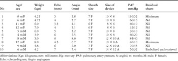
We attempted to occlude large ducts in 10 very sick infants. Their minimal diameters ranged from 4.1 to 8.6 millimetres, with a mean of 6.3 millimetres. We classified 6 of the ducts as having an ampulla at their aortic end, and for these infants, we deployed one 8/6 and five 10/8 modified occluders. In the remaining 4 infants, we judged the ducts to be tubular, and without narrowings along their length. For one of these infants, we used a 10/8 angled occluder, using a 12/10 angled occluder in 2, and a 14/12 angled occluder in the other. The youngest patient, 4 months old, weighed just 3.9 kilograms. In this patient, we closed a duct of 7.5 millimetres diameter with a 10/8 modified occluder. This device was not sufficiently small to fit snugly in the duct, and moved partially into the aorta on releasing the screw. It was safely pushed back into the duct by gently inflating a 6 millimetre balloon in the aorta. Following this procedure, there was no gradient across the aorta on pullback tracings. Colour Doppler echocardiogram also failed to show any evidence of obstruction within the aorta. In another patient, the angiogram taken prior to releasing of a 10/8 device showed that it appeared to overlap the left pulmonary artery (Fig. 5a). But, as there is no polyester material at the pulmonary end of this device, angiography showed no obstruction to the flow through the left pulmonary artery. Pullback tracings confirmed absence of any gradient across the left pulmonary artery, so we released the device. Echocardiography confirmed absence of obstruction to the left pulmonary artery (Fig. 5b). The largest duct closed was 8.6 millimetres in diameter, bigger than the aorta measured at 8.2 millimetres in this infant of 8 months weighing 5 kilograms. The duct was closed successfully with a 12/10 angled occluder.

Figure 5. In Panel (a), the modified 10/8 occluder appears to overlap the left pulmonary artery, but there was no gradient produced, since there is no polyester in the pulmonary end to obstruct the flow. The echocardiogram (b) showed the device to be appropriately positioned, with no turbulence shown in the left pulmonary artery on Colour Doppler.
Table 1. Results of the study group.

The overall times required for the procedure ranged from 30 to 80 minutes, with a mean of 40.5 minutes. Fluoroscopic time varied from 3 to 28 minutes, with a mean of 13 minutes, and we used from 16 to 20 millilitres of contrast material. Deployment was successful in 9 cases. In one attempt, a 14/12 angled occluder slipped into the pulmonary artery during positioning. We repositioned the device, and deployed it, but it embolised into the pulmonary trunk after a few hours, from where it was retrieved. During the retrieval, however, fluoroscopy revealed a right-sided hydronephrosis and hydroureter, and hypoplasia of the left kidney (Fig. 6). This very sick infant sadly did not recover from anaesthesia, and died later. All the remaining patients showed significant improvement, and gained weight remarkably after closure of their ducts. The youngest infant, 4 months old, and weighing 3.9 kilograms, gained 2 kilograms within 2 months.

Figure 6. Abdominal fluoroscopy shows hydronephrosis and hydroureter of the right kidney, with an unascended and hypoplastic left kidney.
The remaining 3 patients in whom we had used an angled occluder had minimal residual shunting revealed by angiography immediately after the procedure. This, however, is to be expected, since there is no polyester material in the device. No residual shunting was found at follow-up echocardiographic examination.
Discussion
Transcatheter closure of large patent arterial duct in very sick infants, especially those of certain morphology, is known to be difficult, or even impossible, due to protrusion of the devices into the left pulmonary artery or descending aorta. To date, such patients have been referred for surgery. Though surgical ligation is safe, and the oldest modality of treatment,6 with mortality now close to zero, it is still associated with higher morbidity compared to non-surgical closure. Complications include recanalisation, palsy of the left recurrent laryngeal and phrenic nerves, pseudoaneurysmal formation, chylothorax, atelectasis of the lung, and the need for prolonged ventilation. In the current era, therefore, transcatheter closure is preferable. Among the available devices, the Amplatzer Duct occluder is the most recent, and is also the most user-friendly. It can be used in infants to close duct having diameters up to 5 millimetres by using 5 or 6 French sheaths. The Amplatzer duct occluder has become the standard device closing those ducts with a big ampulla at their aortic end. For those ducts that are tubular, with no ampulla, the right-angled flat retention disk of the regular occluder is less than ideal, because it can protrude into the descending aorta. Such protrusion occurs due to the acute angle of insertion of the duct in relation to the long axis of the aorta. This angle has been reported, on average, to be 32 degrees. In 12 such infants, Fischer et al.7 attempted closure of large ducts with regular Amplatzer occluders. They abandoned 2 procedures, and faced technical problems in 9 additional patients, mainly in manoeuvring the device across the sheath. They concluded that device closure should not be attempted in infants weighing less than 5 kilograms. Ewert et al.8 closed a duct of 5 millimetres diameter in a child weighing 11 kilograms using an angled device, had reported some problems in manoeuvring the device in the descending aorta. Masura et al.9 had attempted closure using the angled occluder in 9 patients, finding the device to be promising. The bigger devices, however, require larger delivery sheaths, and carry the possible risk of obstruction to the aorta or the left pulmonary artery. Modifying the occluder by removing the Gore-Tex Polyester material from the retention disk and the pulmonary end of the device reduces its profile to the extent that it can be introduced through 5 or 6 French sheath. It also avoids the obstruction to the aorta and left pulmonary artery. This is because the nitinol, which has memory, takes shape and, as there is no polyester material sewn on either end, there is no obstruction to flow in either the aorta or the left pulmonary artery.
When the large device is employed, it is difficult, if not impossible, to negotiate around the curve of the right ventricular outflow tract and through the pulmonary trunk, this angle being almost at right angles in infants. Because of this, the sheath gets kinked, thereby increasing the procedural and fluoroscopic times. The specially braided kink-resistant sheath, combined with the pusher system, has a distinct advantage in these areas, as they help in the smooth delivery of devices even as big as 14/12 without any hitch. We achieved excellent results, with complete closure, in nine of our 10 cases. Our one disappointing experience was in a very sick infant who was found to have hydronephrosis and hydroureter, with an unascended and hypoplastic left kidney. This patient failed to recover from anaesthesia, and died after retrieval of an embolised 14/12 angled occluder.
We recognize that number of cases reported is small, but they represent our initial experience using angled and modified occluder. Such occluders, furthermore, are custom-made, and not yet commercially available. Despite these limitations, nonetheless, our experience shows that transcatheter closure of very large patent arterial ducts is feasible even in very sick infants. The likely complications of aortic and left pulmonary arterial stenosis can be avoided with suitable modifications in the device used for closure. The low profile of these modified devices, combined with use of the kink-resistant sheath and the special pusher system, make the procedure easy and safe, at the same time reducing considerably the procedural and fluoroscopic times. We suggest that our initial experience shows that very large tubular ducts can now be safely and effectively closed in very sick malnourished infants, and that use of the modified and angled Amplatzer duct occluders may replace shortly surgery in this subset of patients.
Acknowledgements
We express our grateful thanks to Kurt Amplatz for his guidance, and for the custom-made modified and angled occluders, along with the innovative and important alterations in the system used for delivery. We also thank Ramesh Arora, former Director of G.B.Panth Hospital, New Delhi, for her kind assistance, and Mohan Nayak, Chief Catheterization Laboratory technician, Sri Jayadeva Institute of Cardiology.