Introduction
Functional endoscopic sinus surgery (FESS) is a well-accepted and effective procedure in treating chronic rhinosinusitis; however, FESS of the frontal sinus is complicated by difficulties in maintaining the patency of the frontal sinus after sinusotomy,Reference Banhiran, Sargi, Collins, Kaza and Casiano1 and it has been shown to have the lowest success rate compared to the other sinuses.Reference Minni, Dragonetti, Sciuto, Rosati, Cavaliere and Ralli2
In order to improve the outcome, frontal sinus stents have been used since the early 1900s;Reference Reeve, Ching, Kim and Schroeder3 numerous materials and techniques have been described, and, so far, there is no consensus.
This paper describes our experience with the use of a modified Montgomery T-tube as frontal sinus stent.
Technical description
Endoscopic sinus surgery with frontal sinusotomy (Draf I, II or III procedure) is performed in each case according to the surgical need of the patient to obtain the maximum diameter of the frontal sinus opening at the end of the procedure; the sinusotomy diameter is measured with the use of instruments.
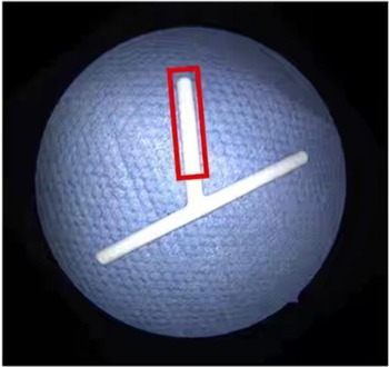
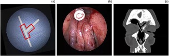
The paediatric Montgomery T-tube stent of the correct diameter (range, 6–9 mm; Figure 1) is cut to shape (‘straight’ if a Draf I, IIa or IIb procedure, or ‘Y-shaped’ if a Draf III procedure; Figures 1 and 2) and introduced intranasally under endoscopic vision; it is then advanced into the frontal recess using the corrugation on the walls of the stent to anchor it to the sinusotomy opening. The lower end of the stent should not be below the level of the middle turbinate.
Paediatric Montgomery T-tube; red lines demonstrate the method of cutting the ‘straight’ stent used for Draf I, IIa and IIb procedures.

‘Y-shaped’ paediatric Montgomery T-tube used for Draf III procedures: (a) method of cutting the stent (indicated by the red lines); (b) endoscopic appearance; and (c) radiological computed tomography appearance in the coronal plane.

Patients are advised to use regular post-operative medical care practices (saline douches, topical steroids and a course of antibiotics). Patients also undergo routine post-operative debridement in the out-patient department.
The duration of stenting depends on the indication for the surgery.
Discussion
The complexity of the surgery for chronic rhinosinusitis of the frontal recess is reflected by the total number of procedures and techniques described.Reference Hoyt4 Regardless of the procedure performed, one-third of the patients will have recurrent disease requiring revision surgery.Reference Reeve, Ching, Kim and Schroeder3,Reference Shahidi, Jama and Ahmed5
The frontal recess is, indeed, prone to re-stenosis and this affects the ventilation of the sinus and the accessibility of topical medical therapy.Reference Minni, Dragonetti, Sciuto, Rosati, Cavaliere and Ralli2 One of the reasons behind these high failure rates seems to be the abnormal scarring, often due to a lack of mucosal preservation during the surgery.Reference Banhiran, Sargi, Collins, Kaza and Casiano1
In order to prevent these problems, the use of sinus stents has long been described, with the aims of preventing synechial formation, occupying space that would otherwise be filled with blood, fibrin and/or mucus, and providing a matrix for epithelial migration at the same time.Reference Mansour6
Numerous types of stent have been described, including absorbable and non-absorbable, self-retaining or non-self-retaining, soft and rigid. However, the different types of stents described have varying results; so far, there are no universally accepted indications.Reference Shahidi, Jama and Ahmed5,Reference Khan, Alshareef, Marglani and Herzallah7 The majority of the authors of previous studies favour six-month stenting in order to allow good and stable re-mucosalisation.Reference Banhiran, Sargi, Collins, Kaza and Casiano1,Reference Minni, Dragonetti, Sciuto, Rosati, Cavaliere and Ralli2,Reference Eifler, Seitz, Weber, Grundke, Reifenrath and Kietzmann8 Authors favouring a stent technique claim a success rate ranging from 80 to 94 per cent.Reference Reeve, Ching, Kim and Schroeder3,Reference Mansour6,Reference Durisin, Seitz, Reifenrath, Weber, Eifler and Maier9
Our technique offers the advantage of a soft stent,Reference Minni, Dragonetti, Sciuto, Rosati, Cavaliere and Ralli2,Reference Shahidi, Jama and Ahmed5 which is well tolerated by the patients; because of its shape, the stent is self-retaining and does not require any sutures. The procedure described here is inexpensive, cost-effective, simple and can be easily performed by experienced endoscopic sinus surgeons.
Although our follow up is normally clinical with flexible nasendoscopy examination, the stent is radiopaque and its position can be easily visualised with computed tomography imaging (Figure 2).
The senior author of this paper has been using the technique described for 10 years and reports no major complications related to it. In our series, the duration of stenting was nine months on average, with most patients (about 80 per cent) having a patent frontal recess on evaluation with flexible nasendoscopy after removal. We found that the need for stenting is actually quite rare (in our series, only about 2 per cent of all the procedures performed in the frontal sinus involved stenting). The stent can be easily removed under local anaesthesia in-office at any time.
Competing interests
None declared