Introduction
Baylisascaris species are large ascarid nematodes. Most of the Baylisascaris species have a similar life cycle, with carnivorous mammals or marsupials serving as definitive hosts and a smaller prey host serving as paratenic (or intermediate) hosts (Sapp et al., Reference Sapp, Gupta, Martin, Murray, Neidringhaus, Pfaff and Yabsley2017). In these hosts, larvae migrate out of the gut, causing extensive pathology, particularly if the central nervous system is involved. Baylisascaris species can have huge impacts on human health, domestic animals or livestock, and can affect species conservation (Bauer, Reference Bauer2013; Kazacos, Reference Kazacos2016). Most documented cases are assumed to be B. procyonis; however a few surveys for other Baylisascaris, and in B. procyonis surveys, larvae often were identified by morphology alone, suggesting that other species could have been overlooked.
Baylisascaris transfuga (Rudolphi 1819) nematodes are the common intestinal parasites of bears (Ursidae) (Rogers and Rogers, Reference Rogers, Rogers, Pelton, Lentfer and Folk1976; De Ambrogi et al., Reference De Ambrogi, Aghazadeh, Hermosilla, Huber, Majnaric, Reljic and Elson-Riggins2011; Catalano et al., Reference Catalano, Lejeune, Tizzani, Verocai, Schwantje, Nelson and Duignan2015, Sapp et al., Reference Sapp, Gupta, Martin, Murray, Neidringhaus, Pfaff and Yabsley2017; Štrkolcová et al., Reference Štrkolcová, Goldová, Šnábel, Špakulová, Orosová, Halán and Mojžišová2018). Experimental inoculations of laboratory animals demonstrated the persistence and viability of B. transfuga larvae in these hosts and their ability to migrate into different organs and tissues (Sprent, Reference Sprent1952; Matoff and Komandarev, Reference Matoff and Komandarev1965; Papini et al., Reference Papini, Renzoni, Lo Piccolo and Casarosa1996; Sato et al., Reference Sato, Matsuo, Osanai, Kamiya, Akao, Owaki and Furuoka2004; Cho et al., Reference Cho, Egami, Ohnuki, Saito, Chinone, Shichinohe, Suganuma and Akao2007). Recently, B. transfuga larvae were discovered in the brain of moose Alces alces (Hoberg et al., Reference Hoberg, Burek-Huntington, Beckmen, Camp and Nadler2018) and it is yet the only case of Baylisascaris larvae presence in the wild animals.
Previous surveys for parasites in the excrements of brown bears (Ursus arctos Linnaeus 1758) collected along the White Sea Coast of the Kola Peninsula yielded numerous Baylisascaris eggs (Bugmyrin et al., Reference Bugmyrin, Tirronen, Panchenko, Kopatz, Hagen, Eiken and Kuznetsova2017). The present study aimed to elucidate whether rodents inhabiting the area can take part in the life cycle of Baylisascaris nematodes.
Material and methods
Small mammals were collected on July 2–15, 2015 at the southern part of the Kola Peninsula (Terskii administrative district of the Murmansk Region, Russia) near Varzuga (66.282170° 36.576518°), Kashkarantsy (66.368415° 35.757145°) and Umba (66.573285° 34.657413°). Snap traps (bread soaked in sunflower oil as bait), were arranged in lines of 25 or 50 across the different types of habitats includes coastal meadows, spruce, pine and small-leaved forests. The sampling effort was 600 trap-nights.
In total, 55 animals of four species were collected and examined, including 15 grey-sided voles Myodes rufocanus (Sundevall 1846), ten northern red-backed voles Myodes rutilus (Pallas 1779), three bank voles Myodes glareolus (Schreber 1780) and 27 tundra voles Microtus oeconomus (Pallas 1776). Standard parasitological techniques were applied for the collection of parasites during dissection (Anikanova et al., Reference Anikanova, Bugmyrin and Ieshko2007). Internal organs (liver, heart, kidney, bladder, spleen, lungs, diaphragm and intercostal muscles) were examined under low magnification (×28) in compressed between glass plates the same day the animal was trapped. The intestines of the hosts were fixed in 70% ethanol. Encapsulated larvae discovered in the course of the dissection on the mesentery and intestinal walls were collected and fixed in 96% ethanol.
To sequence parasites, larvae were excised from capsules, heads and tails were removed with a blade, and the middle part of the larva body was homogenized and transferred to 1.7 mL Eppendorf tube. DNA was extracted with QIAamp DNA Mini Kit from QIAGEN according to the manufacturer's protocol. About 1–1.4 µL of the extracted DNA solution was used as template for PCR. PCR reactions were performed with the EncycloPlus PCR kit (Evrogen, Moscow, Russia) according to the manufacturer's protocol. A portion of 18S rDNAwas amplified using the primer pair Nem18S_F (5′-CGC GAA TRG CTC ATT A CA ACA GC-3′) and 26R (5′-CAT TCT TGG CAA ATG CTT TCG-3′) (Floyd et al., Reference Floyd, Rogers, Lambshead and Smith2005). The reaction parameters included primary denaturation at 94 °C for 5 min followed by 34 cycles at 94 °C for 45 s, 54 °C for 60 s, and 72 °C for 1 min, followed by postamplification extension at 72 °C for 3 min. The pair of primers Vrain_F (5′-TTG ATT ACG TCC CTG CCC TTT-3′) and AB28 (5′-ATA TGC TTA AGT TCA GCG GGT-3′) was used to amplify ITS rDNA region (Tranbenkova and Spiridonov, Reference Tranbenkova and Spiridonov2017). PCR cycling parameters included primary denaturation at 94 °C for 5 min followed by 35 cycles at 94 °C for 30 s, 52 °C for 35 s, and 72 °C for 70 s. The resultant amplicon contained partial 18S rDNA, complete ITS1 and 5.8S and partial ITS2 rDNA. Two pairs of primers were used to amplify the sequence of the CoxI mitochondrial gene. JB3_F (5′-TTT TTT GGG CAT CCT GAG GTT TAT-3′) and JB7GED_R (5′-ATC AGG ATA ATC CAA ATA YTT WCG WGG-3′) were used to amplify 650 bp long 3′ portion of the CoxI gene of mtDNA (Bowles et al., Reference Bowles, Blair and McManus1992; Derycke et al., Reference Derycke, VanAverbeke, Rigaux, Backeljau and Moens2010). PCR cycling parameters included primary denaturation at 95 °C for 5 min followed by 35 cycles at 94 °C for 45 s, 56 °C for 60 s, and 72 °C for 70 s. Primers COI_F1 and COI_R2 (Kanzaki and Futai, Reference Kanzaki and Futai2002) were used to amplify a portion of CoxI mtDNA partially overlapping the previous one (in approx. 200 bp) but situated closer to the 5′ end of the gene. The PCR products were visualized in 1% agarose gel. Bands of amplified DNA were excised from preparative 0.8% agarose gel. The DNA was extracted using the Wizard SV Gel and PCR Clean-Up System (Promega, Madison, USA) and sequenced with the same primers as used for amplification. The sequencing with each primer was repeated twice. BLAST-search for similar sequences was performed in NCBI GenBank (Altschul et al., Reference Altschul, Gish, Miller, Myers and Lipman1990). Clustal X software was used to obtain alignment of the compared sequences (Thompson et al., Reference Thompson, Gibson, Plewniak, Jeanmougin and Higgins1997) and GeneDoc 2.7 (Nicholas et al., Reference Nicholas, Nicholas and Deerfield1997) to remove the flanking parts of the sequences with different lengths and obtain a rectangular data matrix. Phylogenetic analyses were performed using MEGA 7.0.14 software (Kumar et al., Reference Kumar, Stecher and Tamura2016). Three methods of analysis were used: maximum parsimony (MP), neighbour joining (NJ), and maximum likelihood (ML). The bootstrap support was calculated after 1000 repeats in MP and NJ, and after 500 repeats in ML. Two resultant sequences were deposited in GenBank as MK496546 for the ITS rDNA sequence and MK558921 for the CoxI mtDNA data. The partial sequence of 18S rDNA was completely identical to the one already present in NCBI GenBank (JN256988) and was not deposited.
To examine larval morphology, the remaining anterior and posterior ends of the larvae studied with molecular methods were examined under scanning electron microscope Tescan Vega TS5130MM after standard preparation (dehydration in ethanol series and acetone and critical point drying).
Results
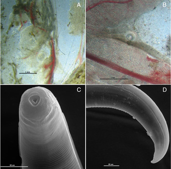
Out of 55 small mammals examined, encapsulated larvae were found in one adult male tundra vole captured from a riparian forbs meadow (N66.278967° E36.566988°) and one adult female grey-sided vole captured from a moss-type pine stand (N 66.380429° E 35.679855°). In total, 78 larvae were found in M. oeconomus and 34 larvae in M. rufocanus. A majority of the larvae (78 in M. oeconomus and 27 spm. in M. rufocanus) were found in the walls of the large intestine and mesentery (Fig. 1A and B), and seven were localized in the small intestine wall in M. rufocanus. The rest of the examined organs (liver, heart, kidney, bladder, spleen, lungs, diaphragm, and intercostal muscles) contained no larvae.
Baylisascaris transfuga larvae. (A, B) Images taken under a light stereo microscope (×28). Larvae in the mesentery and large intestine wall of grey-sided vole (Myodes rufocanus). (C, D) images taken under a scanning electron microscope. Morphology of Baylisascaris transfuga larvae from tundra voles (Microtus oeconomus) from the Kola Peninsula: anterior end, dorsal view (С) and tail end, lateral view (D).

The larvae were first identified as belonging to the genus Baylisascaris Sprent 1968 based on the following morphological characters (Fig. 1C and D). The anterior end of larvae is rounded, with four slightly protruding elevations, probably corresponding to the cephalic sensillae, and with two circular spots in the lateral position (amphids?). A narrow ridge-like line on the surface divides the cephalic capsule from the remaining surface of the anterior end. Cuticular annulations are well discernible on the anterior end. Stomatal cavity is shallow, with three-radial openings of the oesophageal lumen on the bottom (Fig. 1C and D). Lateral alae start within a short distance behind the anterior end and end in close proximity to the tail terminus.
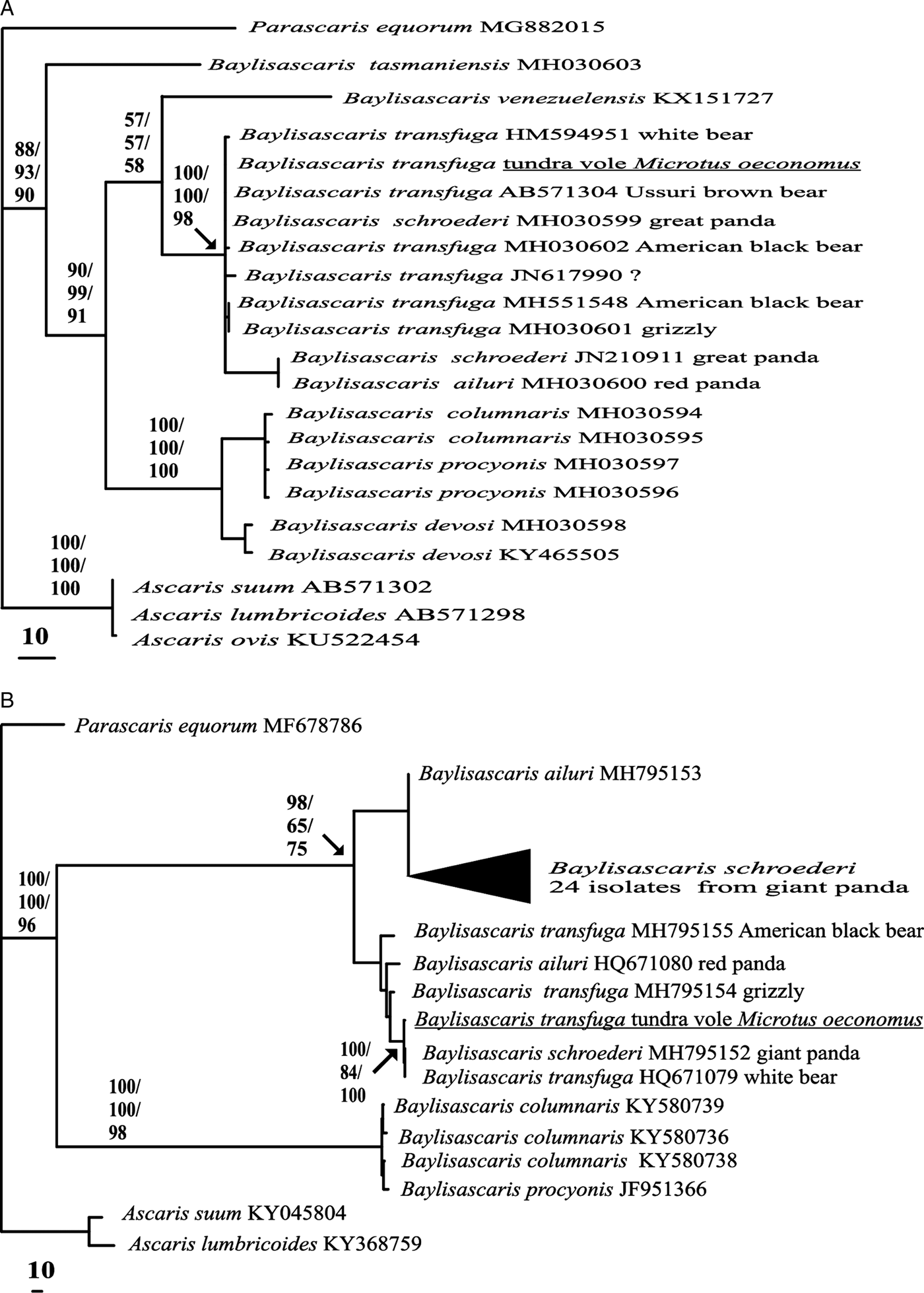
The obtained partial 830 bp long sequence of 18S rDNA of the larvae studied from the tundra vole exactly matched of Baylisascaris transfuga (accession number – U94369), as well as with that of B. schroederi. The 723 bp long ITS rDNA sequence of the larvae was found to be very similar to various Baylisascaris specimens: mainly B. transfuga, but also B. schroederi and B. ailuri from pandas (Fig. 2A). Bootstrap support for the clade of these sequences was always high (98–100%). Bootstrap support for the group B. columnaris + B. procyonis + B. devosi was also high, and the monophyly of the entire genus Baylisascaris was supported at 88–93% depending of the analysis method.
Phylogenetic relationships of Baylisascaris transfuga from tundra voles (Microtus oeconomus) from the Kola Peninsula with other ascaridid species. (A) Tree inferred from ITS rDNA analysis; (B) tree inferred from CoxI mtDNA analysis. Bootstrap support values are given in MP/NJ/ML format.

Partial sequences of the CoxI mitochondrial gene (580 bp long for CoxI_F1-CoxI_R2 primers and 520 bp for JB3-JB7_GED primers) were assembled into a single 986 bp long sequence. In the tree inferred from CoxI mtDNA analysis, the sample from the tundra vole from the Kola Peninsula clustered with sequences from B. transfuga and B. schroederi with strong or moderate bootstrap support (100% in MP and ML, and 83% in NJ analysis). The monophyly of Baylisascaris was strongly supported in the CoxI-tree, whereas some inner nodes of the Baylisascaris phylogeny were only weakly supported (Fig. 2B).
Discussion
It was expected that small rodents were involved in the Baylisascaris transfuga life cycle. Other Baylisascaris species naturally infect a variety of birds and mammals, and experimental studies have shown than B. transfuga can infect chickens, jirds, and mice, etc. (Papini et al., Reference Papini, Cavicchio and Casarosa1993, Reference Papini, Renzoni, Lo Piccolo and Casarosa1996; Sato et al., Reference Sato, Matsuo, Osanai, Kamiya, Akao, Owaki and Furuoka2004; Cho et al., Reference Cho, Egami, Ohnuki, Saito, Chinone, Shichinohe, Suganuma and Akao2007; Kazacos, Reference Kazacos2016). Following experimental inoculation of laboratory mice, B. transfuga larvae encapsulated within the walls of the large intestine (Sprent, Reference Sprent1952; Matoff and Komandarev, Reference Matoff and Komandarev1965).
In the described case, the nucleotide sequences of the studied larvae were completely identical (18S rDNA) or very similar (ITS rDNA and CoxI mtDNA) to those of Baylisascaris transfuga and two other Baylisascaris species (B. schroederi, B. ailuri). These three species from bears were forming well-supported clades under different methods of analysis, and only the polyphyletic nature of B. schroederi in some trees contradicted that phylogeny (Fig. 2A and B). An additional sequencing of Baylisascaris samples could clarify this controversy.
An alternative explanation for the observed inconsistencies in the relationship between B. transfuga and B. schroederi sequences is an intensive exchange of Baylisascaris parasites between different species of bears. Indeed, one can see that nearly identical haplotypes of Baylisascaris spp. have been detected in a wide range of Ursidae genera and species (Fig. 2A and B). We cannot exclude situation when Baylisascaris specimens were identified up to the species level with respect to host species. In some Baylisascaris studies, the DNA material was obtained from animals kept in zoos, where the exchange of parasites is possible.
The area of our field surveys is noted for a locally high density of the brown bear population (Tirronen et al., Reference Tirronen, Panchenko and Kuznetsova2015), which likely entails a high rate of the nematode B. transfuga infection in bears. Based on the limited material examined, is it hard to judge whether observed infection patterns of wild rodents are a local phenomenon or if they can be expected in other areas. Larval Baylisascaris transfuga infection might be underreported. During previous surveys in Karelia, larvae with similar morphology were reported in rodents. For example, at a Gomselga Village field station in the Republic of Karelia (N 62.069723, E 33.961420, Russia), the examination of nearly 1500 rodents revealed four cases of encapsulated and unidentified nematode larvae (three in May 2003 and one in May 2011), all associated with overwintered adult voles (unpublished). In one of the infested voles, 78 encapsulated larvae were recorded in walls of the large intestine, 16 larvae in the small intestine and 38 larvae within the liver. Unfortunately, no fixation of this material was done.
We believe that the same presence of B. transfuga infection in rodents reflects trophic relationships between bears and rodents in this area. Thus, while analysing brown bear feces, we quite often encountered tapeworm eggs of the family Anoplocephalidae, specific parasites of voles (Bugmyrin et al., Reference Bugmyrin, Tirronen, Panchenko, Kopatz, Hagen, Eiken and Kuznetsova2017). In our opinion, this is a result of the bears feeding on rodents. In the same time the feeding of voles on bear feces, containing seeds and other undigested plant material is also plausible.
It is not clear how long encapsulated larvae of B. transfuga can survive in a rodent, but their presence in the overwintered voles indicates that they stay alive at least for six or seven months. Sprent (Reference Sprent1953) previously demonstrated that the encapsulated third-stage larvae of B. devosi remained alive for at least six months within mice tissues. We cannot claim that larvae migrations lethal to the host do not occur in the population of small mammals. We believe, however, that in a majority of infected wild voles, B. transfuga would not cause fatal pathologies, and that larvae are primarily localised in intestine walls, as has been shown for laboratory animals.
The study sample size being so small, we were unable to draw any conclusions regarding the host-, season- or habitat-specific patterns of B. transfuga distribution. In our results, only 3.6% (2 out of 55) of the examined rodents were infected by B. transfuga larvae, and the Abundance was near 2. Since we did not do a full-body digest to cover the brain and muscles, and therefore, could have missed the larvae localised there, these values may be somewhat underestimated. In the literature containing data on the occurrence of other Baylisascaris species (e.g. B. procyonis), infection rates in small mammals are much higher, depending both on the host species (Weinstein, Reference Weinstein2017) and the habitat (Page et al., Reference Page, Swihart and Kazacos2001; Beasley et al., Reference Beasley, Eagan, Page, Hennessy and Rhodes2013).
The presence of Baysliascaris transfuga in rodents suggests that these rodents might act as intermediate or paratenic hosts in the B. transfuga life cycle. It appears that the voles survive the B. transfuga infection without visible debilitation and can serve as paratenic hosts for this nematode species. As some Baylisascaris species (e.g. B. procyonis) are known to be harmful for humans, the possible threat caused by the related nematode species requires further research.
Acknowledgements
The study utilized the equipment of the Core Facilities of the Severtsov Institute of Ecology and Evolution (Moscow) and the Karelian Research Centre RAS (Petrozavodsk). The authors are grateful to Drs K.F. Tirronen and D.V. Panchenko (IB KarRC RAS) for their help with field surveys in the Kola Peninsula. We thank two anonymous reviewers for improving the quality of the manuscript.
Financial support
The study was carried out under state order (projects No. 0218-2019-0075; AAAA-A18-118042490057).
Conflict of interest
None.
Ethical standards
All procedures followed were in accordance with the European Convention for the Protection of Vertebrate Animals used for Experimental and other Scientific Purposes (ETS No. 123). All necessary permits for sampling and observational field studies were obtained by the authors from the competent authorities.