Introduction
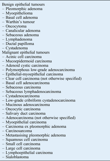
Primary salivary gland tumours are rare entities, comprising 3–5 per cent of all head and neck cancers. The most recent 2005 World Health Organization (WHO) classification of salivary gland tumours recognised 10 benign and 24 malignant primary epithelial neoplasms (Table I).Reference Eveson, Auclair, Gnepp, El-Naggar, Ellis, Simpson, Barnes, Eveson, Reichart and Sidransky1 Additional to this are a variety of non-epithelial tumour-like growths, lymphoproliferative disorders and malignancies that metastasise to salivary glands. This histopathological variety is unique to the salivary gland which can make diagnosis challenging.Reference Johnson and Myers2
Who classification of epithelial salivary gland tumours (2005)Reference Eveson, Auclair, Gnepp, El-Naggar, Ellis, Simpson, Barnes, Eveson, Reichart and Sidransky1

WHO = World Health Organization
The parotid is the most common salivary gland affected by tumour. Tumours in the parotid gland contribute to 60–75 per cent of all salivary gland tumours. Benign primary tumours are more common than malignant tumours, with the ratio approximately 3:1. However, this pattern is site specific; 75–80 per cent of parotid tumours are benign, whereas 75 per cent of sublingual tumours are malignant.Reference Johnson and Myers2–Reference Westra4 Pleomorphic adenoma is the most common benign tumour of the salivary gland, accounting for 45–75 per cent of all salivary gland tumours, followed by Warthin's tumour.Reference Johnson and Myers2, Reference Carlson and Webb3 Although incidence varies between studies, the most commonly encountered primary malignancy of the parotid gland is mucoepidermoid carcinoma, accounting for around 10 per cent of malignant salivary neoplasms. This is followed by acinic carcinoma and adenoid cystic carcinoma.Reference Johnson and Myers2, Reference Pinkston and Cole5–Reference Bjørndal, Krogdahl, Therkildsen, Overgaard, Johansen and Kristensen7
Skin malignancies of the sun-exposed face and scalp also metastasise to intra- or para-parotid lymph nodes, and can present as a parotid mass. Cutaneous squamous cell carcinoma (SCC) of the head and neck metastasises in up to 5 per cent of patients, parotid lymph nodes being the most common site for spread.Reference Veness, Porceduu, Palme and Morgan8 Lymphoma may also arise in the intra- or para-parotid lymph nodes, and, rarely, distant tumours may also metastasise to these sites.Reference Johnson and Myers2 The WHO estimates that metastatic disease accounts for 5 per cent of all malignant tumours of salivary gland masses, though this varies between populations.Reference Eveson, Auclair, Gnepp, El-Naggar, Ellis, Simpson, Barnes, Eveson, Reichart and Sidransky1 Eighty per cent of these are to the parotid gland from head and neck neoplasms, the majority being SCC, followed by melanoma. Lymphoma is almost exclusively non-Hodgkin's lymphoma, and accounts for 2 per cent of all salivary gland tumours.
Cutaneous metastases to the parotid gland may be of particular importance in Australia. Australia has the highest world incidence of skin cancers, especially non-melanoma. The annual incidence of non-melanoma skin cancer in Australia is almost 1200 per 100 000 population, with Queensland having the largest number of skin cancer cases.Reference Saleh, Lam, Gertraud Buettner, Glasby, Raasch and Ho9 Nevertheless, the prevalence of parotid tumours, especially in terms of cutaneous metastases, has not been studied in depth in Queensland. Our study aimed to identify the pathology patterns of parotidectomies performed at the Gold Coast health district so as to gain understanding of the likely diagnosis of patients presenting with a clinical parotid mass suspicious of malignancy.
Materials and methods
A retrospective review was performed of consecutive parotidectomy cases under the care of the ENT Department at the Gold Coast University Hospital from 2006 to 2013. The Gold Coast University Hospital is the only tertiary referral centre in the Gold Coast in Queensland, Australia, covering a population of around 550 000 people at Gold Coast and also serving a population in the surrounding areas of northern New South Wales. Low-risk ethics clearance was obtained.
A search of the Operating Room Management Information System (‘ORMIS’) database was conducted to identify patients, using the key words ‘parotidectomy’ and ‘ENT’. Clinical data from patient medical records and histopathology reports were reviewed.
In total, 158 operations were performed between July 2006 and December 2013. All pathological specimens were examined at the Gold Coast health district pathology department. Histological outcomes were descriptively broken down into four groups; namely, benign tumours, primary parotid malignancies, metastatic or infiltrative malignancies, and benign miscellaneous diseases.
Subgroup analysis was performed to determine the prevalence of different pathologies of patients presenting with a clinical parotid mass. In order to maintain epidemiological accuracy, cases of recurrent disease were excluded, as were elective parotidectomies for primary cutaneous malignancy without a clinical parotid mass. A total of 14 cases were excluded. Ten parotidectomies performed for primary cutaneous malignancy were considered elective or without a clinical parotid mass. Three cases were excluded for recurrent disease; two myoepithelial carcinoma recurrence in the same patient and one recurrent case of SCC from skin primary. One case of pre-auricular sinus extending into the parotid gland was also excluded. Histological outcomes were grouped as described above.
Results
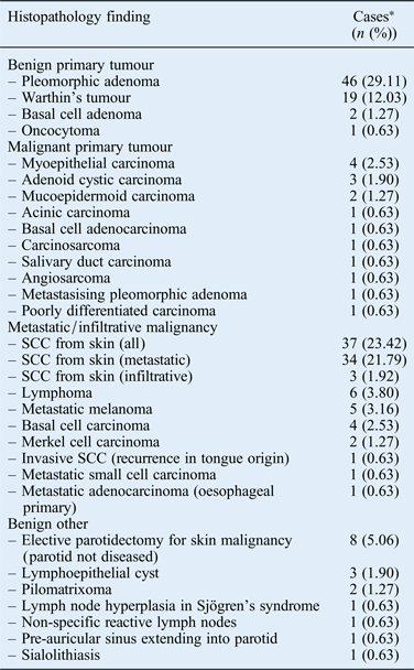
In the whole population in this study (n = 158) (Table II), 53.80 per cent of the pathologies observed in the parotid gland were benign and 46.20 per cent were malignant. These covered 28 different histological patterns. Pleomorphic adenoma was the most common tumour, accounting for 29.11 per cent of cases (Figure 1). This was followed by cutaneous SCC (23.42 per cent) (Figure 2) and Warthin's tumour (12.03 per cent) (Figure 3). Patients' ages ranged from 8 to 94 years, with an average age of 60 years (median, 63 years).
Histology of pleomorphic adenoma, which is composed of ducts in a chondroid myxoid matrix. (H&E; ×7)
Parotid gland is infiltrated by well-differentiated squamous cell carcinoma, indicated by keratin (right side). (H&E; ×5)
Histology of Warthin's tumour, which is composed of oncocytic epithelium with lymphoid stroma. (H&E; ×5)
Histopathology findings of all parotidectomies
*Total n = 158. SCC = squamous cell carcinoma
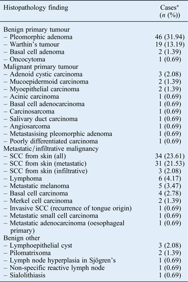
In the subgroup analysis of the parotid masses (n = 144) (Table III), pleomorphic adenoma remained of the highest prevalence at 31.94 per cent, followed by metastatic or infiltrative SCC at 23.61 per cent and Warthin's tumour at 13.19 per cent. Other primary benign tumours included basal cell adenoma (two cases) and oncocytoma (one case).
Histopathology findings of patients presenting with parotid mass
*Total n = 144. SCC = squamous cell carcinoma
There were 14 cases of primary parotid malignancy. Adenoid cystic carcinoma was the most common, with three cases (2.03 per cent), followed by mucoepidermoid and myoepithelial carcinoma, both with two cases (1.39 per cent). The remaining primary malignancies had varying histological patterns, with one case of each.
Forty-five parotidectomies were performed for metastatic or invasive cutaneous malignancies. In the SCC group, average patient age was 74 years (range, 40–94 years; median, 74 years). Three cases were infiltrative advanced lesions located immediately anterior to the ear, overlying the parotid gland. These ranged in size from 2.7 to 7.2 mm in the longest dimension at the skin, resulting in a clinically evident lesion suggestive of malignancy. In these three cases, the parotid lymph nodes were not involved on histological evaluation. The remaining 31 cases were metastatic cutaneous SCC to the intra- and para-parotid lymph nodes. Of these, two cases had unknown primaries. Four individuals presented with concurrent cutaneous SCC, which was excised at the time of parotidectomy; the remainder had previous known SCC, excised from the face, ear or scalp region.
Of the other secondary cutaneous tumours, five cases were metastatic melanoma to the parotid, four were cases of infiltrative basal cell carcinoma from skin overlying the parotid gland (Figure 4) and two were cases of metastatic Merkel cell carcinoma (Figure 5). There were six cases of lymphoma, one case of recurrent SCC of tongue origin invading into the parotid, and one case each of metastatic small cell carcinoma and metastatic adenocarcinoma from the oesophagus.
Strands of basal cell carcinoma in myxoid stroma replacing nearly the entire parotid gland. (H&E; ×8)
Parotid gland is infiltrated by Merkel cell carcinoma, indicated by small round tumour cells with hyperchromatic nuclei (right side). (H&E; ×8)
Eight parotidectomies were performed for benign disease including lymphoepithelial cyst, pilomatrixoma, reactive lymph nodes (including Sjögren's syndrome) and sialolithiasis.
Discussion
Results from this study show that metastatic cutaneous SCC to the parotid accounted for a large group of patients presenting with a parotid mass in our health district in Queensland, Australia. Our observed prevalence of metastatic SCC of 21.53 per cent is more than double the 9.17 per cent in the New South Wales based study by O'Brien et al.,Reference O'Brien, Malka and Mijailovic10 and is significantly higher than the world incidence of 5 per cent of malignant salivary gland tumours.Reference Eveson, Auclair, Gnepp, El-Naggar, Ellis, Simpson, Barnes, Eveson, Reichart and Sidransky1 Previous large case series of salivary gland tumours mostly focus exclusively on primary epithelial salivary gland tumours, excluding metastatic malignancies.Reference Pinkston and Cole5, Reference Bjørndal, Krogdahl, Therkildsen, Overgaard, Johansen and Kristensen7 Therefore, we cannot directly compare our data in this regard. The aforementioned O'Brien study assessed 242 parotidectomies at a single centre from 1987 to 1992, and histological outcomes included metastatic disease.
The large number of metastatic SCC in our study is a reflection of Australia's, and in particular Queensland's, high rate of skin cancer. We expect that other centres around the world would have differing trends. The findings may also, in part, be a result of the referral of head and neck cancer patients from the private to the public sector. Socioeconomic status and limited access to healthcare for rurally based individuals may also contribute.
Veness et al. reported 70–80 per cent of patients with an identifiable cutaneous SCC who subsequently presented with metastatic disease, after treatment of the cutaneous lesion, rather than presenting with a concurrent primary.Reference Veness, Porceduu, Palme and Morgan8 The median time for developing local nodal metastases was 12 months following treatment, with late relapses of up to 3 years occurring. Our results support this; 25 out of 31 patients with metastatic SCC to the parotid had previously treated facial or scalp SCC. The time from initial treatment to presentation was not known and could not be included in the retrospective data collection. In addition, many patients had multiple SCCs previously excised, making identification of the index lesion extremely difficult. Only four patients in the metastatic SCC group presented with concomitant primary cutaneous SCC at the time of parotidectomy. Two out of 31 patients with metastatic SCC (6.45 per cent) had no identifiable primary cutaneous SCC in their history, which is far lower than the 20–30 per cent reported by Veness et al.Reference Veness, Porceduu, Palme and Morgan8 The reported risk factors for developing nodal metastases to the parotid include: tumour size (larger than 2 cm), tumour thickness (greater than 4–5 mm), recurrent disease, high histological grade, peri-neural and lymphovascular invasion, and tumour location close to the parotid gland (ear, cheek, temple or lateral forehead).Reference Veness, Porceduu, Palme and Morgan8
When primary parotid tumours are isolated, our results are similar to those of other case series.Reference Eveson, Auclair, Gnepp, El-Naggar, Ellis, Simpson, Barnes, Eveson, Reichart and Sidransky1, Reference Pinkston and Cole5, Reference Ellis and Auclair6 For instance, Pinkston and Cole studied the incidence of salivary gland tumours over a six-year period in Alabama USA, excluding metastatic disease.Reference Pinkston and Cole5 In that study, 212 parotid cases were identified; 53.3 per cent of these were pleomorphic adenoma, followed by 28.3 per cent Warthin's tumour. We had 82 primary parotid tumours presenting as a parotid mass over the 7-year period. Sixty-eight of these were benign and 14 were malignant. The most frequently occurring primary tumour was pleomorphic adenoma (56.10 per cent), followed by Warthin's tumour (23.17 per cent).
Primary epithelial malignancy was much less common, as expected.Reference Johnson and Myers2, Reference Westra4, Reference Pinkston and Cole5 Of these, 21.43 per cent were adenoid cystic carcinoma, 14.29 per cent were mucoepidermoid carcinoma and 14.29 per cent were myoepithelial carcinoma. If we include recurrent disease, myoepithelial carcinoma was the most common, at 25 per cent. These results differ to other case series, though we acknowledge low numbers make it difficult to ascertain trends.Reference Pinkston and Cole5, Reference Bjørndal, Krogdahl, Therkildsen, Overgaard, Johansen and Kristensen7 Pinkston and Cole reported 31 malignant tumours out of 212 cases. Of these, mucoepidermoid carcinoma was the most common, accounting for 61.3 per cent of malignancies, followed by adenocarcinoma at 9.7 per cent.Reference Pinkston and Cole5
• The parotid gland is the most common salivary gland affected by tumour; histopathological patterns are vast and unique to the salivary gland
• Australia, particularly Queensland, has the highest world incidence of skin cancer
• Over 20 per cent of parotid masses were metastatic squamous cell carcinoma, much higher than the world incidence of 5 per cent of salivary malignancies
• This reflects the raised incidence of non-melanoma skin cancer in Queensland and Australia
• The information presented aids appropriate triage and timely management
• Primary parotid tumour incidence rates were similar to other case series, with pleomorphic adenoma most common, followed by Warthin's tumour
Fine needle aspiration (FNA) biopsy can be performed in the investigation of salivary gland tumours, and is routinely performed at our centre to aid in diagnosis, triage to surgery and pre-operative planning. The multitude of parotid gland pathologies can make interpretation of FNA difficult: the parotid gland is known for having the highest rate of error in biopsies of the head and neck.Reference Eveson, Auclair, Gnepp, El-Naggar, Ellis, Simpson, Barnes, Eveson, Reichart and Sidransky1, Reference Johnson and Myers2 Thus, diagnostic accuracy of FNA was beyond the scope of this study.
A limitation of our study to evaluate total population incidence of parotid pathology is attributable to our case identification. We investigated a population of people with parotid disease who underwent parotidectomy exclusively. We therefore missed cases of patients with benign disease who were managed conservatively because of poor fitness for surgery and those with unresectable malignant disease who underwent palliative treatment only. We estimate that there were very few such cases, with little consequence for overall outcomes.
Our study is unique, including metastatic parotid disease cases so as to gain an understanding of the likely pathology of patients presenting with a parotid mass in our health district. In our case series, primary parotid neoplasms had similar prevalence rates to those of other studies, with benign pleomorphic adenomas being most common. Squamous cell carcinoma from a cutaneous primary accounted for nearly a quarter of parotid masses, a result which no doubt reflects the high incidence of non-melanoma skin cancer in Queensland and Australia in general.
This information, when considered in context with the clinical scenario, can indicate the likely histopathological diagnosis of a patient presenting to our centre with a parotid mass. Furthermore, this aids appropriate triage and timely management. Comprehensive patient history-taking must cover previous head and neck skin cancers. Initial clinical examination should include a careful examination of the face and scalp skin for skin cancers, and biopsy of any suspicious skin lesions should be performed.
Acknowledgement
The authors would like to thank Ms Melissa Leung for her assistance in the laboratory in acquiring the histological images.