Implications
Parthenogenetic activation of mammalian oocytes is commonly used in reproductive biotechnologies, for example, cloning using nuclear transfer, and its success depends on an adequate artificial stimulus. Nitric oxide (NO) has been described as an oocyte activator but it does not induce a cortical reaction. Protein kinases C (PKC) play an important role in the cortical reaction but the PKC cascade is not triggered after oocyte activation by an NO-donor. There is a possibility of improving the results of oocyte activation by using an NO-donor together with PKC stimulation. We verified that this treatment induces meiosis resumption accompanied by a cortical reaction in pig oocytes. These results might lead to the optimization of oocyte activation using NO.
Introduction
Mammalian oocyte activation involves a sequence of morphological and biochemical events including an increase in intracellular calcium levels, cortical granule (CG) exocytosis and release from the second meiotic arrest. Under physiological conditions, oocyte activation is induced by sperm penetrating the oocyte (reviewed by Williams, Reference Williams2002). The sperm contains a sperm-specific phospholipase C isoform (PLCζ; Saunders et al., Reference Saunders, Larman, Parrington, Cox, Royse, Blayney, Swann and Lai2002), which stimulates the production of diacylglycerol (DAG) and inositol 1,4,5-trisphosphate (IP3), a common Ca2+-releasing compound, from phosphatidyl-inositol 4,5-bisphosphate (PIP2). Elevation in intracellular calcium causes the fusion of CGs with oolema. The enzymes released from the CGs into the perivitelline space modify the zona pellucida to prevent additional sperm penetration (Bleil et al., Reference Bleil, Beall and Wassarman1981; reviewed by Williams, Reference Williams2002). Calcium oscillations also induce cyclin B degradation and cytostatic factor inactivation, allowing the female chromatin entry into anaphase II (Lorca et al., Reference Lorca, Cruzalegui, Fesquet, Cavadore, Méry, Means and Dorée1993).
Under in vitro conditions, various artificial stimuli increase the intracellular calcium levels, for example, electric pulses (Sun et al., Reference Sun, Hoyland, Huang, Mason and Moor1992; Koo et al., Reference Koo, Jang, Kwon, Kang, Kwon, Park, Kang and Lee2008), calcium ionophore (Jílek et al., Reference Jílek, Hüttelová, Petr, Holubová and Rozinek2000 and Reference Jílek, Hüttelová, Petr, Holubová and Rozinek2001; Che et al., Reference Che, Lalonde and Bordignon2007) and chemicals that modulate the intracellular turnover of Ca2+ (Petr et al., Reference Petr, Rozinek, Jílek and Urbánková2000 and Reference Petr, Urbánková, Tománek, Rozinek and Jílek2002) and are used for the parthenogenetic activation of porcine oocytes.
Protein kinase C (PKC) is one of the transducers of the calcium signal system. Several studies have demonstrated the possible involvement of PKC in the oocyte activation process (Colonna et al., Reference Colonna, Tatone, Francione, Rosati, Callaini, Corda and Difrancesco1997; Green et al., Reference Green, Kim, Wang, Day and Prather1999; Sedmíková et al., Reference Sedmíková, Rajmon, Petr, Švestková, Chmelíková, Bantirgu, Rozinek and Jílek2006) and the exocytosis of CGs (Bement and Capco, Reference Bement and Capco1989; Fan et al., Reference Fan, Tong, Li, Lian, Chen, Schatten and Sun2002). PKC belongs to the serine/threonine kinase, and 12 isoforms are known, being classified into three groups. Conventional isoforms (PKC-α, -βI, -βII and -γ) are activated by free Ca2+ ions and DAG. Novel PKCs (nPKCs), represented by PKC-δ, -ε, -η, -μ, -θ isoforms, are independent of Ca2+ but can be activated by DAG. Finally, the PKC-ζ, -λ, -τ isotypes belong to the atypical PKCs (aPKCs) and are independent of both Ca2+ and DAG (Liu and Heckman, Reference Liu and Heckman1998). The role of PKC activation in the exit from metaphase II of porcine oocytes has been studied using PKC-activators such as phorbol-12-myristate-13-acetate (PMA: an activator of all isotypes of PKC; Sun et al., Reference Sun, Wang, Hosoe, Taniguchi, Chen and Shioya1997; Ito et al., Reference Ito, Shimada and Terada2003) and 1-oleoyl-2-acetyl-glycerol (OAG: an activator of calcium-dependent PKC isotypes; Sun et al., Reference Sun, Wang, Hosoe, Taniguchi, Chen and Shioya1997). After oocyte treatment and their cultivation for an additional 20 h, the female pronucleus was not observed either with PMA or with OAG (Sun et al., Reference Sun, Wang, Hosoe, Taniguchi, Chen and Shioya1997). Regarding the effect of PKC-activators on CG exocytosis, pig oocytes were treated with PMA (Sun et al., Reference Sun, Wang, Hosoe, Taniguchi, Chen and Shioya1997; Fan et al., Reference Fan, Tong, Li, Lian, Chen, Schatten and Sun2002) and OAG, resulting in no exit from metaphase II but in CG exocytosis (Sun et al., Reference Sun, Wang, Hosoe, Taniguchi, Chen and Shioya1997).
Oocyte activation can also be triggered by nitric oxide (NO), an important intracellular signaling molecule (Kuo et al., Reference Kuo, Baxter, Thompson, Stricker, Patton, Bonaventura and Epel2000; Petr et al., Reference Petr, Rajmon, Rozinek, Sedmíková, Ješeta, Chmelíková, Švestková and Jílek2005 and Reference Petr, Chmelíková, Krejčová, Řehák, Novotná and Jílek2010). In pig oocytes, NO-donors such as S-nitroso-N-acetylpenicillamine (SNAP) or sodium nitroprusside (SNP) are able to induce parthenogenetic activation after 24 h (78.0% activation). However, despite this high rate of exit from metaphase II, this activation does not involve the complete spectrum of events observed in sperm-activated oocytes, since NO-donor-activated oocytes do not exhibit CG exocytosis (Petr et al., Reference Petr, Rajmon, Rozinek, Sedmíková, Ješeta, Chmelíková, Švestková and Jílek2005).
Several studies have demonstrated that oocyte activation and CG exocytosis are two independent processes (Sun et al., Reference Sun, Wang, Hosoe, Taniguchi, Chen and Shioya1997; Petr et al., Reference Petr, Rajmon, Rozinek, Sedmíková, Ješeta, Chmelíková, Švestková and Jílek2005). However, there are no data comparing CG quantification and nuclear status after activation in the same oocyte and no data showing the combined effect of PKC-activators together with an NO-donor on the cortical reaction and meiosis resumption. In pig oocytes, a cortical reaction can be triggered by several artificial stimuli (Sun et al., Reference Sun, Hoyland, Huang, Mason and Moor1992; Okada et al., Reference Okada, Miyano and Miyake2003), although the observed reaction is weaker when induced by a calcium ionophore and an electrical pulse (75.7% and 76.9% of CG release, respectively) than the one triggered by sperm penetration (86.3% CG exocytosis; Wang et al., Reference Wang, Abeydeera, Prather and Day1998). Although many studies have shown that CG exocytosis is a PKC-dependent event, phorbol esters do not mimic the release of CGs exactly as in a fertilized mouse egg and CG exocytosis shows an atypical pattern (Ducibella and LeFevre, Reference Ducibella and LeFevre1997). Furthermore, there is evidence that a cortical reaction can be triggered independently either by an increase in Ca2+ (Sun et al., Reference Sun, Wang, Hosoe, Taniguchi, Chen and Shioya1997; Eliyahu et al., Reference Eliyahu, Tsaadon, Shtraizent and Shalgi2005) or by PKC (Eliyahu et al., Reference Eliyahu, Tsaadon, Shtraizent and Shalgi2005).
The aim of this study was to verify the hypothesis that an NO-donor together with a specific PKC-activator induces porcine oocyte activation accompanied by CG exocytosis.
Material and methods
Culture media
Unless otherwise indicated, all chemicals and reagents were purchased from Sigma-Aldrich Química S.A. (Madrid, Spain). The medium used for oocyte maturation was NCSU-37 (Petters and Wells, Reference Petters and Wells1993) supplemented with 0.57 mM cysteine, 1 mM dibutyryl cAMP (dbcAMP), 5 mg/ml insulin, 50 mM β-mercaptoethanol, 10 IU/ml eCG (Foligon, Intervet International B.V., Boxmeer, Holland), 10 IU/ml hCG (Veterin Corion, Divasa Farmavic, Barcelona, Spain) and 10% porcine follicular fluid (v/v) (Romar et al., Reference Romar, Coy, Gadea and Rath2005).
Oocyte collection and in vitro maturation
Within 30 min of slaughter, ovaries from gilts were transported to the laboratory in saline containing 100 mg/ml kanamycin sulfate at 38.5°C, washed once in a 0.04% cetrimide solution and twice in saline. Oocyte–cumulus cell complexes (COCs) were collected from the antral follicles (3 to 6 mm diameter) and washed twice with Dulbecco's phosphate-buffered saline (DPBS) supplemented with 1 mg/ml polyvinyl alcohol (PVA) and 0.005 mg/ml red phenol and twice more in maturation medium previously equilibrated for a minimum of 3 h at 38.5°C under 5% CO2 in the air. Only COCs with a complete and dense cumulus oophorus were used for the experiments. Groups of 50 to 55 COCs were cultured in 500 μl maturation medium for 22 h at 38.5°C under a 5% CO2 atmosphere. After culture, COCs were washed twice in a fresh maturation medium without dbcAMP, eCG and hCG, and cultured for an additional 20 to 22 h (Funahashi et al., Reference Funahashi, Cantley and Day1997).
Cultivation of oocytes with PKC-activators and an NO-donor
The PKC-activators used were phorbol-12-myristate-13-acetate (PMA; Calbiochem, Germany), an activator of all isotypes of PKC, 1-oleoyl-2-acetyl-sn-glycerol (OAG; Calbiochem, Germany), an activator of calcium-dependent PKC isotypes, and l-α-phosphatidylinositol-3,4,5-trisphosphate dipalmitoyl heptaammonium salt (DPAM; Calbiochem, Germany), an activator of calcium-independent PKC-δ, -ε, -η. As an NO-donor, S-nitroso-N-acetylpenicillamine (SNAP; Axxora, Germany) was used. All PKC-activators were dissolved in dimethylsulfoxide (DMSO) in a stock solution (10 mM PMA, 40 mM OAG, 200 μM DPAM) and then diluted to the desired concentration for the experiments. The stock of SNAP (2 mM) was freshly prepared every day. The final DMSO concentration in all cases was 1%. After the in vitro maturation (IVM) period (42 to 44 h), cumulus cells were removed by pipetting, and denuded oocytes were treated for 10 h with an NO-donor or PKC-activators in fresh supplemented NCSU-37 without dbcAMP or hormones, at 38.5°C, under a 5% CO2 atmosphere.
Assessment of oocyte activation
After treatment, the oocytes were fixed for 30 min (0.5% glutaraldehyde in DPBS), stained for 15 min (1% Hoechst 33342 in DPBS), washed in PBS containing 1 mg/ml polyvinylpyrrolidone and mounted on glass slides. The samples were examined under an epifluorescence microscope at 200× and 400× magnifications. The germinal vesicle and metaphase I oocytes were discarded from the study. Oocytes at metaphase II were not considered as activated. Activation was considered to have occurred when the oocytes reached the anaphase II, the telophase II or the pronuclear stage.
Assessment of CGs
The methods of CG visualization were based on those described by Yoshida et al. (Reference Yoshida, Cran and Pursel1993), with modifications in the fluorescein isothiocyanate-lectin concentration (Coy et al., Reference Coy, Gadea, Romar, Matas and Garcia2002; Romar et al., Reference Romar, Coy, Gadea and Rath2005). Briefly, after treatment with an NO-donor and PKC-activators, oocytes were washed three times in DPBS without calcium chloride (DPBS-w/o). Thereafter, the cells were fixed with freshly prepared 3.7% paraformaldehyde in DPBS-w/o (30 min, room temperature) and washed in DPBS-w/o. This was followed by treatment with freshly prepared 0.1% Triton X-100 in DPBS-w/o for 5 min and three washes in DPBS-w/o. Oocytes were then incubated in the dark with 10 μg/ml FITC-PNA (lectin from Arachis hypogaea conjugated to fluorescein isothiocyanate) in DPBS-w/o for 30 min, followed by three washes in DPBS-w/o. Finally, the oocytes were incubated with 5 μg/ml Hoechst 33342 for 10 min to stain DNA. After staining, the oocytes were gently washed in DPBS-w/o and an equilibration buffer medium (Slowfade® Antifade kit, Molecular Probes, Inc./Life Technologies Czech Republic s.r.o., Prague, Czech Republic). An adhesive ring was attached to a clean slide, 5 to 7 oocytes were transferred inside the ring with 4 μl of antifade mounting medium and finally protected with a coverslide sealed with nail polish. The samples were kept in the dark at 4°C until confocal assessment in <48 h.
The slides were examined under a 63× oil objective of a modular confocal microscope (Eclipse C1 Plus, Nikon, Tokyo, Japan) equipped with a 408 nm diode laser and a 488 nm Argon ion laser. All oocytes were observed under the same setting parameters (14% laser intensity, 87 gain and 1 μm thickness section) from the outer zona pellucida in one oocyte's pole to the opposite pole. The nuclear stage was recorded by means of Hoechst fluorescence. The fluorescent emissions from the oocytes were recorded as TIFF files using the software attached to the microscope. The largest diameter section (equator of the oocyte) was used for further image analysis using MIP-4.5 Microm Image Processing software (Consulting de Imagen Digital S.L., Microm, Barcelona, Spain). The quantitative measurement of CG density (FITC fluorescence intensity) was performed by selecting the FITC-PNA-stained portion (area occupied by CG) in the space immediately below the oolema, as we have previously described (Coy et al., Reference Coy, Gadea, Romar, Matas and Garcia2002; Romar et al., Reference Romar, Coy, Gadea and Rath2005). Briefly, on each captured image, the oocyte's cortical area was selected by drawing two concentric circles. The selected cortical area was processed for further image analysis and the area occupied by CG was calculated as the FITC-PNA-labeled area divided by the entire cortical selected area x 100. In each oocyte, the data of cortical area occupied by CG (%) were calculated and saved for further analysis. The degree of CG exocytosis was evaluated based on cortical area occupied by CG (%) and oocytes were classified into four different groups: complete exocytosis of CG (oocytes with 0% to 5% area occupied by CG), effective CG exocytosis (oocytes with 5% to 8% area), initial CG exocytosis (oocytes with 8% to 15% area) and no cortical reaction (oocytes over 15% area). This classification is based on previous studies with porcine oocytes showing that just after the IVM cortical area occupied by CG is around 20%, whereas in fertilized oocytes assessed 18 h post insemination, it is around 5% (Coy et al., Reference Coy, Gadea, Romar, Matas and Garcia2002; Romar et al., Reference Romar, Coy, Gadea and Rath2005). Penetrated pig oocytes assessed at 4 h post insemination show 8% of the cortical area occupied by CG (Romar et al., 2012).
Experimental design
Experiment 1: effect of PKC-activators and an NO-donor on oocyte activation
Immediately after IVM, all the oocytes were denuded by pipetting and an aliquot (n = 10) was fixed to assess the nuclear status (IVM group). The remaining oocytes were randomly incubated for 10 h in a medium containing 2 μM DPAM (DPAM group), 400 μM OAG (OAG group), 100 nM PMA (PMA group), 2 mM SNAP (SNAP group) or the medium alone (Control group). To study the combined effect of an NO-donor and PKC-activators, some oocytes were also incubated for 10 h with SNAP (0.5 mM) together with PKC-activators at the same concentration as above: SNAP–PMA, SNAP–OAG and SNAP–DPAM groups. The concentrations of PKC-activators and the NO-donor were selected based on previous results (Petr et al., Reference Petr, Rajmon, Rozinek, Sedmíková, Ješeta, Chmelíková, Švestková and Jílek2005 and Reference Petr, Krejčová, Rajmon and Jílek2011). Since DMSO has been reported to induce a loss of electrodensity in pig CGs (Campagna et al., Reference Campagna, Bailey, Sirard, Ayotte and Maddox-Hyttel2006), an aliquot of oocytes was also incubated for 10 h in a medium supplemented with 1% DMSO (DMSO group). As a positive control of oocyte activation, an aliquot of denuded IVM oocytes was exposed to calcium ionophore A23187 (25 μM in DMSO, 5 min; Jílek et al., Reference Jílek, Hüttelová, Petr, Holubová and Rozinek2001). After ionophore treatment, the oocytes were washed and incubated for 10 h in NCSU-37 as with the rest of the groups (Ca-Io group). All oocytes in all the experimental groups were incubated for the same time. After treatment, oocytes from all groups were fixed and stained to observe the nuclear status and to evaluate oocyte activation. This experiment was replicated 4 times with 15 oocytes per group, except the IVM group (10 oocytes per replicate).
Experiment 2: effect of PKC-activators and an NO-donor on cortical reaction
The experimental groups (IVM, Control, DMSO, Ca-Io, DPAM, OAG, PMA, SNAP, SNAP–DPAM, SNAP–OAG, SNAP–PMA) were the same as in Experiment 1. Both after IVM and after the different treatments, the oocytes were processed for a confocal study. In each oocyte, the nuclear status was recorded and the rate of area occupied by CG was quantified. To study CG exocytosis, three repetitions (5 oocytes/group) were performed.
Statistical analysis
The data are presented as mean ± s.d., and all rates were modeled according to the binomial model of parameters. The data were analyzed by a one-way ANOVA with the treatment as a fixed factor. When ANOVA revealed a significant effect, differences among the experimental groups were determined using the post hoc Tukey test. The P-value < 0.05 was considered to indicate statistical significance.
Results
Experiment 1: effect of PKC-activators and an NO-donor on oocyte activation
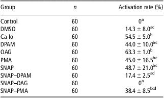
The results showed that both after IVM (IVM group) and after 10 h in NCSU-37 medium (Control group), 100% of oocytes remained at metaphase II and any oocyte exited from this stage (Table 1). The addition of 1% DMSO to the medium did not affect the activation of oocytes (only 14.3 ± 8.0% were activated), but treatment with calcium ionophore did, and 54.5 ± 5.0% of the oocytes exited from MII. Dipalmitoyl (DPAM), which activates calcium-independent PKC isoforms, induced parthenogenetic activation in 44.0 ± 10.0% of pig oocytes. Similarly, PMA, an activator of all PKC isotypes, activated 45.0 ± 16.5% of the oocytes. Oleoyl (OAG), an activator of calcium-dependent PKC isoforms, was the most effective in the activation of oocytes, inducing exit from MII in 63.3 ± 1.0% of the oocytes. The NO-donor alone activated 48.7 ± 21.0% of the oocytes. A combination of PKC-activators and an NO-donor did not lead to oocyte activation. Only 17.4 ± 2.5% of oocytes treated with dipalmitoyl and SNAP exited from the MII stage. The combination of PMA and SNAP activated 38.4 ± 8.5% of the oocytes, but the combined treatment of an NO-donor with OAG did not induce oocyte activation (0%).
Oocyte activation results in in vitro matured pig oocytes after treatment with different PKC-activators and an NO-donor
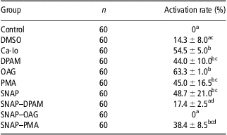
PKC = protein kinase C; NO = nitric oxide; DMSO = dimethylsulfoxide; Ca-Io = calcium ionophore A23187; DPAM = l-α-phosphatidylinositol-3,4,5-trisphosphate dipalmitoyl heptaammonium salt; OAG = 1-oleoyl-2-acetyl-sn-glycerol; PMA = phorbol-12-myristate-13-acetate; SNAP = S-nitroso-N-acetylpenicillamine; n = number of oocytes assessed.
The oocytes were treated with DMSO, calcium ionophore, PKC-activators (DPAM, OAG or PMA) or NO-donor (SNAP) for 10 h in NCSU-37 medium. Activation was considered to have occurred when the oocytes reached the anaphase II, the telophase II or the pronuclear stage. The data are presented as mean ± s.d.
Different lettersa,b,c,d indicate differences among groups (P < 0.05).
Experiment 2: effect of PKC-activators and an NO-donor on cortical reaction
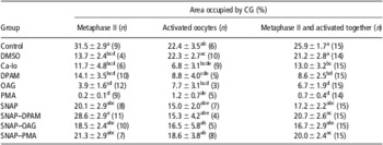
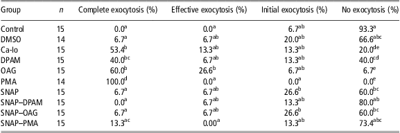
All oocytes in all the experimental groups were treated as in Experiment 1. The individual analysis of nuclear status and CG quantification revealed that after activation treatment, a cortical reaction had occurred in some oocytes at the metaphase II stage, and some oocytes that had reassumed meiosis still had their CGs under oolema (Table 2). These data confirm that nuclear progression and CG exocytosis can occur as independent events in pig oocytes. Because of this observation and in order to accurately analyze the likely differences in the cortical reaction among treatments, all oocytes from each group were considered together independent of their nuclear status. The results showed that after 42 to 44 h IVM, 17.8 ± 2.4% of the cortical area was occupied by CG. Matured oocytes incubated for 10 h in NCSU-37 medium (Control group) showed 25.9 ± 1.7% of their area occupied by PNA and the presence of 1% DMSO in the medium did not have any effect on the rate of CG area (21.2 ± 2.8%; Table 2). However, activation with a calcium ionophore-induced CG exocytosis, and the cortical area occupied by CG decreased significantly compared with the control group (25.9 ± 1.7% v. 13.0 ± 3.2%). PKC-activators (DPAM, OAG and PMA) significantly reduced the cortical area occupied by CG (Table 2), and all PKC-activators induced the exocytosis of CG (Figure 1). The area occupied by CG decreased after incubation with PKC-activators compared with those oocytes incubated for 10 h in the same medium without activators (8.6 ± 2.5, 6.7 ± 1.9 and 0.7 ± 0.4 for DPAM, OAG and PMA groups v. 25. 9 ± 1.7% in the Control group). When oocytes were classified according to the degree of cortical reaction, the results showed that PMA, which activates all isoforms of PKC, induced the greatest cortical reaction of all the PKC-activators used, whereas the weakest cortical reaction was induced by dipalmitoyl (DPAM), an activator of calcium-independent PKC isotypes (Figure 1; Table 3).
Cortical area occupied by CG (%) based on nuclear status in in vitro matured pig oocytes after treatment with different PKC-activators and an NO-donor

CG = cortical granule; PKC = protein kinase C; NO = nitric oxide; DMSO = dimethylsulfoxide; Ca-Io = calcium ionophore A23187; DPAM = l-α-phosphatidylinositol-3,4,5-trisphosphate dipalmitoyl heptaammonium salt; OAG = 1-oleoyl-2-acetyl-sn-glycerol; PMA = phorbol-12-myristate-13-acetate; SNAP = S-nitroso-N-acetylpenicillamine; n = number of assessed oocytes.
The oocytes were treated with DMSO, calcium ionophore, PKC-activators (DPAM, OAG or PMA) or NO-donor (SNAP) for 10 h in NCSU-37 medium. The area occupied by CG was calculated from images of the cortical area at the equatorial level using image analysis. The data are presented as mean ± s.e.m.
Different lettersa,b,c,d,e in the same column indicate differences among groups (P < 0.05).
Cortical granule (CG) distribution in in vitro matured porcine oocytes after treatment with different protein kinase C (PKC)-activators and a nitric oxide (NO)-donor. The matured oocytes were treated with dimethylsulfoxide (DMSO), calcium ionophore, PKC-activators (l-α-phosphatidylinositol-3,4,5-trisphosphate dipalmitoyl heptaammonium salt (DPAM), 1-oleoyl-2-acetyl-sn-glycerol (OAG) or phorbol-12-myristate-13-acetate (PMA)) and an NO-donor (S-nitroso-N-acetylpenicillamine (SNAP)) for 10 h in NCSU-37 medium. The images show the oocyte equatorial level under a confocal microscope. (a) Oocyte after 10 h in medium – Control. (b) Oocyte incubated with DMSO. (c) Oocyte activated by a calcium ionophore. (d) Oocyte treated with DPAM. (e) Oocyte treated with OAG. (f) Oocyte treated with PMA. (g) Oocyte treated with SNAP. (h) Oocyte treated with SNAP–DPAM. (i) Oocyte treated with SNAP–OAG. (j) Oocyte treated with SNAP–PMA. The scale bar represents 50 μm.

Degree of CG exocytosis in in vitro matured pig oocytes after treatment with different PKC-activators and an NO-donor
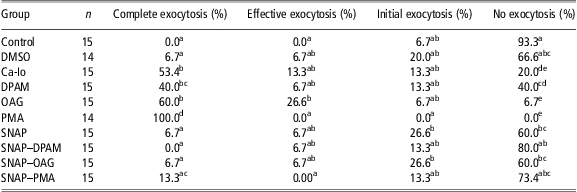
PKC = protein kinase C; NO = nitric oxide; DMSO = dimethylsulfoxide; Ca-Io = calcium ionophore A23187; DPAM = l-α-phosphatidylinositol-3,4,5-trisphosphate dipalmitoyl heptaammonium salt; OAG = 1-oleoyl-2-acetyl-sn-glycerol; PMA = phorbol-12-myristate-13-acetate; SNAP = S-nitroso-N-acetylpenicillamine; n = number of assessed oocytes; CG = cortical granule.
The degree of CG exocytosis was evaluated based on the cortical area occupied by CG. Complete exocytosis (oocytes with a 0% to 5% area occupied by CG), effective CG exocytosis (oocytes with a 5% to 8% area), initial CG exocytosis (oocytes with an 8% to 15% area) and no cortical reaction (oocytes over the 15% area). The data are presented as % oocytes of the total number of oocytes in the group, which occurred at a given level of CG exocytosis.
Different lettersa,b,c,d,e in the same column indicate differences among groups (P < 0.05).
Incubation with an NO-donor alone (SNAP group) or combined with any PKC-activator (SNAP–DPAM, SNAP–OAG and SNAP–PMA) prevented the occurrence of a cortical reaction. Treatment with SNAP alone did not induce a cortical reaction in the oocytes (Figure 1g), which reached an area stained by PNA-FITC similar to that of oocytes kept in culture (17.2 ± 2.2% v. 25.9 ± 1.7%; Table 2). The combination of an NO-donor with PKC-activators prevented CG exocytosis (Figure 1).
Discussion
Our results demonstrated a different role of PKC and an NO-donor in the parthenogenetic activation of porcine oocytes. Despite oocyte activation using these chemicals, in which a high rate (over 78%) is achieved, a full cortical reaction pattern is not always observed (Su et al., Reference Su, Xia, Byskov, Fu and Yang1999; Shimada et al., Reference Shimada, Maeda and Terada2001; Petr et al., Reference Petr, Rajmon, Rozinek, Sedmíková, Ješeta, Chmelíková, Švestková and Jílek2005). It has been shown in pigs that different events in egg activation may be unrelated to each other (Sun et al., Reference Sun, Wang, Hosoe, Taniguchi, Chen and Shioya1997), leading to some conflicting results in the literature. In the present study, both exit from MII and the cortical reaction were analyzed after the treatment of eggs with a calcium ionophore, an NO-donor and PKC-activators.
We observed the effect of individual PKC-activators on oocyte activation and CG exocytosis. PKC is involved in regulating the exit from metaphase II in porcine oocytes (Su et al., Reference Su, Xia, Byskov, Fu and Yang1999; Shimada et al., Reference Shimada, Maeda and Terada2001), but it is not entirely clear how PKC participates in the activation of oocytes. Our results show that oleoyl was the most effective in the activation of oocytes among the PKC-activators. This PKC-activator was able to induce exit from MII in 63% of the oocytes. The treatment of oocytes by dipalmitoyl or PMA induced oocyte activation only in 44% and 45% of the oocytes, respectively. In contrast to our results, Sun et al. (Reference Sun, Wang, Hosoe, Taniguchi, Chen and Shioya1997) did not observe exit from the metaphase II stage and completion of meiosis after the stimulation of PKC in mature pig oocytes. Green et al. (Reference Green, Kim, Wang, Day and Prather1999) determined that PKC inhibition leads to the activation of porcine oocytes, but Sedmíková et al. (Reference Sedmíková, Rajmon, Petr, Švestková, Chmelíková, Bantirgu, Rozinek and Jílek2006) found that inhibition of the calcium-independent isoform PKC-δ inhibits the activation of pig oocytes by a calcium ionophore. However, inhibition of PKC isoforms that are calcium-dependent is unable to suppress the activation of porcine oocytes. In our experiments, we observed 63% of activation of porcine oocytes by the activator of calcium-dependent PKC isotypes. The activator of calcium-independent PKC isoforms activated only 44% of the oocytes. The different results obtained can be attributed to the drug concentration, the duration of oocyte treatment and time for assessment of oocyte activation. The role of PKC in the activation of matured oocytes was also confirmed in Xenopus and mice oocytes, where there is a typical process for the activation of oocytes after treatment with PKC-activators (Bement and Capco, Reference Bement and Capco1991; Colonna et al., Reference Colonna, Tatone, Francione, Rosati, Callaini, Corda and Difrancesco1997). However, PKC activation does not induce a complete spectrum of typical processes for the full activation of oocytes (Moore et al., Reference Moore, Kopf and Schultz1995; Ducibella and LeFevre, Reference Ducibella and LeFevre1997). In our study, we examined the influence of individual PKC-activators on the cortical reaction in porcine oocytes. The strongest effect on CG exocytosis was achieved with PMA (activator of all PKC isoforms), which is in agreement with the results of Sun et al. (Reference Sun, Wang, Hosoe, Taniguchi, Chen and Shioya1997), who recorded CG exocytosis in porcine oocytes treated 2 h with PKC-activators (OAG and PMA) independent of the Ca2+ ion concentration. In contrast, in our experiments, DPAM, an activator of calcium-independent PKC isoforms, showed the weakest influence on CG exocytosis. Incubation of oocytes for 10 h with 100 nM PMA led to a cortical reaction in 100% of pig oocytes. Wu et al. (Reference Wu, Zhang, Li, Cheng, Kuai, Wang and Guo2006) reported similar results using 3.2 μM PMA for 15 min, with considerable CG exocytosis in 80% of treated human oocytes. However, in pigs, Sun et al. (Reference Sun, Wang, Hosoe, Taniguchi, Chen and Shioya1997) reported a full cortical reaction only in 33% of the oocytes after treatment with 100 nM PMA for 2 h. Even in this case, similar factors such as the different results of activation could have played a role.
In agreement with our previous results (Petr et al., Reference Petr, Rajmon, Rozinek, Sedmíková, Ješeta, Chmelíková, Švestková and Jílek2005), we demonstrated that the activation of pig oocytes by an NO-donor did not induce CG exocytosis. However, exit from the metaphase II stage was achieved in 48% of the oocytes. It was shown that a relatively long exposure (10 h) is needed for the activation of pig oocytes with 2.0 mM SNAP (Petr et al., Reference Petr, Rajmon, Rozinek, Sedmíková, Ješeta, Chmelíková, Švestková and Jílek2005), but activation cannot be considered complete, since this treatment does not induce a full cortical reaction. We did not observe CG exocytosis in those oocytes treated with SNAP alone, not even in combination with individual PKC-activators. The participation of individual PKC isoforms in the activation of porcine oocytes significantly differs in calcium ionophore-induced activation and activation induced by NO. When oocytes were activated by the NO-donor SNAP, there was no significant effect of PKC inhibitors on oocyte activation and CG exocytosis. This suggests that the oocyte activation events induced by the NO-donor depend on PKC to a very limited extent or are completely independent of PKC, as it has been previously suggested (Petr et al., Reference Petr, Chmelíková, Dörflerová, Ješeta and Kuthanová2007). Despite this, we hypothesize that the combined effects of an NO-donor and PKC-activator lead to an inhibition in the cortical reaction, suppressing the individual effect of PKC-activators on CG exocytosis. In pigs, CGs are exocyted within 6 h after insemination (Wang et al., Reference Wang, Sun, Hosoe, Shioya and Day1997; Coy et al., Reference Coy, Gadea, Romar, Matas and Garcia2002). However, we could not observe CG exocytosis even after 10 h of the combined treatment with NO-donor + PKC-activator. These results indicate for the first time that combined treatment is unable to induce CG exocytosis. It is possible that functional PKC pathways and NO block each other. Perhaps the activation of porcine oocytes by an NO-donor proceeds at least partly through the activation of soluble guanylyl cyclase (sGC). The role of the cyclic guanosine monophosphate (cGMP)-dependent signaling cascade is apparently specific for the parthenogenetic activation of oocytes by NO-donors (Petr et al., Reference Petr, Rajmon, Chmelíková, Tománek, Lánská, Pribanová and Jílek2006). The linkage of NO- and Ca2+-dependent cascades may be mediated by cGMP and cGMP-dependent protein kinase G (PKG). PKG may regulate the amount of endogenous agonists of IP3 receptors, thus inhibiting the activity of phospholipase C, which is part of the activation cascade of PKC (Ruth et al., Reference Ruth, Wang, Boekhoff, May, Pfeifer, Penner, Korth, Breer and Hofmann1993). Intracellular Ca2+, which the oocyte utilizes during the activation, is mobilized through the inositol trisphosphate or ryanodine receptors. In sea urchin eggs, the increase in the intracellular NO level induces an increase in the intracellular levels of free Ca2+ ions through de novo synthesis of cyclic ADP ribose (Sethi et al., Reference Sethi, Empson and Galione1996; Willmott et al., Reference Willmott, Sethi, Walseth, Lee, White and Galione1996), which is a known agonist of ryanodine receptors (Sitsapesan and Williams, Reference Sitsapesan and Williams1995). Cyclic ADP ribose alone is able to induce parthenogenetic activation in porcine oocytes. However, other mechanisms may be involved in the NO-dependent activation of pig oocytes. NO activates many molecules through their nitrosylation, for example ryanodine receptors, which, after activation, are able to induce parthenogenesis in matured pig oocytes (Petr et al., Reference Petr, Urbánková, Tománek, Rozinek and Jílek2002).
In conclusion, PKC-activators induce CG exocytosis to the same degree as a calcium ionophore. However, the combination of PKC-activators with NO-donors inhibits the stimulus, leading to exit from the metaphase II stage and a cortical reaction in pig oocytes.
Acknowledgments
We thank the staff from El Pozo slaughterhouse for supplying the ovaries. We thank Mrs Lois Russell and Mr Chris Thomas for their editorial assistance with this manuscript. This work was supported by grants GAČR 502/10/0794 and GAČR 523/08/H064 and MEC and FEDER (AGL2009-12512-C02-01). L. Tůmová's stay in Murcia was supported by an MŠMT grant.