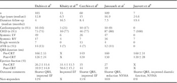
Cardiac re-synchronisation therapy is a well-established treatment option for adults with symptomatic heart failure refractory to pharmacotherapy. In patients with left ventricular dysfunction and electrical dyssynchrony secondary to left bundle branch block, cardiac re-synchronisation therapy has produced both acute and chronic improvements in left ventricular myocardial performance, exercise endurance, and quality-of-life metrics;Reference Abraham, Fisher and Smith 1 – Reference Cazeau, Leclercq and Lavergne 4 however, extrapolating such results to children has been fraught with the heterogeneity of cardiomyopathies and CHD, as well as the fact that most paediatric heart failure patients have right bundle branch block as opposed to left bundle branch block. In fact, right bundle branch block accounted for only 10% of adults in cardiac re-synchronisation therapy trials.Reference Bristow, Saxon and Boehmer 2 The absence of paediatric prospective blinded and crossover studies makes it difficult to assess the true efficacy of cardiac re-synchronisation therapy; yet, despite these limitations, a number of recent publications have affirmed cardiac re-synchronisation therapy to be beneficial in certain paediatric populations. Table 1 summarises four retrospective US and European CT studies in children and adult CHD patients.Reference Dubin, Janousek and Rhee 5 – Reference Khairy, Fournier, Thibault, Dubuc, Therien and Vobecky 9 The Heart Rhythm Society in partnership with the American College of Cardiology and American Heart Association recently proposed recommendations for cardiac re-synchronisation therapy in adult CHD patients.Reference Khairy, Van Hare and Balaji 10 In addition, advanced imaging modalities have been gaining momentum to identify patients with dyssynchrony and heart failure who may be suitable candidates for cardiac re-synchronisation therapy.Reference Friedberg and Mertens 11 Industry advancements as well as creative implantation methodologies by electrophysiologists have expanded the number of patients who may receive such therapy. Nevertheless, despite these advances, much remains unknown about cardiac re-synchronisation therapy efficacy in children. The goals of this manuscript are to review the published clinical experience with cardiac re-synchronisation therapy in children with cardiomyopathies and patients with CHD, as well as identify areas for future research.
CRT retrospective studies in children and adult CHD patients.

AVB=atrio-ventricular block; CRT=cardiac re-synchronisation therapy; EF=ejection fraction; LV=left ventricle; RV=right ventricle
Principles of cardiac re-synchronisation therapy for ventricular dyssynchrony
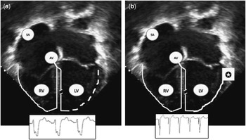
It has been known for nearly 20 years that QRS duration is directly related to heart failure.Reference Dhingra, Pencina and Wang 12 Prinzen et alReference Prinzen, Hunter, Wyman and McVeigh 13 described the deleterious consequences of dyssynchronous activation on local myocardial work with a decrease in the early sites contracting and an increase in the late sites whose pre-load was increased by the preceding stretch (Fig 1). These dyssynchronous changes may result in a combination of any of the following: reduced atrial kick, pre-systolic atrioventricular valve regurgitation, paradoxical myocardial stretch, disturbed relaxation, and increased myocardial oxygen demand.Reference Prinzen, Hunter, Wyman and McVeigh 13 Published data have shown that restoration of a normal mechanical contractile pattern, normalisation of the QRS, will increase contraction efficiency;Reference Nelson, Berger and Fetics 14 however, the relationship between electrical conduction delay and mechanical dyssynchrony is not straightforward, and QRS duration to predict the response to cardiac re-synchronisation therapy may be sub-optimal with a specificity and sensitivity around 54%.Reference Mollema, Bleeker, van der Wall, Schalij and Bax 15 In paediatric biventricular reports, a QRS>120 ms and ejection fraction ⩽35% were observed in <60% of the patients.Reference van der Hulst, Delgado and Blom 16 Pham et alReference Pham, Balaji, Shen, Ungerleider, Li and Sahn 17 observed an acute increase in ejection fraction with biventricular pacing in 19 CHD patients with a narrow QRS (96±18 ms). The extent of this technology clearly extends beyond a one-time QRS measurement and likely encompasses a better understanding of mechanical dyssynchrony with inter- and/or intra-ventricular dyssynchrony.
Scheme of re-synchronisation therapy – left bundle branch block. ( a ) During native LBBB conduction, the activation of the LV free wall (dotted lines) is late and results in electrical and mechanical dyssynchrony. ( b ) CRT is initiated with fusion between the spontaneous native septal conduction and the paced free wall (star), resulting in a narrow QRS complex and resolution of dyssynchrony. AV=atrioventricular; CRT=cardiac re-synchronisation therapy; LBBB=left bundle branch block; LV=left ventricle; RV=right ventricle; SN=Sinus Node.

Cardiac re-synchronisation therapy for chronic right ventricular pacing
Conventional anti-bradycardia pacing is easily achieved by right ventricular apical pacing, given the ease of implanting in the venous ventricle and the proven long-term lead stability; however, chronic right ventricular apical pacing results in both inter- and intra-ventricular dyssynchrony and may in fact be the least favourable place to pace.Reference Prinzen and Peschar 18 Long-term studies in the young have shown that 5–10% of patients with complete atrioventricular block and chronic right ventricular apical pacing develop heart failure symptoms, altered myocardial performance, and a dilated cardiomyopathy.Reference Breur, Udink Ten Cate and Kapusta 19 – Reference Karpawich, Rabah and Haas 22 Neonates presenting with congenital complete atrioventricular block are at a higher risk for developing a dilated cardiomyopathy, and the cumulative probability of freedom from dilated cardiomyopathy at 10 years has been demonstrated to be 59%.Reference Kurosaki, Miyazaki, Watanabe and Echigo 23 Between 45 and 78% of paediatric and CHD patients in large paediatric retrospective cardiac re-synchronisation therapy studies are under biventricular pacing essentially as an upgrade to traditional pacing-related dyssynchrony and heart failure.Reference Dubin, Janousek and Rhee 5 – Reference Janousek, Gebauer and Abdul-Khaliq 7 Marked improvement in left ventricular function and reverse re-modelling have been reported in children with atrioventricular block and a dilated cardiomyopathy, in whom conventional right ventricular apical pacing was upgraded to cardiac re-synchronisation therapy.Reference Moak, Hasbani and Ramwell 21 , Reference Janousek, Tomek and Chaloupecky 24 In fact, Janousek et alReference Janousek, Gebauer and Abdul-Khaliq 7 in the large European multi-centre study demonstrated that the cohort of patients upgraded from conventional pacing to biventricular pacing secondary to dyssynchrony had the best response, demonstrating clinical improvement, reverse re-modelling, and near-normalisation of the QRS duration. Infants and young children with atrioventricular block requiring ventricular pacing should be considered for pro-synchronisation via a left ventricular apical approach. Both animal and human data have shown left ventricular apical pacing to result in the lowest decrease in maximum left ventricular dP/dt.Reference Gebauer, Tomek and Salameh 25 – Reference Vanagt, Verbeek and Delhaas 27 Individuals with heart failure and a dilated cardiomyopathy secondary to chronic right ventricular apical pacing should be considered for upgrade to cardiac re-synchronisation therapy before more advanced heart failure therapies including heart transplantation.
Cardiac re-synchronisation therapy for dilated cardiomyopathy – non-pacing induced
The initial positive response to cardiac re-synchronisation therapy in adults with non-ischaemic dilated cardiomyopathy unfortunately has not translated to similar results in children with dilated cardiomyopathy, excluding those patients with dilated cardiomyopathy secondary to isolated chronic right ventricular pacing;Reference Chen, Hsiao and Wang 28 however, anecdotal case reports of patients with dilated cardiomyopathy responding to cardiac re-synchronisation therapy have been noted. Ascertaining which patient with idiopathic dilated cardiomyopathy will respond to cardiac re-synchronisation therapy remains a challenge. Mechanical dyssynchrony is highly prevalent in those with QRS prolongation, but is also frequently observed in patients without QRS prolongation.Reference Friedberg, Roche and Balasingam 29 In a recent large multi-centre study, 12% of children with idiopathic dilated cardiomyopathy were identified as having classic-pattern dyssynchrony – mechanical dyssynchrony with underlying electrical activation delay.Reference Forsha, Slorach and Chen 30 This small but important sub-population of children with dilated cardiomyopathy may identify as ideal candidates for cardiac re-synchronisation therapy. Future multi-centre studies potentially as an offshoot from heart failure transplant centres will need to delve into the small number of patients with CPD.
Cardiac re-synchronisation therapy for the pulmonary right ventricle
The group of tetralogy of Fallot patients is one of the most studied sub-populations in CHD history from the paradigm of a pressure–volume load CHD defect. Right ventricular dysfunction in the sub-pulmonary right ventricular CHD population may be secondary to a chronic pressure overload, volume overload, and myocardial scarring. Furthermore, surgically induced right bundle branch block may result in electrical dyssynchrony and more advanced heart failure.Reference Vogel, Sponring, Cullen, Deanfield and Redington 31 , Reference Abd El Rahman, Hui and Yigitbasi 32 Diastolic dysfunction is also more prevalent in children with right bundle branch block after a ventricular septal defect closure as opposed to children without right bundle branch block after ventricular septal defect closure.Reference Pedersen, Andersen, Knudsen, Christensen, Sorensen and Hjortdal 33 Similar to left ventricular pacing for left bundle branch block, it seems logical that right ventricular pacing for right bundle branch block with an appropriately timed atrioventricular interval would allow merging of the native conduction through the left bundle with that of the paced right ventricle. A number of acute trials conferred short-term benefits of such an application, although interestingly, despite the appearance of classic right bundle branch block, the site that resulted in the narrowest QRS varied between the right ventricular apex, septum, and the outflow tract. Reference Dubin, Feinstein, Reddy, Hanley, Van Hare and Rosenthal 34 , Reference Janousek, Vojtovic and Hucin 35 Although single-site right ventricular pacing is technically feasible, the continued need to adjust right ventricular pacing as the intrinsic atrioventricular interval changes limits acceptable fusion. Given the constraints of variations in atrioventricular intervals over a broad range of heart rates, Thambo et alReference Thambo, Dos Santos and De Guillebon 36 proposed biventricular cardiac re-synchronisation therapy for tetralogy of Fallot patients with right ventricular dysfunction. Over a period of 6 months, cardiac re-synchronisation therapy in the sub-pulmonary right ventricular demonstrated ventricular synchrony and increased myocardial function and exercise performance.Reference Thambo, Dos Santos and De Guillebon 36 Left ventricular dysfunction has also been reported in 5–10% of patients after repair of tetralogy of Fallot, increasing the prevalence of heart failure but also a potential harbinger for ventricular tachycardia and sudden cardiac death.Reference Ghai, Silversides, Harris, Webb, Siu and Therrien 37 , Reference Broberg, Aboulhosn and Mongeon 38 In a small series of adult tetralogy of Fallot patients with ventricular dysfunction, cardiac re-synchronisation therapy increased the left ventricular ejection fraction from 24±10.5 to 36.7±13% during mid-term follow-up – 9 months. In addition, the left ventricular end-diastolic volume and left ventricular end-systolic volume were also significantly reduced over the same time period.Reference Merchant, Kella, Book, Langberg and Lloyd 39 Longer-term follow-up with a larger number of patients will be required to truly assess cardiac re-synchronisation therapy after tetralogy of Fallot repair with right ventricular, left ventricular, or biventricular dysfunction. Despite promising results in the adult CHD population with a systemic left ventricular, there remain little data on the effectiveness of non-invasive imaging to guide the implanter to the optimal site. We do not understand non-responders. Does it relate to the selected patient? Does it matter whether the dyssynchrony relates to a peripheral or central right bundle branch block? How should we think about cardiac re-synchronisation therapy in the patient who has electrical dyssynchrony with right bundle branch block and left fascicular block?
Cardiac re-synchronisation therapy for the systemic right ventricle
Patients with systemic right ventricles as part of a two-ventricle circulation may have congenitally corrected levo-transposition of the great arteries or transposition of the great arteries s/p an atrial baffle procedure such as the Mustard or Senning repairs. Patients with systemic right ventricles have an unfavourable long-term history and may develop right ventricular failure.Reference Hraska, Duncan, Mayer, Freed, del Nido and Jonas 40 In addition, patients with levo-transposition of the great arteries may develop atrioventricular block secondary to the unusual course of the conduction axis further exposing the myocardium to electrical and mechanical dyssynchrony with lifelong pacing.Reference Huhta, Maloney, Ritter, Ilstrup and Feldt 41 The results of cardiac re-synchronisation therapy in the systemic right ventricular population are mixed. A total of five studies have reported cardiac re-synchronisation therapy results in 68 patients with all studies noting a reduction in QRS duration, but clinical improvement ranged from 25 to 100% over a 4- to 19-month median duration.Reference Dubin, Janousek and Rhee 5 – Reference Jauvert, Rousseau-Paziaud and Villain 8 , Reference Janousek, Tomek and Chaloupecky 24 Some of the discrepancy in response may relate to the age of the patient at cardiac re-synchronisation therapy implantation with less favourable responses seen in older patients.Reference Cecchin, Frangini and Brown 6 The sub-optimal response to cardiac re-synchronisation therapy in the systemic right ventricle may be secondary to changes in myocardial fibre arrangement and contraction patterns compared with the left ventricle. In a recent study of carefully pre-selected patients with both inter- and intra-ventricular dyssynchrony by tissue tracking, all patients responded favourably to cardiac re-synchronisation therapy; however, this small group accounted for only 10% of the right ventricular systemic population followed-up at the investigators’ institutions.Reference Jauvert, Rousseau-Paziaud and Villain 8 In many of the initial cardiac re-synchronisation therapy trials, the degree of mitral regurgitation improved with biventricular pacing; however, in the systemic right ventricular population, there is good evidence that the degree of systemic atrioventricular valve regurgitation does not change with cardiac re-synchronisation therapy. As such, cardiac re-synchronisation therapy should not be used to address systemic atrioventricular valve regurgitation in patients with a systemic right ventricle. Adults undergoing systemic atrioventricular valve, tricuspid valve, surgery with right bundle branch block (QRS⩾150 ms) should be considered for cardiac re-synchronisation therapy at the time of valve surgery.Reference Khairy, Van Hare and Balaji 10 , Reference Janousek, Tomek and Chaloupecky 24 Recently, there has been concern over the possibility of cardiac re-synchronisation therapy resulting in sub-pulmonary left ventricular dysfunction in patients with a failing systemic right ventricle.Reference Kiesewetter, Michael, Morgan and Veldtman 42 A follow-up registry will be critical to help answer some of these questions in adult CHD patients with a systemic right ventricle who undergo biventricular pacing.
Cardiac re-synchronisation therapy for the single ventricle
Myocardial dysfunction is common sequelae after the Fontan operation. A number of small studies have reported positive short-term outcomes in children with single ventricle physiology and heart failure-related symptoms.Reference Dubin, Janousek and Rhee 5 – Reference Janousek, Gebauer and Abdul-Khaliq 7 , Reference Bacha, Zimmerman and Mor-Avi 43 , Reference Senzaki, Kyo and Matsumoto 44 There have been no dedicated long-term studies evaluating cardiac re-synchronisation therapy following the Fontan operation; however, the response across all single ventricle populations has been inconsistent. The disparity in response to cardiac re-synchronisation therapy may be related to the changes in myocardial dyssynchrony versus electrical dyssynchrony. Motonaga et alReference Motonaga, Miyake, Punn, Rosenthal and Dubin 45 evaluated 11 patients with hypoplastic left heart syndrome using three-dimensional electrical mapping, tissue-Doppler imaging, and vector velocity imaging. The results of this study showed that hypoplastic left heart syndrome patients with preserved right ventricular systolic function had mechanical dyssynchrony, but not necessarily electrical dyssynchrony. As such, the presence of mechanical dyssynchrony, although frequently observed in single ventricle patients, in and of itself, does not identify individuals who will respond to cardiac re-synchronisation therapy.Reference Motonaga, Miyake, Punn, Rosenthal and Dubin 45 In the future, identifying single ventricle patients with heart failure who have both mechanical and electrical dyssynchrony may best identify responders.
Imaging issues
The multitude of tools now available to the imager for cardiac functional analysis is impressive. Left ventricular ejection fraction, although frequently used as the gold standard, is limited by the fact that left ventricular end-diastolic diameter and left ventricular end-systolic diameter are simply traced endocardial borders and may not equate to myocardial performance. Tissue-Doppler imaging and color-coded tissue-Doppler imaging measuring myocardial velocities may allow for the assessment of left ventricular synchrony; two-dimensional strain and speckle-tracking strain permits multi-directional and angle-independent assessment of left ventricular deformation. Recently, 3D speck track echocardiography has produced a new insight into dyssynchrony.Reference Seo, Ishizu, Atsumi, Kawamura and Aonuma 46 The various anatomical substrates may have patterns of dyssynchrony independent of the bundle branch block and improve selection of children with 3D speckle track imaging.
Technical issues
Biventricular pacing in patients with CHD can be technically challenging. The implanting physician must not only understand the reparative or palliative surgery performed, but the patent vascular routes and the inaccessible vascular routes. Careful review of any previous angiography, CT, or MRI imaging is mandatory. Cardiac re-synchronisation therapy in patients with CHD may be approached purely endocardially, epicardially, or by some hybrid version of the two. Apart from vascular patency, the presence of a prosthetic tricuspid valve or residual intra-cardiac shunt, especially right-to-left, may make an epicardial approach more desirable. In patients with levo-transposition of the great arteries, the size, length, and number of tributaries can be different compared with normal hearts. Anatomical variations in the lateral veins may be seen in patients with levo-transposition of the great arteries, although veins in the inter-ventricular groove may be normal.Reference Manchanda, McLeod, Killu and Asirvatham 47 In addition, review of echo imaging to pre-identify the most dyssynchronous areas may be worthwhile, although such areas may be impossible to reach or result in electrical standstill when attempting to pace. Questions of immediate vascular patency are just as important in the growing child as long-term vascular patency concerns. Coronary sinus drainage may be an issue related to the underlying heart disease or the surgical approach that was taken. In dextro-transposition of the great arteries following an atrial baffle procedure, the coronary sinus may drain into either the systemic or the pulmonary neo-atrium and 20% may in fact have an atretic coronary sinus ostium.Reference Ebert, Gay and Engle 48 , Reference Bottega, Kapa and Edwards 49
Future thoughts
Although adult heart failure trials and guidelines use a prolonged QRS duration as criteria for cardiac re-synchronisation therapy, a number of trials have shown that mechanical dyssynchrony may be more predictive of a positive response than an isolated QRS duration.Reference Yu, Chan and Zhang 50 – Reference Achilli, Sassara and Ficili 52 Nevertheless, extrapolating those same criteria to a paediatric or CHD population has not been adjudicated. Understanding the contributory effects of electrical and mechanical dyssynchrony on myocardial performance and heart failure in a diverse paediatric population is critical so as to identify which patients will respond to advanced technologies. Moving forward, electrophysiologists and imagers will need to abandon traditional silos and approach these challenging patients in a collaborative and multi-disciplinary model. Paediatric heart failure specialists should consider prospective multi-centre registries to follow-up children with heart failure secondary to dilated cardiomyopathy and CHD who receive cardiac re-synchronisation therapy.
Acknowledgement
I would like to acknowledge Drs. Jeff Jacobs, Marshall Jacobs, and Alfred-Assante Korang for their support during the Heart Failure Summit.
Conflicts of Interest
None.