Introduction
Coarctation of the aorta is a common CHD in children, characterised by localised narrowing of the aortic lumen. It accounts for 6% to 8% of all CHDs Reference Kenny and Hijazi1 with an incidence of approximately 3 to 4 per 10,000 live births. Reference Kim, Andrade and Cook2 The clinical manifestations in affected children are closely related to the degree of narrowing and the presence of associated intracardiac anomalies: mild cases may be asymptomatic, while severe cases are prone to hypertension and tissue ischaemia. Currently, echocardiography and CT are the primary diagnostic modalities for this condition. Reference Ganigara, Doshi, Naimi, Mahadevaiah and Buddhe3,Reference Gong, Zhang and Feng4
Surgical intervention is the only curative treatment for coarctation of the aorta. Reviewing the history of surgical development, the rate of postoperative restenosis was as high as over 50% with early end-to-end anastomosis techniques. Reference Kvitting and Olin5,Reference Cangussú, Lopes and Barbosa6 With improvements in surgical techniques and the application of monofilament sutures, multiple multicentre studies have confirmed that the incidence of postoperative restenosis has significantly decreased to between 11% and 25%. Reference Kvitting and Olin5 Among these techniques, simple resection with end-to-end anastomosis and resection with extended end-to-end anastomosis, validated by several meta-analyses, have become the most widely used mainstream surgical procedures. Reference Padalino, Bagatin and Bordin7 Additionally, other commonly employed interventions include Gore-tex patch aortoplasty, subclavian flap aortoplasty, and end-to-side anastomosis.
Since the 1990s, transcatheter intervention has been widely utilised in the treatment of aortic coarctation. While it is highly effective for older children and adults, the reintervention rate of transcatheter therapy in younger children (age ≤ 12 years) is significantly higher than that of surgical repair (43% vs. 15%). Reference Wu, Jin and Kuang8,Reference Hlebowicz, Cardenas and Jeremiasen9 Given its relative advantage in reducing the long-term risk of reintervention, surgery currently remains the primary treatment modality for children aged 12 years and under. Reference Hysko, Bertram, Bobylev, Horke and Hansmann10
Despite continuous advancements in surgical techniques, postoperative restenosis remains a critical challenge. Following surgery, structural and functional abnormalities—such as the fragmentation of elastic fibres, medial smooth muscle hyperplasia, and collagen deposition in the vessel wall—can trigger luminal renarrowing through inflammatory responses, excessive proliferation of vascular smooth muscle cells, and extracellular matrix remodelling. Reference Salciccioli and Zachariah11– Reference Ma, Yan and Li12 Furthermore, residual pressure gradients (e.g., >20 mm Hg), increased vascular stiffness, and endothelial dysfunction further accelerate the restenosis process. Reference Ghorbannia, Jurkiewicz and Nasif13 The optimal surgical approach remains highly controversial, particularly for complex cases involving aortic arch hypoplasia. In such instances, our centre primarily employs the end-to-side anastomosis technique to achieve superior clinical outcomes.
Identifying high-risk children for postoperative restenosis and optimising clinical management is a key focus of current research. Previous studies have mostly adopted traditional statistical methods such as logistic regression and Cox proportional hazards models and have identified risk factors including low body weight and aortic arch hypoplasia. However, these methods rely on linear assumptions and fixed functional forms, making it difficult to capture complex nonlinear relationships and high-order interactions among multiple factors, resulting in limited predictive accuracy. Merchain learning provides a new approach to overcoming this limitation. As a data-driven core technology of artificial intelligence, machine learning algorithms such as random forest, gradient boosting, and multilayer perceptron do not require strong assumptions and can automatically mine hidden risk patterns in multi-dimensional data. They have demonstrated superior predictive performance compared to traditional models in the cardiovascular field: for instance, machine learning models based on routine clinical data can accurately identify in-stent restenosis risk. Reference Sampedro-Gómez, Dorado-Díaz and Vicente-Palacios14 Machine learning models integrating cardiovascular magnetic resonance parameters and clinical variables can predict ventricular function deterioration in patients who underwent Tetralogy of Fallot repair. Reference Ishikita, McIntosh and Hanneman15
Although previous studies have applied decision trees to predict postoperative restenosis in coarctation of the aorta patients, Reference Weismann, Grell, Odermarsky, Mellander and Liuba16 a high-precision predictive model that systematically compares multiple mainstream machine learning algorithms and integrates baseline data, preoperative imaging findings, and intraoperative parameters has not been reported. Given the multifactorial determinism of postoperative restenosis in coarctation of the aorta, this study is designed to fill this research gap: by collecting detailed clinical data of coarctation of the aorta children who underwent definitive surgery, we aim to develop and validate postoperative restenosis prediction models based on multiple machine learning algorithms, screen the optimal algorithm to achieve accurate quantification of individual risk, and provide clinicians with decision support for early identification of high-risk children, formulation of individualised intervention strategies, and optimisation of surgical plans. Ultimately, this study seeks to reduce the incidence of postoperative restenosis.
Materials and methods
Study objective
This study aims to develop and validate a machine learning model using preoperative and intraoperative clinical data to predict the risk of postoperative restenosis in children with congenital aortic arch coarctation after undergoing direct repair so as to provide data support for individualised postoperative follow-up strategies.
Study design and patients
This was a single-centre retrospective cohort study. Consecutive paediatric patients aged under 12 years who underwent their first-time direct aortic arch repair in the Department of Cardiac Surgery at Beijing Children’s Hospital from June 2016 to December 2024 were enrolled. All patients received routine postoperative follow-up for 1 year. Inclusion criteria: (1) confirmed diagnosis of congenital aortic coarctation and underwent one of the following surgeries: coarctation segment resection with resection with end-to-end anastomosis, resection and extended end-to-end anastomosis, or end-to-side anastomosis; (2) completed regular follow-up for 1 year or longer after surgery, with complete preoperative and postoperative imaging data available. Exclusion criteria: (1) complicated with severe complex congenital heart defects (e.g., single ventricle, transposition of the great arteries, Shone’s syndrome); (2) underwent concurrent complex cardiovascular surgeries during the same operation period; (3) died from any cause during the 1-year postoperative follow-up period. Ultimately, a total of 117 paediatric patients were included in the analysis, among whom 81 were male (69.2%) and 36 were female (30.8%). The mean age of the patients was [21.5] months (standard deviation, [32.98] months), with a median age of 5.53 months (interquartile range: 2.25 – 21.27 months). This study was approved by the Institutional Review Board of Beijing Children’s Hospital (IRB number: [2025]-E-076-R), with a waiver of informed consent from the patients.
Selection of surgical techniques for aortic coarctation
All patients received general intravenous combined anaesthesia. Following internal team discussions within each surgical group, the corresponding surgical approach and anastomotic technique were selected based on the type of aortic coarctation, the presence of concomitant intracardiac malformations, and the complexity of such malformations. For patients with isolated aortic coarctation or those complicated with simple intracardiac malformations (e.g., patent foramen ovale, small atrial septal defect, patent ductus arteriosus, or clinically insignificant cardiac variants such as aberrant subclavian artery or bicuspid aortic valve malformation), thoracotomy was typically performed via a left posterolateral incision under normothermic non-cardiopulmonary bypass. Depending on the length of the coarctation segment, either resection with end-to-end anastomosis or resection and extended end-to-end anastomosis was performed.
In cases of aortic arch coarctation complicated with aortic arch hypoplasia but without other complex intracardiac malformations, the surgeon could choose to perform resection and extended end-to-end anastomosis combined with resection with end-to-end anastomosis under normothermic non-cardiopulmonary bypass with selective cerebral perfusion or end-to-side anastomosis under cardiopulmonary bypass for aortic arch reconstruction, based on personal preference. For patients with aortic arch coarctation complicated with mildly complex intracardiac malformations requiring concurrent correction (e.g., atrial septal defect and ventricular septal defect), if there was no accompanying aortic arch hypoplasia, resection with end-to-end anastomosis, resection and extended end-to-end anastomosis, and end-to-side anastomosis could be performed. If aortic arch hypoplasia was also present, resection and extended end-to-end anastomosis or end-to-side anastomosis was selected for aortic arch reconstruction at the surgeon’s discretion. All concomitant intracardiac malformations were corrected during the same operation.
Data collection and definitions
Definition of restenosis
Postoperative restenosis was defined as a peak pressure gradient ≥20 mmHg at the aortic isthmus or anastomosis site, as indicated by three consecutive echocardiographic examinations.
Echocardiographic measurements
Preoperative echocardiography was independently performed by two physicians with more than 5 years of experience in diagnosing CHDs, using a double-blind method. To assess the consistency of the measurements, we calculated the intraclass correlation coefficient between the two physicians’ measurements, which yielded intraclass correlation coefficient values greater than 0.85 (95% confidence interval: 0.80–0.95) for all indicators, indicating good inter-rater reliability. The average of the two physicians’ measurements was used as the final measurement value for subsequent model training. The aortic arch was divided into three segments: proximal, distal, and isthmic. The proximal aortic arch refers to the segment from the origin of the brachiocephalic artery to the left common carotid artery; the distal aortic arch refers to the segment from the left common carotid artery to the origin of the left subclavian artery; the isthmus refers to the segment connecting the distal aortic arch and the juxtaductal region of the descending aorta. The transverse arch diameter was defined by the echocardiographers as the diameter of the proximal aortic arch. The diagnostic criterion for aortic arch hypoplasia was a transverse arch Z-score <−2. All Z-score data used in this study were obtained from the Congenital Heart Disease Knowledge Repository of Children’s Hospital of Zhejiang University School of Medicine (https://hdb.nbscn.org/zscore).
Statistical analysis
Comparison of baseline data
Patients were divided into two groups according to the presence or absence of postoperative restenosis. The normality of continuous variables was assessed using the Shapiro–Wilk test. Normally distributed variables were expressed as mean ± standard deviation and compared using the independent samples t-test. Non-normally distributed variables were expressed as median (interquartile range) and compared using the Mann–Whitney U test. Categorical variables were expressed as n (percentage) and compared using the chi-square test or Fisher’s exact test. A p value < 0.05 was considered statistically significant. This analysis was performed using SPSS Statistics 29.0 software.
Construction and evaluation of machine learning models
Feature Selection and Preprocessing: All potential predictive variables (including demographic characteristics, perioperative indicators, and imaging Z-scores) were initially included, and recursive feature elimination with cross-validation was used for feature selection. Algorithms and Model Building: We preselected six commonly used algorithms that cover different types of machine learning methods, namely logistic regression, random forest, decision tree, support vector machine, gradient boosting, and multilayer perceptron. Among them, logistic regression serves as a traditional linear baseline; decision tree, random forest, and gradient boosting are tree-based ensemble methods known for robustly handling nonlinear interactions; support vector machine is included for its geometric margin maximisation mechanism; and multilayer perceptron represents an artificial neural network capable of capturing complex high-order patterns. All models were built on the training set using five-fold cross-validation, with hyperparameters optimised via random search. Performance Evaluation and Interpretation: The performance of the models was comprehensively evaluated on the validation set using the area under the curve, accuracy, precision, recall, and F1 score. The SHapley Additive exPlanation method was used to conduct interpretability analysis of the optimal model. The machine learning analysis was completed based on the Python 3.12.7 environment.
Results
Patient characteristics
During postoperative follow-up, restenosis occurred in 21 cases (17.9%), while the remaining 96 cases (82.1%) had no restenosis. Among the restenosis cases, 16 (76.2%) occurred within 1 year after surgery. The postoperative therapeutic interventions were distributed as follows: 5 patients underwent percutaneous balloon angioplasty, 1 patient received patch augmentation angioplasty, and 1 patient underwent anastomotic resection and reanastomosis. The remaining patients with restenosis underwent no active intervention due to the mild degree of narrowing, absence of significant clinical symptoms, and parental concerns regarding reoperation. Notably, 1 patient with velocardiofacial syndrome and 1 patient with Kawasaki disease also developed restenosis during postoperative follow-up. All patients were treated for aortic arch coarctation using one of the following surgical procedures: resection with end-to-end anastomosis in 54 cases (46.2%), resection and extended end-to-end anastomosis in 24 cases (20.5%), and end-to-side anastomosis in 29 cases (24.8%). Among them, 43.5% of patients (51 cases) underwent median sternotomy, while 56.5% (66 cases) underwent lateral thoracotomy.
Factors identified by univariate analysis
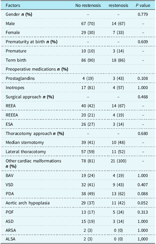
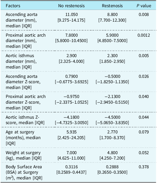
Univariate analysis included 20 influencing factors, which were divided into categorical variables and continuous variables (see Tables 1 and 2 for details). All continuous variables were tested for normality using the Shapiro–Wilk test, which revealed a non-normal distribution. Significant differences were observed between the two groups in the ascending aortic diameter (cm)/Z-score, aortic isthmic diameter (cm)/Z-score, and ascending aortic diameter (cm)/Z-score (P < 0.05). This suggests that these factors are all potentially important predictive indicators for restenosis after resection with end-to-end anastomosis.
Patient characteristics and postoperative outcomes (categorical variables)

Table 1. Long description
The table presents a comparison of patient characteristics and postoperative outcomes between two groups: no restenosis and restenosis. It includes various factors such as gender, prematurity at birth, preoperative medications, surgical approach, thoracotomy approach, and other cardiac malformations. The table has 12 rows and 5 columns, with column headers labeled as Factors, No restenosis, restenosis, and P value. Each row provides data on the percentage of patients in each group for specific factors. For example, under gender, 70 percentage of patients in the no restenosis group are male, compared to 67 percentage in the restenosis group. The table also includes data on preoperative medications such as prostaglandins and inotropes, surgical approaches like REEA and ESA, and thoracotomy approaches like median sternotomy and lateral thoracotomy. Additionally, it lists other cardiac malformations such as BAV, VSD, PDA, aortic arch hypoplasia, POF, ASD, ARSA, and ALSA. The P values indicate the statistical significance of the differences between the two groups for each factor.
BAV = bicuspid aortic valve; VSD = ventricular septal defect; PDA = patent ductus arteriosus; AAH = aortic arch hypoplasia; POF = patent foramen ovale; ASD = atrial septal defect; ARSA = aberrant right subclavian artery; ALSA = aberrant left subclavian artery.
Continuous clinical characteristics of patients

Table 2. Long description
The table presents a comparison of clinical characteristics between patients with and without restenosis. It includes factors such as ascending aorta diameter, proximal aortic arch diameter, aortic isthmus diameter, and their respective Z-scores. The table has 10 rows and 5 columns, with columns for factors, no restenosis, restenosis, and P value. Each row provides median values and interquartile ranges for these factors. Notable trends include significant differences in ascending aortic diameter, proximal aortic arch diameter, aortic isthmus diameter, and their Z-scores between the two groups, indicating potential predictive indicators for restenosis after resection with end-to-end anastomosis.
Data are presented as median (interquartile range). P-values were calculated using the Mann-Whitney U test.
Predictive feature selection and model construction
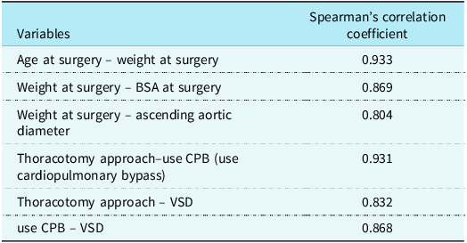
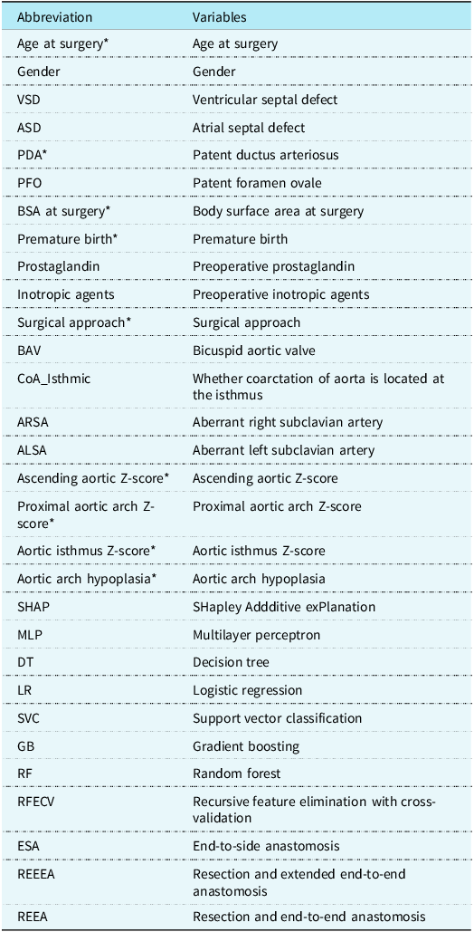
Based on the preoperative and intraoperative baseline characteristics of patients, this study developed a machine learning predictive model for postoperative restenosis in patients with coarctation of the aorta. During the feature preprocessing stage, Spearman’s correlation analysis revealed strong correlations among multiple variables (Spearman’s correlation coefficient > 0.8) (Table 3). Therefore, age at surgery, body surface area at surgery, and ventricular septal defect were selected as representative variables for inclusion in the subsequent analysis. Furthermore, recursive feature elimination with cross-validation was adopted for feature dimensionality reduction, which screened out 9 potential predictive variables from the initial 18 features for final model construction. These variables were “Age at Surgery,” “PDA,” “BSA at Surgery,” “Premature Birth,” “Surgical Approach,” “Ascending aortic Z-score,” “Proximal aortic Z-score,” “Isthmus Z-score,” and “Aortic arch hypoplasia” (Table 4).
Top Spearman’s rank correlation coefficients among variables

Table 3. Long description
The table presents Spearman's rank correlation coefficients among different surgical variables. It includes six rows and two columns. The first column lists variable pairs such as Age at surgery – weight at surgery, Weight at surgery – BSA at surgery, and others. The second column provides the corresponding Spearman's correlation coefficients. Notable high correlations include Age at surgery – weight at surgery with a coefficient of 0.933 and Thoracotomy approach – use CPB with a coefficient of 0.931. The table highlights significant relationships between these variables.
This table lists variable pairs with Spearman‘s correlation coefficients |ρ| > 0.8.
Features and abbreviations

Table 4. Long description
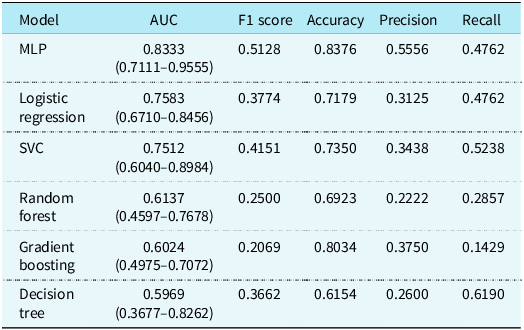
The table presents a comparison of performance metrics for different machine learning models used in predicting postoperative restenosis after aortic arch coarctation. It includes six models: MLP, Logistic Regression, SVC, Random Forest, Gradient Boosting, and Decision Tree. The metrics evaluated are AUC, F1 score, Accuracy, Precision, and Recall. The table has six rows, each representing a different model, and six columns for the metrics. Notable trends include the MLP model having the highest AUC at 0.8333, while the Decision Tree model has the lowest AUC at 0.5969. The Logistic Regression and MLP models share the highest Recall value of 0.4762. The Gradient Boosting model shows the highest Accuracy at 0.8034 but the lowest F1 score and Recall values. The Random Forest model has the lowest Precision at 0.2222. Each model's performance varies significantly across the different metrics, highlighting the importance of selecting the right model for accurate prediction.
* Variables selected for inclusion in the model following refevc screening.
Comparison of model performance
Based on the aforementioned 9 features, a 5-fold cross-validation framework was adopted. In each fold, hyperparameter tuning was performed using randomised search cross-validation combined with 5-fold internal validation. Six different algorithms were employed, and additional hyperparameter optimisation was conducted via grid search to obtain the optimal model parameters.
The area under the curve was used as the primary evaluation metric, with accuracy, F1-score, recall, and precision as secondary metrics. After internal validation, the multilayer perceptron model achieved the highest area under the curve of 0.8333 (95% confidence interval [CI]: 0.7111−0.9555) (Figure 1), indicating superior predictive discriminative ability. The multilayer perceptron model also demonstrated the highest accuracy (0.8376), resulting in a lower misclassification rate in practical applications. Additionally, the multilayer perceptron model exhibited balanced and relatively high precision (0.5556) and recall (0.4762), avoiding the issue of imbalance in individual metrics (Table 5).
Considering the comprehensive predictive performance and clinical applicability, the multilayer perceptron model was selected as the final model for predicting the risk of postoperative restenosis in aortic arch coarctation.
ROC curve comparison of machine learning models for postoperative restenosis prediction after aortic arch coarctation surgery.

Figure 1. Long description
A line graph compares the performance of various machine learning models for predicting postoperative restenosis after aortic arch coarctation surgery. The x-axis represents the false positive rate, ranging from 0.0 to 1.0, while the y-axis represents the true positive rate, also ranging from 0.0 to 1.0. The graph includes multiple lines, each representing a different machine learning model: Random Forest, Decision Tree, Logistic Regression, Support Vector Classifier (SVC), Gradient Boosting, and Multi-Layer Perceptron (MLP). Each model's performance is measured by the Area Under the Curve (AUC) and the 95% confidence interval (CI). The Random Forest model has an AUC of 0.614 with a 95% CI of 0.460 to 0.768. The Decision Tree model has an AUC of 0.597 with a 95% CI of 0.368 to 0.826. The Logistic Regression model has an AUC of 0.758 with a 95% CI of 0.671 to 0.846. The SVC model has an AUC of 0.751 with a 95% CI of 0.604 to 0.898. The Gradient Boosting model has an AUC of 0.602 with a 95% CI of 0.498 to 0.707. The MLP model has an AUC of 0.833 with a 95% CI of 0.711 to 0.955. A dashed line represents the performance of a random classifier. All values are approximated.
Performance metrics of machine learning models for postoperative restenosis prediction after aortic arch coarctation

Table 5. Long description
The table presents data on three surgical approaches for treating aortic arch hypoplasia in children, focusing on the incidence of postoperative restenosis. It includes three rows for each surgical approach and a total row, with columns for no restenosis percentage, restenosis percentage, and total percentage. The surgical approaches are REEA, REEEA, and ESA. REEA shows 7 cases with no restenosis and 7 with restenosis out of 14 total cases. REEEA has 6 cases with no restenosis and 2 with restenosis out of 8 total cases. ESA presents 16 cases with no restenosis and 2 with restenosis out of 18 total cases. Overall, there are 29 cases with no restenosis and 11 with restenosis out of 40 total cases.
Model result interpretation
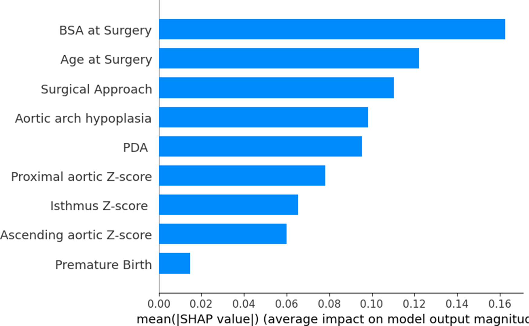
Due to the “black-box effect” of machine learning, conventional methods are challenging for interpreting such models. We employed SHapley Additive exPlanation analysis to interpret the multilayer perceptron model. The results revealed that age at surgery, body surface area at surgery, and surgical approach were identified as the three relatively most important factors (SHapley Additive exPlanation values > 0.1). The next four factors of secondary importance included patent ductus arteriosus, ascending aortic Z-score, proximal aortic Z-score, isthmus Z-score, and aortic arch hypoplasia—while premature birth was of relatively low importance for postoperative restenosis in aortic arch coarctation.
As indicated by the arrows, patients with a significant contribution from surgical age (highlighted in red in the corresponding column) often exhibit a low body surface area (shown as blue data points). This suggests that “advanced age” and “low BSA” frequently co-occur, resulting in a compounded risk.
Discussion
Postoperative restenosis remains a highly challenging complication following the repair of coarctation of the aorta. In our centre, the overall incidence of post-repair restenosis is 17.9%, a figure that aligns with existing domestic and international literature. Reference Dias, Barros, Leite-Moreira and Miranda17 The onset of this complication significantly compromises patients’ growth, development, and overall quality of life. Consequently, the early identification of patients susceptible to restenosis is of paramount importance for guiding clinical decision-making and facilitating family counselling. Despite this need, current literature and predictive tools for postoperative coarctation of the aorta restenosis remain largely limited. To address this gap, we retrospectively reviewed patients who underwent direct coarctation of the aorta repair between June 2016 and December 2024. By leveraging their baseline characteristics, we developed six machine learning models—logistic regression, decision tree, random forest, multilayer perceptron, support vector classification, and gradient boosting—to predict the likelihood of restenosis. Notably, the multilayer perceptron model exhibited the highest area under the curve. Our findings indicate that a low body surface area, aortic arch hypoplasia, concomitant patent ductus arteriosus, the specific surgical modality, and a smaller aortic arch diameter serve as significant risk factors. Utilising this predictive model can facilitate the early identification of postoperative restenosis in coarctation of the aorta patients.
In early studies on the surgical treatment of coarctation of the aorta, scholars generally believed that age at surgery, body weight, and preterm status might be associated with the risk of postoperative restenosis. Reference Dias, Barros, Leite-Moreira and Miranda17–Reference Thomson19 However, multiple recent studies have failed to confirm a significant correlation between younger age, lower body weight, and increased restenosis risk. Reference Zhao, Pan and Wu20–Reference Burch, Cowley and Holubkov22 The results of our centre’s study indicated that age at surgery and body surface area at surgery are important risk factors for postoperative restenosis, with a trend suggesting “higher restenosis risk in children with older age at surgery” (Figure 2). This finding, although seemingly inconsistent with previous research conclusions, has a reasonable explanation: further analysis using SHapley Additive exPlanation summary plots revealed that in cases where age at surgery contributed significantly to the prediction results, low body surface area often exerted a synergistic effect. This suggests that at the same age at surgery, children with a smaller body surface area (i.e., lower body weight) may have a higher risk of restenosis. We hypothesise that low body weight at the same age usually reflects more significant growth retardation in children, which is often associated with more severe aortic coarctation lesions. This further confirms the key role of body surface area in risk stratification for postoperative restenosis in with children coarctation of the aorta.
In the results of this study, surgical approach was also identified as one of the significant factors influencing postoperative restenosis (SHapley Additive exPlanation value > 0.1). SHapley Additive exPlanation analysis revealed differences in the degree of contribution of various anastomotic techniques to restenosis risk: compared with resection and extended end-to-end anastomosis and end-to-side anastomosis, resection with end-to-end anastomosis showed a higher contribution to the risk of postoperative restenosis, suggesting that this technique may be associated with a relatively higher risk of restenosis. In contrast, resection and extended end-to-end anastomosis and end-to-side anastomosis had a relatively smaller impact on restenosis (Figures 3 and 4).
SHAP summary plot for the MLP model, showing the contribution of preoperative variables to the prediction of postoperative restenosis.

Feature importance of recurrent coarctation based on SHAP values.

Figure 3. Long description
The bar graph illustrates the feature importance of recurrent coarctation based on SHAP values. The x-axis represents the mean SHAP value, indicating the average impact on model output magnitude, ranging from 0.00 to 0.16. The y-axis lists the factors influencing recurrent coarctation, including BSA at Surgery, Age at Surgery, Surgical Approach, Aortic arch hypoplasia, PDA, Proximal aortic Z-score, Isthmus Z-score, Ascending aortic Z-score, and Premature Birth. BSA at Surgery has the highest impact, followed by Age at Surgery and Surgical Approach. The bars are colored blue and vary in length according to their impact values. All values are approximated.
SHAP plot of predictors for recurrent coarctation after surgery.

Figure 4. Long description
A heat map visualizes the contribution of preoperative variables to the prediction of postoperative restenosis using an MLP model. The heat map features a grid layout with instances on the x-axis and preoperative variables on the y-axis. The variables include BSA at Surgery, Age at Surgery, Surgical Approach, Aortic arch hypoplasia, PDA, Proximal aortic Z-score, Isthmus Z-score, Ascending aortic Z-score, and Premature Birth. The color scale ranges from blue to red, indicating SHAP values from -0.3892 to 0.3892, which represent the impact on model output. Higher values are shown in red, and lower values are shown in blue. Notable patterns include varying contributions of each variable across different instances, with some variables showing more significant impacts than others.
This finding is consistent with existing literature: for instance, a study by Massimo A. Padalino et al. supported the advantage of resection and extended end-to-end anastomosis in reducing restenosis, Reference Padalino, Bagatin and Bordin7 and Lehnert et al. reached similar conclusions. Reference Dias, Barros, Leite-Moreira and Miranda17–Reference Thomson19 Additionally, multiple studies have indicated that end-to-side anastomosis also performs well in reducing restenosis. Reference Dharmapuram, Ramadoss, Verma, Vejendla and Ivatury23,Reference Tulzer, Mair, Kreuzer and Tulzer24 Therefore, integrating the results of this study with previous evidence, the sole use of resection with end-to-end anastomosis should be avoided as much as possible in clinical surgical approach selection, and techniques such as resection and extended end-to-end anastomosis or end-to-side anastomosis, which may be more conducive to reducing restenosis risk, should be prioritised.
Numerous studies by Melanie R. F. Gropler et al. have confirmed that aortic arch hypoplasia is an independent risk factor for postoperative restenosis in children with CHD. Reference Rakhra, Lee and Lyengar25–Reference Hager, Schreiber, Nützl and Hess28 Consistent with these findings, results from our centre indicated that concurrent aortic arch hypoplasia increases the risk of postoperative restenosis in paediatric patients. However, quantitative analysis using SHapley Additive exPlanation values revealed that this factor has a relatively low impact weight (SHapley Additive exPlanation value < 0.1) and is not a key risk factor affecting prognosis.
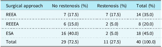
Among children with aortic arch hypoplasia treated at our centre, the distribution of surgical approaches and the incidence of postoperative restenosis are summarised in Table 6: end-to-side anastomosis was performed in 18 cases (45%), with only 2 cases developing postoperative restenosis; resection and extended end-to-end anastomosis was utilised in 8 cases (20%), with 2 cases of restenosis; and resection with end-to-end anastomosis was adopted in 14 cases (35%), with 7 cases of restenosis. End-to-side anastomosis, which reconstructs the aortic arch, significantly reduced the risk of postoperative restenosis in children with aortic arch hypoplasia (P < 0.05). This conclusion is corroborated by the findings of Tulzer et al., collectively confirming the favourable safety and efficacy of end-to-side anastomosis in the treatment of aortic arch hypoplasia. Reference Tulzer, Mair, Kreuzer and Tulzer24
Comparison of different surgical approaches for postoperative restenosis in paediatric patients with aortic arch hypoplasia

Table 6. Long description
The table presents abbreviations and their corresponding variables used in a study developing a machine learning predictive model for postoperative restenosis in patients with coarctation of the aorta. It includes 18 rows and 2 columns. The first column lists abbreviations such as Age at surgery, Gender, VSD, ASD, PDA, PFO, BSA at surgery, Premature birth, Prostaglandin, Inotropic agents, Surgical approach, BAV, CoA Isthmic, ARSA, ALSA, Ascending aortic Z-score, Proximal aortic arch Z-score, Aortic isthmus Z-score, Aortic arch hypoplasia, SHAP, MLP, DT, LR, SVC, GB, RF, RFECV, ESA, REEEA, and REEA. The second column provides the full forms of these abbreviations, such as Age at surgery, Gender, Ventricular septal defect, Atrial septal defect, Patent ductus arteriosus, Patent foramen ovale, Body surface area at surgery, Premature birth, Preoperative prostaglandin, Preoperative inotropic agents, Surgical approach, Bicuspid aortic valve, Whether coarctation of aorta is located at the isthmus, Aberrant right subclavian artery, Aberrant left subclavian artery, Ascending aortic Z-score, Proximal aortic arch Z-score, Aortic isthmus Z-score, Aortic arch hypoplasia, SHapley Additive exPlanation, Multilayer perceptron, Decision tree, Logistic regression, Support vector classification, Gradient boosting, Random forest, Recursive feature elimination with cross-validation, End-to-side anastomosis, Resection and extended end-to-end anastomosis, and Resection and end-to-end anastomosis. The table helps in understanding the variables selected for the predictive model and their abbreviations.
In this study, machine learning models combined with SHapley Additive exPlanation value analysis identified that the ascending aortic Z-score, proximal aortic Z-score, and isthmus Z-score were all negatively correlated with the risk of postoperative restenosis in children with CHD. Specifically, the smaller these Z-scores, the higher the likelihood of restenosis. However, quantitative SHapley Additive exPlanation value results indicated that these Z-score indicators had relatively low impact weights (SHapley Additive exPlanation values < 0.08), which were significantly lower than those of other key risk factors in the model (e.g., surgical approach and body surface area). Thus, they are not the primary driving factors influencing restenosis prognosis.
This finding shares partial consistency with the 2023 study by Zhenjiang Zhao’s team: they confirmed that the ascending aortic Z-score is an independent risk factor for postoperative restenosis, suggesting that abnormalities in aortic luminal anatomical structure may participate in the pathophysiological process of restenosis by affecting haemodynamics, vascular endothelial repair, and other mechanisms. Reference Zhao, Pan and Wu20 Furthermore, the present study further confirmed that low proximal aortic Z-scores and isthmic Z-scores are also associated with an increased risk of restenosis. Although the aforementioned Z-score indicators are not the primary predictive factors for restenosis, their correlation with risk still holds clinical reference value. In preoperative comprehensive assessment, Z-scores of various aortic segments should be incorporated into the anatomical risk stratification system. When Z-scores are significantly low, postoperative follow-up and imaging monitoring should be strengthened to facilitate early detection of restenosis signs.
Machine learning denotes a computer-aided generative process for sophisticated analytical models anchored in structured learning frameworks, with the core objective of refining and optimising predictive accuracy. Amid the rapid advancement of computer science, this paradigm has been seamlessly integrated across multifaceted medical disciplines, enabling the development of robust diagnostic and prognostic models via rigorous interrogation of real-world patient datasets. In contradistinction to conventional regression methodologies (e.g., logistic regression), which are inherently constrained by pre-specified linearity assumptions between covariates and outcomes, machine learning algorithms autonomously delineate non-linear interactive effects among heterogeneous risk factors. Moreover, traditional statistical approaches typically yield population-level odds ratios that merely delineate aggregate trends, whereas machine learning architectures generate individualised restenosis probabilities. Compounded by interpretive tools such as SHapley Additive exPlanation analysis, these models further elucidate the dominant driving forces governing risk stratification for each individual patient. This analytical framework does not supplant the irreplaceable value of conventional regression analysis in aetiological inference; instead, it furnishes a novel quantitative instrument and complementary perspective to optimise postoperative risk stratification and tailor individualised longitudinal follow-up protocols.
Notably, the present study represents the pioneering application of machine learning models for risk prediction of restenosis following repair of coarctation of the aorta. A panel of state-of-the-art algorithms, encompassing logistic regression, decision tree, support vector classification, random forest, multilayer perceptron, and gradient boosting, was deployed to construct predictive models for post-coarctation of the aorta repair restenosis based on baseline patient characteristics. Among these, the multilayer perceptron model achieved the highest mean area under the curve, indicative of enhanced reproducibility and predictive reliability. Subsequent SHapley Additive exPlanation-based interpretive analysis of the optimal multilayer perceptron model identified body surface area body surface area as the paramount predictive feature for restenosis onset. While the key risk factors delineated herein—including diminished body surface area and reduced aortic calibre—are consistent with prior published findings, our machine learning-driven framework uniquely quantifies the complex non-linear interplay between these determinants. The capacity of this model to deliver individualised risk stratification confers substantial translational potential for clinicians to identify high-risk patients with early restenosis following aortic arch coarctation repair, thereby establishing a rigorous foundation for the implementation of precision medicine strategies tailored to the unique clinical profiles of coarctation of the aorta patients.
Limitation
Although we have attempted to overcome all the shortcomings of previous meta-analyses and updated the latest relevant literature, there were still a few limitations in our study.
Firstly, this study adopted a single-centre retrospective design, and its inherent limitations are unavoidable. The quality and completeness of the data are limited by the accuracy of medical records. Additionally, all study participants were recruited from Beijing Children’s Hospital, which serves as a National Children’s Medical Center of China. Patients admitted to our hospital generally present with severe cardiac malformations and significant symptoms, while the consultation rate of mild asymptomatic children is relatively low, potentially leading to a certain degree of selection bias.
Second, our model relies primarily on morphological and geometric echocardiographic parameters, as well as operative details. Functional haemodynamic measurements, such as stroke volume or preoperative cuff-based blood pressure gradients, were not included in the predictive models. In our predominantly infant cohort, obtaining standardised and reliable cuff pressures is often limited by patient non-compliance, and functional metrics were inconsistently documented in the retrospective medical records. Future prospective studies incorporating comprehensive functional haemodynamics could provide additional predictive value.
Thirdly, the choice of surgical approach was determined by the operating surgeons based on preoperative anatomical assessment and institutional experience. Although not entirely randomised, these decisions were made following internal team discussions within each surgical group, which reflects real-world clinical practice. Moreover, some key indicators rely on the subjective judgement of sonographers, which may result in inter-rater variability. Regarding the definition of restenosis, our centre uses a peak gradient >20 mmHg as the criterion. This cutoff value is a widely accepted surrogate marker for haemodynamically significant restenosis and helps ensure an adequate study sample size. However, future research should validate the findings of this study by using re-intervention as the clinical endpoint in order to further confirm its clinical robustness. Furthermore, despite the use of cross-validation methods, the current sample size remains relatively limited for constructing machine learning models, and there is still a risk of overfitting. Therefore, the model requires external validation through multi-centre, large-scale prospective cohorts before being applied in clinical practice.
Finally, the inherent “black-box” nature of machine learning models also poses challenges for clinical interpretation and practical application. Future studies will further explore how to effectively translate the model’s prediction results into clinical decision support tools.
Conclusions
This study established a machine learning model based on baseline data of patients with coarctation of the aorta to predict postoperative restenosis in children who underwent direct coarctation of the aorta repair. We found that factors such as age at surgery, body surface area at surgery, and surgical approach exerted a significant impact on the occurrence of early postoperative adverse events in paediatric patients. Based on the interpretation of SHapley Additive exPlanation analysis for the machine learning model, the use of resection with end-to-end anastomosis should be minimised. Furthermore, sufficient attention should be paid to children with low body surface area after surgery to detect the occurrence of adverse events at an early stage.
Data availability statement
The datasets used and analysed during the current study are available from the corresponding author on reasonable request.
Acknowledgements
We would like to express our sincere gratitude to Hongxia Wang for the profound inspirations and valuable ideas they have provided in the field of machine learning.
Author contributions
DRG conceived the study, collected the data, developed the model, performed the analysis, and drafted the paper. WZX collected the data. LZQ and YKX reviewed and edited the manuscript. LZQ, DN, WZX, CYG, YHL, ZYB, BS, LXF, and TF contributed to the surgical technique sections. All authors read and approved the final manuscript.
Financial support
Not applicable.
Competing interests
The authors declare no competing interests.
Ethical standard
The study protocol was approved by the Ethics Committee of Beijing Children’s Hospital, Capital Medical University (IRB number: [2025]-E-076-R) and conducted in accordance with the ethical principles of the Declaration of Helsinki. Due to the retrospective design of this study, which involved only anonymised data and posed minimal risk to participants, the Ethics Board waived the requirement for informed consent.
Consent for publication
Not applicable.