Autism spectrum disorder (ASD) is a life-long developmental disorder characterized by qualitative impairments in social and communication behavior and a restricted range of activities and interests. ASD is estimated to affect 1 in 150 persons; thus, it is no longer considered a rare disorder (Kuehn, Reference Kuehn2007).
During the past three decades, conceptualizations of ASD have changed dramatically. Whereas autism previously was considered a disorder with an extremely poor prognosis with only 50% of individuals developing spoken language (see Dawson, Reference Dawson1989), it has now been demonstrated that 75–95% of children who receive early intensive behavioral intervention develop useful speech by age 5 (Lovaas, Reference Lovaas1987; McGee, Morrier, & Daly, Reference McGee, Morrier and Daly1999; for a review, see Rogers, Reference Rogers1998). Three separate groups have now reported that a significant proportion of children receiving intensive intervention early in life make outstanding progress, with autism symptoms diminishing and developmental outcomes improving such that these children no longer have evidence of disability (Howard, Sparkman, Cohen, Green, & Stanislaw, Reference Howard, Sparkman, Cohen, Green and Stanislaw2005; McEachin, Smith, & Lovaas, Reference McEachin, Smith and Lovaas1993; Sallows & Graupner, Reference Sallows and Graupner2005).
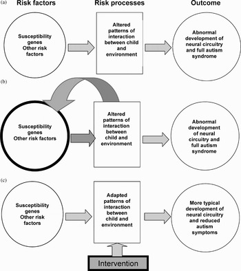
Rapid advances in the fields of cognitive and affective developmental neuroscience, developmental psychopathology, neurobiology, genetics, and applied behavior analysis have contributed to a more optimistic outcome for individuals with ASD. These advances have led to new methods for early detection and more effective treatments. For the first time, prevention of ASD is plausible. Prevention will entail detecting infants at risk before the full syndrome is present and implementing treatments designed to alter the course of early behavioral and brain development. To provide a framework for understanding early brain plasticity in ASD and its role in prevention of the disorder, Dawson (Dawson & Faja, in press; Dawson, Sterling, & Faja, in press) has proposed a developmental model of risk, risk processes, symptom emergence, and adaptation in ASD. This model posits that there are genetic, environmental, and phenotypic risk indices that ultimately will allow very early identification of infants who are vulnerable to developing ASD. Identification of such risk indices is a focus of current research in the field. Early genetic and environmental risk factors contribute to an atypical trajectory of brain and behavioral development that is manifest in altered patterns of interaction between the child and his/her environment. An important aspect of this altered interaction is a failure on the part of the child to actively engage in early social interaction. Such altered interactions, referred to as risk processes, are hypothesized to preclude normal social and prelinguistic input that normally promotes the development of social and linguistic brain circuitry during early sensitive periods, thus serving as mediators of the effects of early susceptibilities on later outcome. Through this mediational process, early susceptibilities contribute to outcome, the full autism syndrome, as illustrated in Figure 1a. Risk processes thus amplify the effects of early susceptibilities. Effective interventions target these risk processes.
A developmental model of risk factors, risk processes, and outcome in autism.

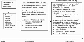
Numerous authors (e.g., Dawson, Carver, et al., Reference Dawson, Carver, Meltzoff, Panagiotides, McPartland and Webb2002; Dawson, Webb, Wijsman, et al., Reference Dawson, Webb, Wijsman, Schellenberg, Estes and Munson2005; Grelotti, Gauthier, & Schultz, Reference Grelotti, Gauthier and Schultz2002; Johnson et al., Reference Johnson, Griffin, Csibra, Halit, Farroni and de Haan2005; Kuhl, Reference Kuhl2007; Kuhl et al., Reference Kuhl, Coffey-Corina, Padden and Dawson2005; Mundy & Neal, Reference Mundy and Neal2001) have described how the development of social and language brain circuitry, its acquisition, organization, and function, results from the interaction between the infant's brain and his or her social environment. Dawson described a developmental model for the normal emergence of social brain circuitry during infancy, stressing the key role of early parent–child interaction in the development of the social brain (Dawson, Webb, & McPartland, Reference Dawson, Webb and McPartland2005; Dawson, Webb, Wijsman, et al., Reference Dawson, Webb, Wijsman, Schellenberg, Estes and Munson2005; see Figure 2). In the context of reciprocal social interactions, engagement with a social partner facilitates cortical specialization and perceptual and representational systems for social and linguistic information. Social engagement is required for the well-documented fine-tuning of perceptual systems (Kuhl, Reference Kuhl2007). Brain regions specialized for the perceptual processing of social stimuli, such as the fusiform gyrus and superior temporal sulcus, become integrated with regions involved in reward (e.g., amygdala, ventromedial prefrontal cortex), as well as regions involved in motor actions and attention (cerebellum, prefrontal/cingulate cortex). Reward mechanisms mediated by the amygdala serve to encode and consolidate memories of social–emotional experiences (LaBar, Reference LaBar2007). Through this integrative process, an increasingly complex social brain circuitry emerges. This supports more complex behaviors, such as disengagement of attention, joint attention, intentional communication, and social imitation, behaviors that are typically impaired in ASD.
The emergence of social brain circuitry in the first years of life: role of social reward. From “Neurocognitive and electrophysiological evidence of altered face processing in parents of children with autism: Implications for a model of abnormal development of social brain circuitry in autism,” by G. Dawson S. J. Webb, E. Wijsman, G. Schellenberg, A. Estes, J. Munson, and S. Faja, Reference Dawson, Webb, Wijsman, Schellenberg, Estes and Munson2005, Development and Psychopathology, 17, p. 691. Copyright 2005 Cambridge University Press.

Altered interactions between the infant and his/her social environment resulting from genetic risk factors might further influence gene expression. Such gene–environment interactions have been demonstrated in animal studies. For example, maternal nursing and grooming behavior by rats early in development produces changes in behavioral and hypothalamic–pituitary–adrenal stress responses that last into adulthood (Caldji et al., Reference Caldji, Tannenbaum, Sharma, Francis, Plotsky and Meaney1998; Liu et al., Reference Liu, Diorio, Tannenbaum, Caldji, Francis and Freedman1997). The mechanism for this change is epigenetic, with maternal behavior directly influencing DNA methylation and chromatin structure (Weaver et al., Reference Weaver, Cervoni, Champagne, D'Alessio, Sharma and Seck2004). Such gene–environment interactions may play a role in ASD as well. Whether and how alterations in early parent–child interaction in ASD influence gene expression is unknown; it is plausible, however, that gene–environment interactions occurring during postnatal life amplify the effects of initial autism susceptibility genes (see Figure 1b).
The model of risk and prevention illustrated in Figure 1 further posits that early intervention can alter the abnormal developmental trajectory of young children with ASD and help guide brain and behavioral development back toward a normal pathway; early intervention targets risk processes involving interaction between the child and his/her social partner (Figure 1c). Brain-based outcome measures will allow us to assess whether such interventions actually result in more normal patterns of brain function and organization.
This article begins by describing the progress that has been made in identifying risk indices for ASD. Studies aimed at discovering genetic and environmental risk factors will be described first; a brief review of studies describing the behavioral, neurophysiological, and other brain-based risk indices will follow. The role of altered social interactions as a risk process affecting the development of the social brain next will be discussed. Next, infant–toddler interventions aimed at reducing and preventing ASD symptoms will be described. Suggestions will be offered for how brain-based measures of outcome can be incorporated into intervention and prevention studies to allow assessment of the impact of early intervention on brain function and organization. Finally, factors hypothesized to account for the tremendous variability in response to early intervention will be discussed.
Risk Indices in ASD
Genetic risk factors
One goal of genetic research is to identify infants at increased risk for ASD at birth so that intervention can begin as soon as possible. Although progress in autism genetics is being made, the heterogeneity and complexity of the ASD phenotype pose considerable challenges. There is strong evidence for the role of genetics in autism. A substantial number of cases of autism have co-occurring medical conditions, some of which can be linked to identifiable genetic disorders, such as fragile X (Rutter, Bailey, Bolton, & LeCouteur, Reference Rutter, Bailey, Bolton and LeCouteur1994). The remaining cases are considered idiopathic and likely involve multiple autism susceptibility genes. A multifactor epistatic model with 2–10 contributing loci (Pickles et al., Reference Pickles, Bolton, Macdonald, Bailey, Le Couteur and Sim1995) has been proposed. Concordance rates for monozygotic (MZ) twins are estimated to be 69–95% (Bailey et al., Reference Bailey, Le Couteur, Gottesman, Bolton, Simonoff and Yuzda1995; Folstein & Rutter, Reference Folstein and Rutter1977a, Reference Folstein and Rutter1977b; Ritvo et al., Reference Ritvo, Jorde, Mason-Brothers, Freeman, Pingree and Jones1989; Ritvo, Freeman, Mason-Brothers, Mo, & Ritvo, Reference Ritvo, Freeman, Mason-Brothers, Mo and Ritvo1985; Steffenburg et al., Reference Steffenburg, Gillberg, Hellgren, Andersson, Gillberg and Jakobsson1989), whereas concordance rates for dizygotic (DZ) twins are much lower (approximately 3–8%). Genetic liability extends to a lesser variant, referred to as the “broader autism phenotype.” When a broader ASD phenotype (e.g., language and/or social impairment) is considered, concordance rates for twins increase (88–91% for MZ, 9–30% for DZ; Bailey et al., Reference Bailey, Le Couteur, Gottesman, Bolton, Simonoff and Yuzda1995; Folstein & Rutter, Reference Folstein and Rutter1977b; Steffenburg et al., Reference Steffenburg, Gillberg, Hellgren, Andersson, Gillberg and Jakobsson1989). Initial estimates of sibling recurrence rates for ASD ranged from 2.8 to 7.0%, significantly higher than the general population (August, Stewart, & Tsai, Reference August, Stewart and Tsai1981; Bailey, Phillips, & Rutter, Reference Bailey, Phillips and Rutter1996; Smalley, Asarnow, & Spence, Reference Smalley, Asarnow and Spence1988). More recent studies of infant siblings, however, have reported much higher recurrence rates (e.g., Landa & Garrett-Mayer, Reference Landa and Garrett-Mayer2006). Bolton et al. (Reference Bolton, MacDonald, Pickles, Rios, Goode and Crowson1994) estimated that 12–20% of siblings exhibit a lesser variant of autism. This study was based on a family history method that likely would yield lower rates than the true rate based on direct assessment. Several studies have documented elevated rates of autism related symptoms in immediate family members (Bailey et al., Reference Bailey, Le Couteur, Gottesman, Bolton, Simonoff and Yuzda1995, Reference Bailey, Phillips and Rutter1996; Folstein & Rutter, Reference Folstein and Rutter1977b; Landa, Folstein, & Isaacs, Reference Landa, Folstein and Isaacs1991; Landa et al., Reference Landa, Piven, Wzorek, Gayle, Chase and Folstein1992; Narayan, Moyes, & Wolff, Reference Narayan, Moyes and Wolff1990; Toth, Dawson, Meltzoff, Greenson, & Fein, Reference Toth, Dawson, Meltzoff, Greenson and Fein2007; Wolff, Naravan, & Moyes, Reference Wolff, Narayan and Moyes1988). In a large sample of parents of children with autism, Dawson et al. (Reference Dawson, Webb and McPartland2005) reported that parents showed a decrement in face recognition ability (performance at an average level) relative to their verbal and visual spatial skills (significantly higher than the norm in both domains). Current autism genetic linkage studies are using quantitative measures of autistic traits (e.g., quantitative trait locus analyses) to better capture the variation in autism broader phenotype (e.g., Sung et al., Reference Sung, Dawson, Munson, Estes, Schellenberg and Wijsman2005).
Several genome-wide linkage studies of autism have been conducted (Auranen et al., Reference Auranen, Vanhala, Varilo, Ayers, Kempas and Ylisaukko-oja2002; Barrett et al., Reference Barrett, Beck, Bernier, Bisson, Braun and Casavant1999; Buxbaum et al., Reference Buxbaum, Silverman, Smith, Kilifarski, Reichert and Hollander2001; Cantor et al., Reference Cantor, Kono, Duvall, AlvarezRetuerto, Stone and Alarcon2005; International Molecular Genetic Study of Autism Consortium [IMGSAC], 1998, 2001a, 2001b; Lamb et al., Reference Lamb, Barnby, Bonora, Sykes, Bacchelli and Blasi2005; Liu et al., Reference Liu, Nyholt, Magnussen, Parano, Pavone and Geschwind2001; McCauley et al., Reference McCauley, Li, Jiang, Olson, Crockett and Gainer2005; Philippe et al., Reference Philippe, Martinez, Guilloudbataille, Gillberg, Rastam and Sponheim1999; Risch et al., Reference Risch, Spiker, Lotspeich, Nouri, Hinds and Hallmayer1999; Schellenberg et al., Reference Schellenberg, Dawson, Sung, Estes, Munson and Rosenthal2006; Shao et al., Reference Shao, Wolpert, Raiford, Menold, Donnelly and Ravan2002; Stone et al., Reference Stone, Merriman, Cantor, Yonan, Gilliam and Geschwind2004; Yonan et al., Reference Yonan, Alarcon, Cheng, Magnusson, Spence and Palmer2003). Although replicability of signals across studies has generally been weak and promising, if not entirely consistent, evidence of linkage has been found at some chromosome sites, including 1p (Auranen et al., Reference Auranen, Vanhala, Varilo, Ayers, Kempas and Ylisaukko-oja2002; Risch et al., Reference Risch, Spiker, Lotspeich, Nouri, Hinds and Hallmayer1999), 2q (Buxbaum, Reference Buxbaum, Silverman, Smith, Kilifarski, Reichert and Hollander2001; Lamb et al., Reference Lamb, Barnby, Bonora, Sykes, Bacchelli and Blasi2005; Liu et al., Reference Liu, Nyholt, Magnussen, Parano, Pavone and Geschwind2001; Shao et al., Reference Shao, Wolpert, Raiford, Menold, Donnelly and Ravan2002), 7q (Barrett et al., Reference Barrett, Beck, Bernier, Bisson, Braun and Casavant1999; IMGSAC, 1998, 2001a, 2001b; Lamb et al., Reference Lamb, Barnby, Bonora, Sykes, Bacchelli and Blasi2005; Schellenberg et al., Reference Schellenberg, Dawson, Sung, Estes, Munson and Rosenthal2006), 17q (Cantor et al., Reference Cantor, Kono, Duvall, AlvarezRetuerto, Stone and Alarcon2005; Lamb et al., Reference Lamb, Barnby, Bonora, Sykes, Bacchelli and Blasi2005; Liu et al., Reference Liu, Nyholt, Magnussen, Parano, Pavone and Geschwind2001; McCauley et al., Reference McCauley, Li, Jiang, Olson, Crockett and Gainer2005), and 19q (Philippe et al., Reference Philippe, Martinez, Guilloudbataille, Gillberg, Rastam and Sponheim1999; Shao et al., Reference Shao, Wolpert, Raiford, Menold, Donnelly and Ravan2002), with the 2q, 7q, and 17q regions giving the strongest signals.
Well over 100 candidate genes have been studied. One promising lead is Engrailed 2 (En-2) located on chromosome 7. Animal studies have shown that EN-2 is expressed in the cerebellum and plays a role in cerebellar development (Cheh et al., Reference Cheh, Millonig, Roselli, Ming, Jacobsen and Kamdar2006; Millen, Wurst, Herrup, & Joyner, Reference Millen, Wurst, Herrup and Joyner1994). Abnormalities in cerebellar development have been consistently demonstrated in individuals with autism, including reduced Purkinje cells in the cerebellar cortex (Bailey et al., Reference Bailey, Luthert, Dean, Harding, Janota and Montgomery1998; Courchesne, Reference Courchesne1997; Reference Courchesne2004; Kemper & Bauman, Reference Kemper and Bauman1998; Ritvo et al., Reference Ritvo, Freeman, Scheibel, Duong, Robinson and Guthrie1986). En-2 knockout mice have a reduction in Purkinje cells and a decreased size of the cerebellar lobes (Kuemerle, Zanjani, Joyner, & Herrup, Reference Kuemerle, Zanjani, Joyner and Herrup1997; Millen et al., Reference Millen, Wurst, Herrup and Joyner1994) and display a number of autistic-like behaviors including reduced social play and increased repetitive behavior (Cheh et al., Reference Cheh, Millonig, Roselli, Ming, Jacobsen and Kamdar2006).
The serotonin transporter gene SLC6A4 also likely has a role in autism genetic susceptibility (reviewed in Devlin et al., Reference Devlin, Cook, Coon, Dawson, Grigorenko and McMahon2005). Elevated levels of platelet serotonin (5-HT) have been found in individuals with autism (Rolf, Haarmann, Grotemeyer, & Kehrer, Reference Rolf, Haarmann, Grotemeyer and Kehrer1993). Pharmacological treatment in ASD often involves selective 5-HT reuptake inhibitors. 5-HT is involved in guiding neuronal development, modulating sensory input and arousal, sleep, mood, aggression, impulsivity, and affiliation (Lucki, Reference Lucki1998). 5-HT innervates the limbic regions involved in social and emotional behavior. Devlin et al. (Reference Devlin, Cook, Coon, Dawson, Grigorenko and McMahon2005) reported an excess transmission of the short allele of 5HTTLPR in individuals with autism. Wassink and colleagues (Wassink et al., Reference Wassink, Hazlett, Mosconi, Epping, Arndt and Schellenberg2007) examined the relationship between variability in 5HTTLPR and early abnormalities in brain growth in autism. Autism has been associated with early enlargement of the brain. In a combined sample from University of Washington and University of North Carolina, Wassink et al. (Reference Wassink, Hazlett, Mosconi, Epping, Arndt and Schellenberg2007) found that the short (S) allele was strongly associated with increased cerebral cortical gray matter. These findings are the first to establish a direct association between a genetic variation and atypical brain development in autism.
Levitt and colleagues (Campbell et al., Reference Campbell, Sutcliffe, Ebert, Militerni, Bravaccio and Trillo2006) analyzed the gene encoding the MET receptor tyrosine kinase and showed a genetic association between the C allele in the promoter region of the MET gene. MET signaling is involved in neocortical and cerebellar development, immune function, and gastrointestinal repair.
Several genetic disorders have been associated with increased risk for ASD or expression of an autistic-like phenotype. These include fragile X syndrome, Rett syndrome, Angelman syndrome, tuberous sclerosis, and neurofibromatosis (see Veenstra-VanderWeele & Cook, Reference Veenstra-VanderWeele and Cook2004, for review). The 15q11–q13 region associated with Angelman syndrome codes for subunits of the gamma-aminobutyric acid A (GABAA) receptor. GABAergic interneurons have a role in establishing the architecture of cortical columns (DeFelipe, Hendry, Hashikawa, Molinari, & Jones, Reference DeFelipe, Hendry, Hashikawa, Molinari and Jones1990; Peters & Sethares, Reference Peters and Sethares1997). The increased prevalence of epilepsy in individuals with autism and 15q11–q13 duplications is consistent with the involvement of GABA. Hippocampal GABA receptor binding in autism is abnormally low (Blatt et al., Reference Blatt, Fitzgerald, Guptill, Booker, Kemper and Bauman2001) as are platelet GABA levels (Rolf et al., Reference Rolf, Haarmann, Grotemeyer and Kehrer1993).
A combined set of results suggests that autism is a disorder of the synapse (Garber, Reference Garber2007; Zoghbi, Reference Zoghbi2003). Zoghbi proposed that autism results from disruption of postnatal or experience-dependent synaptic plasticity. Rare mutations in the neuroligin 3 and neuroligin 4 genes have been found individuals with autism (Jamain et al., Reference Jamain, Quach, Betancur, Rastam, Colineaux and Gillberg2003). Neuroligins are proteins expressed on the surface of the postsynaptic neuron that bind to proteins on the presynaptic neuron, neurexin, thus forming the synapse. SHANK3 is another protein that is involved in the neuroligin pathway; SHANK3 mutations have also been found in individuals with autism, accounting for about 1% of cases (Durand et al., Reference Durand, Betancur, Boeckers, Bockmann, Chaste and Fauchereau2007). More evidence for involvement of this pathway comes from the findings of the Autism Genome Project (Szatmari et al., Reference Szatmari, Paterson, Zwaigenbaum, Roberts, Brian and Liu2007) involving collaboration among 50 institutions that pooled genetic data from 1,200 multiplex families. This group found evidence that autism was associated with neurexin 1, which binds to neuroligin at the synapse, and is part of a family of genes that plays a role in the neurotransmitter, glutamate. Glutamate is involved in both synaptogenesis and learning.
New evidence suggests that many individuals with autism have novel deletions and duplications in their genome, most likely arising during meiosis. Sebat et al. (Reference Sebat, Lakshmi, Malhotra, Troge, Lese-Martin and Walsh2007) use comparative genomic hybridization on DNA collected from individuals with autism and a control sample, and found that autism was associated with de novo copy number variants (CNVs). CNVs were found in about 10% of the individuals with autism who were from families in which only one person had autism. Zhao and colleagues (Reference Zhao, Leotta, Kustanovich, Lajonchere, Geschwind and Law2007) have proposed a genetic model of autism in which two genetic types exist: a small minority of cases for whom the risk of autism in males is nearly 50%, and the larger majority of cases for whom male offspring have low risk. In the latter case, sporadic autism is possibly caused by a spontaneous mutation with high penetrance in males and poor penetrance in females. High-risk families, in contrast, are from those offspring (most typically female) who carry a mutation but are unaffected. They are hypothesized to transmit the mutation in dominant fashion to their offspring.
Environmental risk factors
Although it is clear that genetic factors contribute to risk for developing ASD, it is likely that such genetic factors interact with environmental factors to confer risk (Newschaffer et al., Reference Newschaffer, Croen, Daniels, Giarelli, Grether and Levy2007). Among the environmental factors that been proposed are toxins (e.g., environmental pollutants, pesticides, thimerosal in vaccinations) and viruses (e.g., measles in the measles, mumphs, rebulla vaccine, prenatal exposure to influenza infection, rubella, and cytomegalovirus), among others (e.g., Miles & Takahashi, Reference Miles and Takahashi2007; Tsuchiya et al., Reference Tsuchiya, Hashimoto, Iwata, Tsujii, Sekine and Sugihara2007). As well, other factors related to the intrauterine environment, including maternal hypothyroxinemia (Roman, in press), maternal influenza (Fatemi et al., Reference Faterni, Earle, Kanodia, Kist, Emamian and Patterson2002; Patterson, Reference Patternson2002; Smith, Garbett, Mirnics, & Patterson, Reference Smith, Li, Garbett, Mirnics and Patternson2007), and exposure to increased levels of sex hormones related to infertility treatment (Croughan et al., Reference Croughan, Schembri, Bernstein, Chamberlain, Purcell and Camarano2006) have also been implicated. Investigators have also reported a statistically significant link between a positive family history for allergic/autoimmune disorders and clinical features of ASD, including regression and larger head sizes, as well as atypical prenatal maternal immune responses, suggesting significant genetic and perhaps prenatal contributions autism related to immune function (Croen, Grether, Yoshido, Odouli, & van de Water, Reference Croen, Grether, Yoshida, Odouli and Van de Water2005; Molloy et al., Reference Molloy, Morrow, Meinzen-Derr, Dawson, Bernier and Dunn2006; Sacco et al., Reference Sacco, Militerni, Frolli, Bravaccio, Gritti and Elia2007; Zimmerman et al., Reference Zimmerman, Connors, Matteson, Lee, Singer and Castaneda2007). Evidence of a worsening developmental trajectory, most dramatically seen in cases of autistic regression (Dawson & Werner, Reference Werner and Dawson2005; Dawson et al., Reference Dawson, Munson, Webb, Nalty, Abbott and Toth2007), also raises the possibility that postnatal environmental exposures may be of etiologic significance in genetically susceptible children, implicating gene–environmental interactions.
Several studies have revealed evidence of abnormal immune function in autism. Indicators of chronic neuroinflammation have been identified in brains of individuals with autism (Vargas, Nascimbene, Krishman, Zimmerman, & Pardo, Reference Vargas, Nascimbene, Krishnan, Zimmerman and Pardo2005) and markers of inflammation and oxidative stress have also been identified in blood and urine of individuals with autism (e.g., Ashwood & Van de Water, Reference Ashwood and Van de Water2004; James et al., Reference James, Cutler, Melnyk, Jernigan, Janak and Gaylor2004). Thus, a potentially useful direction in future candidate gene research is to examine genes related to environmental responsiveness, such as those related to cell cycle, DNA repair, and immune and inflammatory response (Herbert et al., Reference Herbert, Russo, Yang, Roohi, Blaxill and Kahler2006).
Summary
In summary, although there is strong evidence for genetic influences in autism, the role of susceptibility genes in autism and the manner in which such genes interact with environmental factors remain an active area of investigation. It has been theorized that, in many instances of ASD, it is likely that multiple genes interact with each other and environmental factors to increase susceptibility to ASD (although see Zhao et al., Reference Zhao, Leotta, Kustanovich, Lajonchere, Geschwind and Law2007, for a different view). As Belmonte et al. (Reference Belmonte, Cook, Anderson, Rubenstein, Greenough and Beckel-Mitchener2004) point out, although the small effect of each gene by itself makes it difficult to identify specific genes, “the advantage in terms of treatment is that intervening to restore regulation to a single gene or to a small set of genes may diminish the multiplicative effect enough to yield large preventative or therapeutic effects” (p. 650). Because the expression and effects of many genes are influenced by environmental factors, it is possible that early treatment can alter genetic expression, brain development, and behavioral outcome in ASD, especially if intervention can begin early during the infant period before the symptoms of autism are fully manifest. The identification of autism susceptibility genes and other biomarkers will allow detection of infants at increased risk for ASD at birth. It is likely that early detection will eventually involve a combination of biomarkers and phenotypic risk indices. Fortunately, detection using early phenotypic risk indices is rapidly improving as will be discussed next.
Behavioral risk indices
The first studies describing how autism emerges during infancy were based on home videotapes recorded before a diagnosis of autism was made (see Palomo, Belinchón, & Ozonoff, Reference Palomo, Belinchón and Ozonoff2006, for review). It was discovered that infants at risk for autism show very few, if any, behavioral symptoms at 6 months; by 12 months, however, core autism symptoms are apparent for many infants (Dawson, Osterling, Meltzoff, & Kuhl, Reference Dawson, Osterling, Meltzoff and Kuhl2000; Osterling & Dawson, Reference Osterling and Dawson1994; Osterling, Dawson, & Munson, Reference Osterling, Dawson and Munson2002). Failure to respond to name is evident by 8 to 10 months (Werner, Dawson, Osterling, & Dinno, Reference Werner, Dawson, Osterling and Dinno2000). By 12 months, infants later diagnosed with autism can be distinguished from typical infants by a failure to respond to name (Baranek, Reference Baranek1999; Osterling & Dawson, Reference Osterling and Dawson1994; Osterling et al., Reference Osterling, Dawson and Munson2002), decreased looking at the faces of others (Osterling & Dawson, Reference Osterling and Dawson1994), and low rates of showing things to others and pointing to request and share interest (Adrien et al., Reference Adrien, Lenoir, Martineau, Perrot, Hameury and Larmande1993; Maestro et al., Reference Maestro, Muratori, Cavallaro, Pei, Stern and Golse2002; Osterling & Dawson, Reference Osterling and Dawson1994; Osterling et al., Reference Osterling, Dawson and Munson2002; Werner & Dawson, Reference Werner and Dawson2005). Poor eye contact and a failure to respond to name also best distinguishes them from infants with developmental delay but without autism (Baranek, Reference Baranek1999; Osterling et al., Reference Osterling, Dawson and Munson2002).
Prospective studies of infant siblings of children with autism have provided new insights into the early development of ASD (e.g., Zwaigenbaum et al., Reference Zwaigenbaum, Bryson, Rogers, Roberts, Brian and Szatmari2005). Estimates of risk rates for autism in siblings range from 3 to 7%; however, the rates in most published studies of infant siblings have been significantly higher (e.g., Landa & Garrett-Mayer, Reference Landa and Garrett-Mayer2006). Zwaigenbaum et al. (Reference Zwaigenbaum, Bryson, Rogers, Roberts, Brian and Szatmari2005) have followed a sample of 150 infant siblings of children with autism and 75 low-risk infants from the age of 6 months or younger. Because children were enrolled prior to onset of symptoms, the sample was based on risk for developing symptoms rather than parental concern about symptoms. Zwaigenbaum et al. (Reference Zwaigenbaum, Bryson, Rogers, Roberts, Brian and Szatmari2005) reported on a sample of 65 high-risk and 23 low-risk siblings that had been followed up to at least 24 months. Infants were assessed using the Autism Observation Scale for Infants (AOSI; Bryson, McDermott, Rombough, Brian, & Zwaigenbaum, Reference Bryson, McDermott, Rombough, Brian and Zwaigenbaum2007), which measures visual attention, response to name, response to a brief still face, anticipatory responses, imitation, social babbling, eye contact and social smiling, reactivity, affect, ease of transitioning, and atypical motor and sensory behaviors. These markers did not distinguish groups at 6 months of age on the basis of their diagnostic classification at 24 months; however, a subset of the children who were later diagnosed exhibited impairments in responding to name or unusual sensory behaviors. By 12 months groups could be distinguished on the basis of having at least seven markers. Only 2 of 58 at risk siblings who did not receive an ASD diagnosis and none of the 23 controls exhibited seven or more markers. Predictive 12-month markers from the AOSI included atypical eye contact, visual tracking, disengaging visual attention, orienting to name, imitation, social smiling, reactivity, social interest, and sensory-oriented behaviors. Parents of children who received an ASD diagnosis at 24 months also reported poor gesture use and understanding of words (Mitchell et al., Reference Mitchell, Brian, Zwaigenbaum, Roberts, Szatmari and Smith2006).
Two risk behaviors that were not as well documented in retrospective home videotape studies were identified in the prospective study by Zwaigenbaum et al. (Reference Zwaigenbaum, Bryson, Rogers, Roberts, Brian and Szatmari2005). First, differences in visual attention that emerged between 6 and 12 months were observed in infants who later developed ASD. Such infants showed a decline in their performance on a visual attention task that required the infant to disengage his/her attention from a previously salient stimulus; in contrast, none of the infants whose performance was similar or better at 12 months relative to their performance at 6 months developed ASD. Second, infants who later developed ASD exhibited differences in temperament characterized by a lower activity level and more frequent and intense distress reactions. They also spent longer fixating on a single object and were less active in their spontaneous visual exploration. Detailed study of the first nine children who developed ASD (Bryson, Zwaigenbaum, et al., Reference Bryson, Zwaigenbaum, Brian, Roberts, Szatmari and Rombough2007) revealed two subgroups based on the presence or absence of cognitive decline between 12 and 24 months. In children with cognitive loss, symptoms emerged earlier or were more severe. Several investigators have now documented a pattern of cognitive and behavioral decline in infants who develop ASD (reviewed in Dawson et al., 2006).
Landa and Garrett-Mayer (Reference Landa and Garrett-Mayer2006) reported a prospective, longitudinal study that described the cognitive development of high-risk infant siblings who later developed ASD, in comparison to high-risk infant siblings who later developed language delay without autism, and unaffected infants. Infants did not differ at 6 months, but by 14 months, the children who developed ASD differed from the unaffected group in gross and fine motor, receptive and expressive language, and overall intelligence on the Mullen scales (Mullen, Reference Mullen1995). Landa, Holman, and Garrett-Mayer (Reference Landa, Holman and Garrett-Mayer2007) recently described patterns of development from 14 to 24 months in children with early and later diagnosis of ASD. They found that the early-diagnosis group differed from later diagnosis children, siblings with broader phenotype, and nonrisk control infants in their social, communication, and play behavior. For the early-diagnosis group, growth trajectories suggested that autism may involve developmental arrest, slowing, or even regression.
Retrospective and prospective behavioral studies have led to the development of assessment measures of autism risk behaviors that can be administered to infants (Bryson, McDermott, et al., Reference Bryson, McDermott, Rombough, Brian and Zwaigenbaum2007). The Autism Observation Scale for Infants was developed by Zwaigenbaum and colleagues (Reference Zwaigenbaum, Bryson, Rogers, Roberts, Brian and Szatmari2005). This scale involves assessment of 18 risk markers for autism within a brief observational assessment. Infants are engaged in semistructured play and systematic presses are designed to assess various target behaviors, including visual tracking, and attentional disengagement, coordination of eye gaze and action, imitation, affective responses, early social–communicative behaviors, behavioral reactivity, and sensory–motor development. The First Year Inventory (Watson et al., Reference Watson, Baranek, Crais, Steven Reznick, Dykstra and Perryman2007) is a parent questionnaire designed to assess behavioral symptoms related to autism in 12-month-olds. Similar to the Modified-Checklist for Autism in Toddlers (Robins, Fein, Barton, & Greene, Reference Robins, Fein, Barton and Green2001), which was developed for children 18–24 months of age, the First Year Inventory is designed to be a screening instrument for autism that can eventually be readily used by pediatricians and other primary health care providers. Validity, sensitivity, and specificity data on these instruments are promising.
Neurophysiological risk indices
New approaches to early detection of infants at risk for ASD are focusing on neurophysiological risk indices (endophenotypes) with the hope that such measures will improve our ability to identify infants who will develop ASD. The identification of endophenotypes, intermediate, quantifiable traits that predict an individual's risk of having a disorder, which can be linked to underlying cause (Castellanos & Tannock, Reference Castellanos and Tannock2002), will accelerate progress in both clinical and basic research. Endophenotypes based on neurobiological markers (Dawson, Webb, et al., Reference Dawson, Webb, Schellenberg, Aylward, Richards and Dager2002; Skuse, Reference Skuse2000) are likely to be especially useful. In other infant risk populations, neurophysiological measures are more sensitive than behavioral measures at detecting infants who developed later developmental problems (e.g., Black, deRegnier, Long, Georgieff, & Nelson, Reference Black, deRegnier, Long, Georgieff and Nelson2004; Hood & Atkinson, Reference Hood and Atkinson1990). In a 6-year longitudinal study of maternal depression involving 160 mother–infant pairs, Dawson et al. (Dawson et al., Reference Dawson, Frey, Panagiotides, Yamada, Hessl and Osterling1999; Dawson, Frey, Panagiotides, Osterling, & Hessl, Reference Dawson, Frey, Panagiotides, Osterling and Hessl1997) found that infants of depressed mothers showed atypical EEG responses in social situations (e.g., playing with mother or an experimenter); these EEG patterns predicted later presence of behavioral and emotional problems.
Event-related potentials (ERPs) to faces
Given the core impairment in social relatedness found in ASD, neurophysiological measures that assess early social brain circuitry might be sensitive indices of risk for ASD. Dawson and Webb have been interested in face processing ability as a potential neural trait marker for susceptibility to ASD. An innate potential for cortical specialization for faces has been proposed, with experience with faces being necessary and driving such specialization (Johnson, Reference Johnson2005; Nelson, Reference Nelson2001). Experience with faces in the first year of life can influence the development of face perception abilities (e.g., Le Grand, Mondloch, Maurer, & Brent, Reference Le Grand, Mondloch, Maurer and Brent2001; Pascalis et al., Reference Pascalis, Scott, Kelly, Shannon, Nicholson and Coleman2005). Typical 6- to 7-month-old infants reliably exhibit different ERPs to familiar versus unfamiliar faces and to different emotional expressions (de Haan & Nelson, Reference de Haan and Nelson1997; Nelson & De Haan, Reference Nelson and De Haan1996).
Behavioral and neuroimaging studies have found consistent evidence for face processing impairments in individuals with ASD (Boucher & Lewis, Reference Boucher and Lewis1992; Boucher, Lewis, & Collis, Reference Boucher, Lewis and Collis1998; Gepner, de Gelder, & de Schonen, Reference Gepner, de Gelder and de Schonen1996; Klin et al., Reference Klin, Sparrow, deBildt, Cicchetti, Cohen and Volkmar1999). Functional magnetic resonance imaging (fMRI) studies conducted with typical individuals indicate that the right fusiform gyrus is more activated during perception of faces than nonface stimuli (e.g., Haxby et al., Reference Haxby, Horwitz, Ungerleider, Maisog, Pietrini and Grady1994, Reference Haxby, Ungerleider, Clark, Schouten, Hoffman and Martin1999; Kanwisher, McDermott, & Chun, Reference Kanwisher, McDermott and Chun1997). Individuals with ASD exhibit irregular and inconsistent patterns of fusiform gyrus activation; some studies have found that areas involved in object processing are activated instead (Pierce, Muller, Ambrose, Allen, & Courchesne, Reference Pierce, Muller, Ambrose, Allen and Courchesne2001; Schultz et al., Reference Schultz, Gauthier, Klin, Fulbright, Anderson and Volkmar2000).
Preschool-aged children with ASD fail to show different ERPs to familiar versus unfamiliar faces (Dawson, Carver, et al., Reference Dawson, Carver, Meltzoff, Panagiotides, McPartland and Webb2002), faces versus objects (Webb, Dawson, Bernier, & Panagiotides, Reference Webb, Dawson, Bernier and Panagiotides2006), and fearful versus neutral faces (Dawson, Webb, Carver, Panagiotides, & McPartland, Reference Dawson, Webb, Carver, Panagiotides and McPartland2004), whereas mental age-matched children with idiopathic developmental delay and typical development (Dawson, Carver, et al., Reference Dawson, Carver, Meltzoff, Panagiotides, McPartland and Webb2002) do show such differences. Adolescents and adults with ASD (McPartland, Dawson, Webb, & Panagiotides, Reference McPartland, Dawson, Webb and Panagiotides2004) as well as parents of children with ASD also show a similar atypical ERP to faces (Dawson et al., Reference Dawson, Webb and McPartland2005) and facial expressions (Dawson, Webb, Estes, Munson, & Faja, Reference Dawson, Webb, Estes, Munson and Faja2008), suggesting that this electrophysiological endophenotype might be a neural trait marker for autism genetic susceptibility. Given that typically developing infants as young as 6 months of age show different ERPs to familiar versus unfamiliar faces (De Haan & Nelson, Reference de Haan and Nelson1997; Webb, Long, & Nelson, Reference Webb, Long and Nelson2005), and to facial expression of emotion (Nelson & De Haan, Reference Nelson and De Haan1996), ERP measures are currently being investigated as an early index of risk for ASD in infants. Promising evidence for this approach comes from a recent study of infant siblings by Carver et al. (McCleery, Burner, Dobkins, & Carver, Reference McCleery, Burner, Dobkins and Carver2006). They found that, in contrast to nonrisk infants, infant siblings failed to show different ERP responses to faces versus objects.
Based on the idea that face-processing impairments in individuals with ASD may arise from abnormal development of a subcortical system involved in face processing that originates in the magnocellular pathway of the visual system, McCleery, Allman, Carver, and Dobkins (Reference McCleery, Allman, Carver and Dobkins2007) measured the sensitivity of the magnocellular pathway in infant siblings of children with autism and low-risk control infants. They used a visual stimulus designed to selectively stimulate the magnocellular pathway (sensitivity to luminance) and found that high-risk infants exhibited sensitivities nearly twofold greater than those of control infants. Although this study showed enhanced (rather than reduced) luminance sensitivity in high-risk infants, the authors argue that this still should be considered to reflect an abnormality of the magnocellular pathway. They further argue that such an abnormality might contribute to the face-processing impairments found in autism. They note that the magnocellular pathway, via the superior colliculus, provides it to the amygdala, which in turn, is involved in rapid subcortical processing of faces. This methodology may eventually be useful in assessing very young infants at risk for ASD.
ERPs to speech sounds
Another promising neurophysiological index of risk for ASD is ERPs to speech sounds. Research suggests that young children with ASD have atypical ERPs to speech, which is correlated with their preference for listening to speech sounds. In a sample of 3- to 4-year-old children with ASD, Kuhl, Coffey-Corina, Padden, and Dawson (Reference Kuhl, Coffey-Corina, Padden and Dawson2004) found that listening preferences in children with ASD differed dramatically from those of typically developing children. Children with ASD preferred listening to mechanical-sounding auditory signals (signals acoustically matched to speech and referred to as “sine-wave analogs”) rather than speech (motherese). The preference for the mechanical-sounding auditory signal was significantly correlated with lower language ability, more severe autism symptoms, and abnormal ERPs to speech sounds. Children with ASD who preferred motherese were more likely to show different ERPs (mismatch negativity) to different phonemes, whereas those who preferred the mechanical-sounding auditory signal showed no differences between ERP waveforms in response to two different syllables. Such ERP measures are currently being studied in infants at risk for ASD to determine whether they are predictive of later ASD and/or language impairment.
In addition to early indices of brain function, structural and chemical brain imaging measures offer another way of assessing risk for ASD. In the next section, studies using such measures during the infant–preschool period are described.
Atypical brain growth
An atypical trajectory of head growth in the first 2 years of life appears to be a phenotypic risk index in ASD (Courchesne & Pierce, Reference Courchesne and Pierce2005; Redcay & Courchesne, Reference Redcay and Courchesne2005). The pattern of growth in head circumference (HC) in ASD is characterized by normal head size at birth followed by an accelerated pattern of growth in HC that appears to begin at about 4 months of age (Dawson et al., Reference Dawson, Munson, Webb, Nalty, Abbott and Toth2007; Gillberg & de Souza, Reference Gillberg and de Souza2002; Hazlett et al., Reference Hazlett, Poe, Gerig, Smith, Provenzale and Ross2005; Webb et al., in press). Courchesne and colleagues (Courchesne, Carper, & Akshoomoff, Reference Courchesne, Carper and Akshoomoff2003) reported an increase in HC of 1.67 SD between birth and 6–14 months. In a meta-analysis using HC (converted to brain volume), brain volume measured from MRI, and brain weight from autopsy studies, Redcay and Courchesne (Reference Redcay and Courchesne2005) found that brain size changes from 13% smaller than controls at birth to 10% greater than controls at 1 year, and only 2% greater by adolescence.
Dawson et al. (Reference Dawson, Munson, Webb, Nalty, Abbott and Toth2007) examined HC growth longitudinally in 28 children with ASD spectrum disorder from birth through 36 months of age, replicating earlier findings of accelerated head growth. Pattern of head growth was not found to vary as a function of subtype of ASD (autism vs. pervasive development disorder, not otherwise specified) or history of autistic regression (Webb, Munson, Brock, Abbott, & Dawson, in press). Children with ASD, on average, did not have significantly larger HC at birth; however, by 1 year of age, HC was nearly 1 standard deviation larger than the national CDC norms. This unusual and rapid increase in head growth from birth to 12 months was reflected in a significant difference in slope in HC Z scores during this period. Of interest, although children's HC was larger than normal by 12 months of age, the rate of growth in HC after 12 months was not significantly different than the normative sample. Thus, the rate of HC growth appears to decelerate in infants with ASD after 12 months of age relative to the rate from birth to 12 months of age, suggesting that the early period of exceptionally rapid head growth is restricted to the first year of life.
The period of accelerated head growth slightly precedes and then overlaps with the onset of noticeable behavioral risk indices. Notably, the period after 12 months of age, during which deceleration of rate of head growth was detected, is associated with a slowing in acquisition or actual loss in skills in infants who develop ASD (Dawson et al., Reference Dawson, Munson, Webb, Nalty, Abbott and Toth2007). In sample of infant siblings of children with ASD, the pattern of rapid growth from birth to 12 months followed by deceleration after 12 months was found to be a risk marker for developing autism symptoms by 24 months of age (Elder, Dawson, Toth, Munson, & Fernandez-Teruel, Reference Elder, Dawson, Toth, Fein and Munson2008).
Structural brain imaging
Results from structural MRI studies are consistent with the results of HC studies. Sparks et al. (Reference Sparks, Friedman, Shaw, Aylward, Echelard and Artru2002) found that 3- to 4-year-olds with ASD have significantly larger total cerebral volume compared with age-matched typically developing children and age- and IQ-matched developmentally delayed children. In another study of 2- to 4-year-olds with ASD, 90% of children with ASD were found to have MRI-based brain volumes larger than normal (Courchesne et al., Reference Courchesne, Karns, Davis, Ziccardi, Carper and Tigue2001). This abnormal brain growth appears to be due primarily to excessive enlargement cerebral white matter and cerebral grey matter. Courchesne et al. (Reference Courchesne, Karns, Davis, Ziccardi, Carper and Tigue2001) suggested that, early on, children with ASD show an anterior–posterior gradient of overgrowth, with the frontal lobe being the largest, although this needs further confirmation.
Sparks et al. reported that the amygdala was proportionally enlarged relative to total cerebral volume, especially in children with more severe symptoms. Enlarged amygdala at age 3 years (but not total cerebral volume) predicted a more severe course from 3 to 6 years of age (Munson et al., Reference Munson, Dawson, Abbott, Faja, Webb and Friedman2006). Autopsy studies of ASD (Pickett & London, Reference Pickett and London2005) have documented cellular abnormalities of the amygdala including reduced numbers of neurons (Schumann & Amaral, Reference Schumann and Amaral2006), or reduced cell size and increased neuronal cell packing density (Bauman & Kemper, Reference Bauman and Kemper1985, Reference Bauman and Kemper2005). Schumann and Amaral (Reference Schumann and Amaral2006) have identified the lateral nucleus as having accentuated pathological features.
Chemical brain imaging
Magnetic resonance spectroscopy imaging (1H-MRSI) provides a noninvasive method for characterizing tissue-based chemistry and cellular features in vivo. Although MRI is sensitive to changes in tissue water characteristics and defining structure at a macroscopic level, it is insensitive to much of cellular level organization. In this regard,1H-MRS has been used to detect abnormalities in brain regions that appear normal in MRI, as well as shed light on pathology underlying MRI-visible abnormalities. Several chemicals can be measured as spectral peaks, including N-acetyl aspartate (NAA), creatine, choline, and myoinositol. Glutamate and glutamine are typically reported as combined peaks. NAA appears to be a sensitive marker for neuronal integrity or neuronal-glial homeostasis.
An MRSI study of 3- to 4-year-old children with ASD conducted by Friedman et al. (Reference Friedman, Shaw, Artru, Richards, Gardner and Dawson2003) revealed regional and global decreases in NAA as well as lower levels of other chemicals and prolonged chemical T2 relaxation times. Analyses further demonstrated a predominately gray matter tissue distribution of these chemical abnormalities (Friedman et al., Reference Friedman, Shaw, Artru, Dawson, Petropoulos and Dager2006). These findings have implications for understanding the mechanism for abnormal brain growth in ASD. One hypothesis is that enlarged brain volume in ASD is related to a failure of apoptosis or synaptic pruning. This hypothesis would predict increased NAA concentrations, reflecting increased or more densely packed neurons or increased synaptic connections. Findings were, however, decreased NAA concentrations and prolonged chemical and water T2 in the 3- to 4-year-old ASD group (Friedman et al., Reference Friedman, Shaw, Artru, Richards, Gardner and Dawson2003). These MSRI findings suggest a pattern of cellular alterations, predominantly affecting gray matter at an early age, that may reflect reduced synapse density perhaps secondary to migratory/apoptotic abnormalities (Fatemi & Halt, Reference Fatemi and Halt2001), column density/packing abnormalities (Casanova, Reference Casanova2004) and/or active processes such as reactive gliosis and edema (Vargas et al., Reference Vargas, Nascimbene, Krishnan, Zimmerman and Pardo2005).
To assess whether measures of structural and chemical brain development can serve as risk indices for ASD, a large collaborative infant sibling brain imaging project involving University of Alberta, University of North Carolina, McGill University, University of Washington, Washington University at St. Louis, and Yale University was recently funded as part of the National Institutes of Health Autism Centers of Excellence Program.
Summary
Progress is being made in identifying genetic and environmental factors that contribute to susceptibility for ASD. Phenotypic risk indices for ASD that can be measured in the first year of life include several behavioral risk indices, with the earliest symptoms being failure to respond to name, abnormal visual attention, and temperamental difficulties. Future studies of early brain development, as measured by neurophysiological responses, such as ERPs to faces and speech sounds, HC trajectory, and structural and chemical brain imaging techniques, will evaluate the usefulness of these measures for early detection of risk for ASD. Collaborative studies that follow large samples of infant siblings of children with autism to document the relation between the emergence of symptoms and early functional, structural, and chemical alterations in brain development offer promise of identifying neural mechanisms that account for ASD, as well as brain-based methods for detection of infants at high risk for developing ASD before the full blown syndrome is manifest.
ASD clearly is not a static brain disorder but rather is characterized by dynamic postnatal changes in the brain and behavior. According to a cumulative risk model, an accumulation of early risk factors lowers the threshold of vulnerability of suboptimal neuronal processes in ASD. It is likely that brain–environment interactions are additional risk processes that contribute to the eventual development of ASD. Environmental contributions to risk processes can include both biological (e.g., inflammation) and experiential factors (altered patterns of social interaction). The next section provides a discussion of how early experiential factors, namely, altered patterns of interaction between the child and his or her social environment, represents one type of risk process associated with the development of ASD.
Early Experience as a Risk Process in the Development of ASD
The social motivation hypothesis
Impairments in social orienting, joint attention, responses to emotions, imitation, and face processing are evident by toddlerhood or preschool age in ASD. To help understand this wide range of impairments, all of which involve reduced engagement with the social world, Dawson and others have proposed the social motivation hypothesis (see Figure 2). This hypothesis posits that some of the social impairments evident in ASD, such as the well-documented impairments in face processing, are not fundamental, but rather are secondary to a primary impairment in social motivation, which results in failure to attend to and affective tag socially relevant stimuli (Dawson, Webb, Wijsma, et al., Reference Dawson, Webb, Wijsman, Schellenberg, Estes and Munson2005; Dawson, Carver, et al., Reference Dawson, Carver, Meltzoff, Panagiotides, McPartland and Webb2002; Grelotti, Gauthier, & Schultz, Reference Grelotti, Gauthier and Schultz2002; Waterhouse, Fein, & Modahl, Reference Waterhouse, Fein and Modahl1996).
Evidence supporting a core impairment in social motivation comes from both clinical and observational studies. One of the earliest indicators of reduced social motivation is a lack of “social orienting” (Dawson et al., Reference Dawson, Toth, Abbott, Osterling, Munson and Estes2004; Dawson, Meltzoff, Osterling, Rinaldi, & Brown, Reference Dawson, Meltzoff, Osterling, Rinaldi and Brown1998). Diagnostic criteria describe “a lack of spontaneous seeking to share enjoyment, interests, or achievements with other people” and “lack of social or emotional reciprocity.” Preschool age children with ASD are less likely to smile when looking at their mothers during social interaction (Dawson, Hill, Galpert, Spencer, & Watson, Reference Dawson, Hill, Galpert, Spencer and Watson1990), especially during joint attention episodes (Kasari, Sigman, Mundy, & Yirmiya, Reference Kasari, Sigman, Mundy and Yirmiya1990). Young children with ASD fail to show normal preferences for speech sounds (Klin, Reference Klin1991, Reference Klin1992; Kuhl et al., Reference Kuhl, Coffey-Corina, Padden and Dawson2004). Sung et al. (Reference Sung, Dawson, Munson, Estes, Schellenberg and Wijsman2005) found evidence that a social motivation trait (e.g., seeking social activities and friendships) was heritable in multiplex autism families.
According to the social motivation hypothesis, because of reduced social motivation, the infant at risk for ASD spends less time spent paying attention to and socially engaged with people. The infant at risk for ASD, instead, has a stronger focus on objects (Zwaigenbaum et al., Reference Zwaigenbaum, Bryson, Rogers, Roberts, Brian and Szatmari2005). Reduced engagement with the social world contributes to a failure to develop expertise in face, language, and other aspects of processing of social information (Dawson, Webb, & McPartland, Reference Dawson, Webb and McPartland2005; Dawson, Webb, Wijsman, et al., Reference Dawson, Webb, Wijsman, Schellenberg, Estes and Munson2005; Grelotti et al., Reference Grelotti, Gauthier and Schultz2002). Because experience drives cortical specialization (Nelson, Reference Nelson2001), reduced attention to people, including their faces, gestures, and speech, also results in a failure of specialization and less efficient function of brain regions that mediate social cognition (e.g., prolonged latency in electrical brain responses to face stimuli; McPartland et al., Reference McPartland, Dawson, Webb and Panagiotides2004). In an ERP study of preschool aged children with ASD, Webb et al. (Reference Webb, Dawson, Bernier and Panagiotides2006) found that ERPs to faces were not only slower, but also more diffusely distributed across the scalp, whereas typical children showed a well-localized right temporal ERP (N170) to faces.
The abnormal trajectory for brain development in ASD cannot be explained by a lack of exposure to people. Parents of infants with ASD, like those of typically developing infants, hold, talk to, and interact with their infant. If such interactions are not inherently interesting or rewarding for the infant, however, s/he might not be actively attending to the face and voice, tagging such information as emotionally relevant, or perceiving the social information within a larger social/affective context. Recent research by Kuhl and colleagues (Kuhl, Reference Kuhl2007; Kuhl, Tsao, & Liu, Reference Kuhl, Tsao and Liu2003) suggests that simple exposure to language does not necessarily facilitate the development of brain circuitry specialized for speech perception. Instead, speech needs to be experienced by the infant within a social interactive context for speech perception to develop normally.
Social motivation impairments in autism might be related to a difficulty in forming and generalizing representations of the reward value of social stimuli (Dawson, Carver, et al., Reference Dawson, Carver, Meltzoff, Panagiotides, McPartland and Webb2002). One of the primary neural systems involved in processing reward information is the dopamine system (Schultz, Reference Schultz1998). Dopaminergic projections to the striatum and frontal cortex, particularly the orbitofrontal cortex, mediate the effects of reward on approach behavior. Formation of representations of reward value in the orbitofrontal cortex relies on input from basolateral amygdala (Schoenbaum, Setlow, Saddoris, & Gallagher, Reference Schoenbaum, Setlow, Saddoris and Gallagher2003). The amygdala is implicated in both the focusing of attention of emotionally relevant stimuli and the learning and consolidation of emotional memories (LaBar, Reference LaBar2007). This dopamine reward system activates in response to social engagement, for example, when making eye contact (Kampe, Frith, Dolan, & Frith, Reference Kampe, Frith, Dolan and Frith2001). Dopamine D2 receptors in the nucleus accumbens have been shown to be involved in social attachment (Gingrich, Liu, Cascio, Wang, & Insel, Reference Gingrich, Liu, Cascio, Wang and Insel2000). In young children with ASD, the severity of joint attention impairments is strongly correlated with performance on tasks tapping the medial temporal lobe–orbitofrontal circuit (e.g., delayed nonmatching to sample, object discrimination reversal; Dawson, Munson, et al., Reference Dawson, Munson, Estes, Osterling, McPartland and Toth2002).
Oxytocin and vasopressin promote a wide range of social behaviors, including social affiliation (Witt, Winslow, & Insel, Reference Witt, Winslow and Insel1992), maternal behavior (Pedersen, Caldwell, Walker, Ayers, & Mason, Reference Pedersen, Caldwell, Walker, Ayers and Mason1994), and social attachment (Insel & Hulihan, Reference Insel and Hulihan1995; Winslow, Hasting, Carter, Harbaugh, & Insel, Reference Winslow, Hastings, Carter, Harbaugh and Insel1993). These peptides operate on social behavior through their influence on the mesocorticolimbic dopamine circuit. A circuit linking the anterior hypothalamus to the ventral tegmental area and the nucleus accumbens may mediate reward sensitivity in the context of social interaction (Insel & Fernald; Reference Insel and Fernald2004). Modahl et al. (Reference Modahl, Green, Fein, Morris, Waterhouse and Feinstein1998) reported that plasma concentration of oxytocin is reduced in children with autism. Kim et al. (Reference Kim, Young, Gonen, Veenstra-VanderWeele, Courchesne and Courchesne2002) found nominally significant transmission disequilibrium between an arginine vasopressin receptor 1A (AVPR1A) microsatellite and autism. AVPR1A is a V1a receptor in the brain that has been shown to mediate action of vasopressin. Studies have also found an association of the oxytocin receptor gene and autism (Jacob et al., Reference Jacob, Brune, Carter, Leventhal, Lord and Cook2007; Wu et al., Reference Wu, Jia, Ruan, Liu, Guo and Chuang2005). Recent psychopharmacological studies have demonstrated that intravenous oxytocin administration reduces repetitive behavior (Hollander et al., Reference Hollander, Novotny, Hanratty, Yaffe, DeCaria and Aronowitz2003) and increases comprehension of affective meaning (Hollander et al., Reference Hollander, Bartz, Chaplin, Phillips, Sumner and Soorya2007) in individuals with ASD.
Given that altered early experience may act as risk processes in the development of ASD, the goal of intervention is to target these risk processes to provide a more enriched environment for the at-risk child. Animal studies have demonstrated that early enrichment can mitigate the effects of genetic and environmental risk factors. These studies will be reviewed next.
Animal Studies Demonstrating the Effects of Early Enrichment
A large body of research has demonstrated the effects of environmental enrichment on brain and behavioral development in animals. As early as 1947, Hebb demonstrated improved memory of rats that were allowed to freely explore his house compared with caged rats. Environmental enrichment has been shown to direct affect brain development and neural plasticity in animals, as measured by the weight and thickness of the cortex, the density or affinity of neurotransmitter receptors, and increased numbers of synapses and density of dendritic branching (Bredy, Humpartzoomian, Cain, & Meaney, Reference Bredy, Humpartzoomian, Cain and Meaney2003; Diamond, Rosenzweig, Bennett, Linder, & Lyon, Reference Diamond, Rosenzweig, Bennett, Lindner and Lyon1972). Changes at the synapse as well as increases in the number of neurons in regions such as the hippocampus have been induced in adult animals (Greenough, Volkmar, & Juraska, Reference Greenough, Volkmar and Juraska1973; Kempermann, Kuhn, & Gage, Reference Kempermann, Kuhn and Gage1997). Enrichment also results in molecular changes, including modulation of the genetic expression of neurotransmitter pathways, differential transcription of neurotransmitter-related target genes, and increased neurotrophic factors (Pham, Winblad, Granholm, & Mohammed, Reference Pham, Winblad, Granholm and Mohammed2002; Rampon et al., Reference Rampon, Jiang, Dong, Tang, Lockhart and Schultz2000). Long-term potentiation of synapses, believed to be a cellular representation of memory, via increased excitatory responses results from enrichment (e.g., Foster, Gagne, & Massicotte, Reference Foster, Gagne and Massicotte1996). In adult primates, increased density of dendritic spines in the hippocampus and prefrontal cortex were found following 1 month of enrichment (Kozorovitskiy et al., Reference Kozorovitskiy, Gross, Kopil, Battaglia, McBreen and Stranahan2005). Environmental enrichment results in improved learning and memory, increased exploration, more rapid habituation, and decreased fearful responding to novelty (e.g., Benaroya-Milshtein et al., Reference Benaroya-Milshtein, Hollander, Apter, Kukulansky, Raz and Wilf2004; Duffy, Craddock, Abel, & Nguyen, Reference Duffy, Craddock, Abel and Nguyen2001; Escorihuela, Tobena, & Fernandez-Teruel, Reference Escorihuela, Tobena and Fernandez-Teruel1995; Schrijver, Bahr, Weiss, & Wurbel, Reference Schrijver, Bahr, Weiss and Wurbel2002; Wong & Jamieson, Reference Wong and Jamieson1968). In contrast, environmental deprivation in primates results in cognitive impairments and differences in brain structure (e.g., Floeter & Greenough, Reference Floeter and Greenough1979; Sackett, Reference Sackett1972).
Animal models of developmental and degenerative disorders have demonstrated the role of early enrichment in mitigating the effects of genetic risk and injury. Such animal studies have varied living conditions, environmental complexity or novelty, and level of sensory, cognitive, motor, or social stimulation to demonstrate how experience can influence brain development and diminish the effects of genetic risk and/or injury (for reviews, see Lewis, Reference Lewis2004; Nithianantharajah & Hannan, Reference Nithianantharajah and Hannan2006). Enrichment offsets the effects of earlier environmental stressors such as reduction of exaggerated stress responses in prematurely weaned pups (Bredy et al., Reference Bredy, Humpartzoomian, Cain and Meaney2003; Francis, Diorio, Plotsky, & Meany, Reference Francis, Diorio, Plotsky and Meaney2002). Enrichment following frontal lobe lesions results in behavioral and anatomical improvements (Hamm, Temple, O'Dell, Pike, & Lyeth, Reference Hamm, Temple, O'Dell, Pike and Lyeth1996; Kolb & Gibb, Reference Kolb and Gibb1991). Enrichment in the form of social and physical stimulation influences recovery following infarct and protect against drug-induced seizures (Faverjon et al., Reference Faverjon, Silveira, Fu, Cha, Akman and Hu2002; Johansson & Ohlsson, Reference Johansson and Ohlsson1996; Young, Lawlor, Leone, Dragunow, & During, Reference Young, Lawlor, Leone, Dragunow and During1999).
Animal models of genetic diseases have demonstrated that enrichment can reduce or delay the onset of the motor impairments associated with both cerebellar degeneration (the lurcher mutation) and Huntington disease (an autosomal dominant disorder; Caston et al., Reference Caston, Devulder, Jouen, Lalonde, Delhaye-Bouchaud and Mariani1999; Glass, van Dellen, Blakemore, Hannan, & Faull, Reference Glass, van Dellen, Blakemore, Hannan and Faull2004). Fmr1-KO mice are commonly used to model fragile X. These mice exhibit cognitive and brain anomalies associated with fragile X; enrichment, however, influences exploratory behavior, dendritic branching, the number of dendritic spines, and expression of glutamate signaling, but does not appear to directly impact the protein implicated in the genetic mutation (Restivo et al., Reference Restivo, Ferrari, Passino, Sgobio, Bock and Oostra2005).
As a result of standard housing conditions, deer mice develop restricted, repetitive motor behaviors, similar to those seen in individuals with ASD. Mice exposed to enriched rather than standard environments early in their development do not develop motor stereotypies, whereas mice exposed later in development do (e.g., Powell, Newman, McDonald, Bugenhagen, & Lewis, Reference Powell, Newman, McDonald, Bugenhagen and Lewis2000; Turner, Lewis, & King, Reference Turner, Lewis and King2003; Turner, Yang, & Lewis, Reference Turner, Yang and Lewis2002). Thus, there appears to be a critical period during which environmental enrichment precludes the development of these behaviors in mice. Furthermore, mice that did not exhibit stereotyped behavior showed several brain changes, including increased oxidative energy metabolism in the motor cortex, basal ganglia, hippocampus, and amygdala, increased dendritic spine density in the motor cortex and basal ganglia, and more brain derived neurotrophic factor expression. Finally, a rat model of autism has been created via exposure to valproic acid on gestation day 12.5 (Rodier, Ingram, Tisdale, & Croog, Reference Rodier, Ingram, Tisdale and Croog1997). Enrichment reversed most behaviors associated with exposure to valproic acid, including the frequency of social behavior and latency to social exploration, sensitivity to sensory input, and anxious behavior during learning tasks (Schneider, Turczak, & Przewlocki, Reference Schneider, Turczak and Przewlocki2006).
Taken together, this body of work demonstrates enrichment can mitigate the effects of genetic and environmental risk factors on brain and behavioral development. This raises the possibility that early interventions aimed at stimulating young infants and toddlers at risk for ASD can substantially change the course of both behavioral and brain development. Presumably, according to the social motivation model, this would occur by enhancing social motivation by either stimulating nascent neural circuitry involved in social reward, or by co-opting neural reward systems that target nonsocial stimuli through classical conditioning (nonsocial reward, such as a toy, being paired consistently with a social stimulus, such as a person, in the context of treatment; Dawson & Zanolli, Reference Dawson, Zanolli, Bock and Goode2003). Next, a brief review of approaches to early interventions for infants at risk for ASD will be provided.
Infant–Toddler Interventions Designed to Prevent or Reduce Autism Symptoms
Early intensive behavioral intervention in young children with ASD
Studies of early intensive behavioral intervention demonstrate that early intensive behavioral intervention initiated at preschool age and sustained for 2–3 years results in substantial improvements for a large subset of children with ASD. Gains are found in IQ, language, and educational placement (Birnbrauer & Leach, Reference Birnbrauer and Leach1993; Cohen, Amerine-Dickens, & Smith, Reference Cohen, Amerine-Dickens and Smith2006; Dawson & Osterling, Reference Dawson, Osterling and Guralnick1997; Fenske, Zalenski, Krantz, & McClannahan, Reference Fenske, Zalenski, Krantz and McClannahan1985; Harris, Handleman, Gordon, Kristoff, & Fuentes, Reference Harris, Handleman, Gordon, Kristoff and Fuentes1991; Howard et al., Reference Howard, Sparkman, Cohen, Green and Stanislaw2005; Lovaas, Reference Lovaas1987; McEachin et al., Reference McEachin, Smith and Lovaas1993; Rogers, Reference Rogers1998; Sallows & Graupner, Reference Sallows and Graupner2005; Sheinkopf & Siegel, Reference Sheinkopf and Siegel1998; Smith, Groen, & Wynn, Reference Smith, Groen and Wynn2000). Common features of successful early intensive behavioral intervention are (a) a comprehensive curriculum focusing on imitation, language, toy play, social interaction, motor, and adaptive behavior; (b) sensitivity to developmental sequence; (c) supportive, empirically validated teaching strategies (applied behavior analysis); (d) behavioral strategies for reducing interfering behaviors; (e) involvement of parents; (f) gradual transition to more naturalistic environments; (g) highly trained staff; (h) supervisory and review mechanisms; (i) intensive delivery of treatment (25 hr/week for at least 2 years); and (j) initiation by 2–4 years (Dawson and Osterling, Reference Dawson, Osterling and Guralnick1997; Green, Brennan, & Fein, Reference Green, Brennan and Fein2002; National Research Council, 2001; Rogers, Reference Rogers1998). When these features are present, results are remarkable for up to 50% of children. Three randomized controlled trials have assessed the efficacy of comprehensive interventions delivered for 20 or more hours per week. Jocelyn, Casiro, Beattie, Bow, and Kneisz (Reference Jocelyn, Casiro, Beattie, Bow and Kneisz1998) randomized 35 preschool aged children to an experimental group versus a control group. The experimental group received developmentally based intervention focused on social and communication skills and applied behavior analysis for behavior problems delivered by specially trained day care workers and parents. After 3 months, the experimental group demonstrated significantly increased language performance, but no difference in autism severity, compared with controls. Smith et al. (Reference Smith, Groen and Wynn2000) randomized 28 children with ASD to an experimental group versus a parent training group. The experimental group received extensive parent training and Lovaas' (Reference Lovaas1987) comprehensive intervention approach for an average of 25 hr per week, delivered in their homes by trained and supervised therapy assistants. The comparison group received parent training, several hours of in home therapy per week for the first few months of the study, and community services. Results after 2 years revealed significant differences in IQ (gain of 15 points in the experimental group vs. loss of 1 point in the control group). Sallows and Graupner (Reference Sallows and Graupner2005) randomized 24 children with autism to a “clinic-directed” group that replicated the intervention provided in Lovaas' original study versus a “parent-directed group” that received intensive hours of treatment but less supervision. After 4 years of treatment, both groups show similar gains in cognitive, language, social, and academic skills. In each group, 48% of children showed rapid learning, achieved IQs and language abilities in the average range, and were placed successfully in a regular education classroom by age 7.
Interventions for infants and toddlers with ASD
With the goal of intervening at the point when symptoms are first detected, intervention approaches for infants and toddlers with ASD are being developed (Chandler, Christie, Newson, & Prevezer, Reference Chandler, Christie, Newson and Prevezer2002; Drew et al., Reference Drew, Baird, Baron-Cohen, Cox, Slonims and Wheelwright2002; Green et al., Reference Green, Brennan and Fein2002; Mahoney & Perales, Reference Mahoney and Perales2003; McGee et al., Reference McGee, Morrier and Daly1999). No published randomized studies of infant–toddler interventions have been published yet. Dawson and Rogers have been developing the Early Start Denver Model, which is based on the Denver Model. The Denver Model is a comprehensive intensive early behavioral intervention for preschool-age children with ASD originally developed and evaluated by Rogers and colleagues (Rogers, Hall, Osaki, Reaven, & Herbison, Reference Rogers, Hall, Osaki, Reaven, Herbison, Handleman and Harris2000; Rogers, Herbison, Lewis, Pantone, & Reis, Reference Rogers, Herbison, Lewis, Pantone and Reis1986; Rogers & Lewis, Reference Rogers and Lewis1989). The Early Start Denver Model (Smith, Rogers, & Dawson, Reference Smith, Rogers, Dawson, Handleman and Harris2008) is designed to address the unique needs of infant and toddlers with ASD as young as 12 months. Early Start incorporates applied behavior analysis techniques that have received empirical support for improving skill acquisition in very young children with ASD (e.g., Green et al., Reference Green, Brennan and Fein2002; McGee et al., Reference McGee, Morrier and Daly1999), but is delivered in a naturalistic, socially and affectively based relationship context. The intervention is provided in a toddler's natural environment, typically the home, within the context of family and therapist–child interactions. As children reach preschool age, play dates that facilitate child–child interaction and collaboration with preschools are incorporated. In 2003, Dawson, in collaboration with Rogers, initiated a National Institute of Mental Health-funded randomized controlled trial of the Early Start Denver Model with toddlers with ASD at 7the University of Washington. Building on the work of Rogers, the University of Washington project involved developing, refining, and testing both the therapist-training procedures and the toddler intervention model, including a treatment manual, curriculum, and fidelity measures.
Forty-eight toddlers with ASD were randomized to one of two groups: one receives 25–30 hr weekly of the Early Start Denver Model intervention for 2 years; the other, a community comparison group, receives standard community-based interventions provided in the greater Seattle region. The effects of the early intervention are predicted to be partially mediated by the quality of parent–child interaction. Parent–child interaction is viewed as a final common pathway that is influenced both by improvements in parental sensitivity and improvements in child behavior.
Integrating biological measures into the design of an early intervention study for ASD
A goal for the future is to demonstrate that early intervention can have an impact on brain function and organization. Thus, it will be important to incorporate brain-based measures of outcome into intervention and prevention studies. In the current randomized early intervention trial for toddlers with ASD, we hope to demonstrate that very early intervention results not only in significant improvements in behavior, including reduced autism symptoms and increased cognitive, language, and social abilities, but also significant changes in brain function, as reflected in neural responses to social and linguistic stimuli. Both before and after treatment, ERPs to faces and speech stimuli are being collected to assess whether the intervention influences the children's ERP responses to faces versus objects and to speech sounds. Influences on cortical organization and specialization will be assessed by examining the scalp distribution of the ERP.
Outcome measures also include EEG coherence. Functional connectivity in brain networks can be measured by EEG coherence, which assesses the statistical relationships among separate neurophysiological signals measured from the scalp. High coherence between two EEG signals reflects synchronized neuronal oscillations suggesting functional integration between neural populations, whereas low coherence suggests independently active populations. EEG coherence is believed to reflect functional cortical connectivity either directly via corticocortical fiber systems or indirectly through networks that include subcortical structures. In humans, the development of EEG coherence from birth into adulthood has been extensively documented by Thatcher and colleagues (Thatcher, Reference Thatcher and Fischer1994; Thatcher, Krause, & Hrybyk, Reference Thatcher, Krause and Hrybyk1986; Thatcher, Walker, & Guidice, Reference Thatcher, Walker and Guidice1987).
EEG coherence is of theoretical relevance to ASD because, as described above (see Figure 2), ASD is associated with abnormalities in connections among distributed neural systems. Impairments in complex behaviors that emerge between 6 and 12 months in ASD, such as joint attention and imitation, are hypothesized to reflect a failure of integration of cortical–cortical and subcortical–cortical systems. Empirical support for reduced connectivity in ASD comes from findings of increased cell dispersion and reduced sizes of cortical minicolumns in brains of individuals with autism (Casanova, Buxhoeveden, Switala, & Roy, Reference Casanova, Buxhoeveden, Switala and Roy2002) and fMRI studies showing reduced functional connectivity during complex tasks (Just, Cherkassky, Keller, & Minshew, Reference Just, Cherkassky, Keller and Minshew2004). Based on his neuropathology studies, Casonova et al. (Reference Casanova, Buxhoeveden, Switala and Roy2002) has argued that autism is associated with disruptions among local and global cortical circuits (also see Belmonte et al., Reference Belmonte, Cook, Anderson, Rubenstein, Greenough and Beckel-Mitchener2004; Courchesne & Pierce, Reference Courchesne and Pierce2005; Rippon, Brock, Brown, & Boucher, in press). Murias et al. conducted a study showing reduced EEG coherence in adults with ASD (Murias, Webb, Greenson, & Dawson, Reference Murias, Webb, Greenson and Dawson2008). They examined coherent oscillatory activity between all pairs of electrodes in a high-density electrode array in the spontaneous EEG of 18 adults with ASD and 18 control adults at quiet rest. They found robust contrasting patterns of over- and underconnectivity at distinct spatial and temporal scales. In the delta and theta (2–6 Hz) frequency range, individuals with ASD showed locally elevated coherence, especially within left hemisphere temporal and frontal regions. In the lower alpha range (8–10 Hz), the ASD group showed globally reduced EEG coherence within frontal regions, and between frontal and all other scalp regions. The frontal lobe was poorly connected with the rest of the cortex in this frequency range. This is consistent with metabolic studies showing reduced correlated blood flow between frontal and other regions individuals with autism (Horowitz, Rumsey, Grady, & Rappoport, Reference Horowitz, Rumsey, Grady and Rappoport1988). Measures of EEG coherence will provide insight into the effects of early intervention on functional connectivity in the brain in ASD.
Prevention studies in ASD
To date, no prevention studies have been conducted with infants at risk for ASD. Both the National Institutes of Health, as part of the newly launched National Institutes of Health Autism Centers of Excellence program, and Autism Speaks, in their recent initiative to fund treatment studies targeting infants and toddlers at risk for ASD, have invested considerable funds in new studies aim at treating and preventing ASD. Many of these studies are exploring intervention methods that enhance social motivation and promote early social engagement and reciprocity. Some investigators are incorporating neurophysiological measures in the design of these intervention studies to assess whether interventions initiated before the full syndrome of autism is present can prevent autism and result in normal patterns of brain function and organization.
The role of early parent–child interactions in prevention studies
Many of the interventions that are currently being tested with infants at risk for ASD focus on enhancing parent–infant interactions. The important role of parents as collaborators in and mediators of intervention was first introduced by Eric Schopler in the 1960s. Schopler's visionary notion that parents' ability to participate in intervention by adapting their styles of interaction to promote social interaction and communication continues to influence the field today. It has been demonstrated that parents who display higher levels of synchronization and contingent responses during interaction have children with ASD who develop superior communication skills over periods of 1, 10, and 16 years (Siller & Sigman, Reference Siller and Sigman2002). Early nonverbal communication, especially joint attention, is strongly related to language outcome for children with ASD and typical development (Brooks & Meltzoff, Reference Brooks and Meltzoff2005; Dawson et al., Reference Dawson, Toth, Abbott, Osterling, Munson and Estes2004; Sigman & Ruskin, Reference Sigman and Ruskin1999; Toth, Munson, Meltzoff, & Dawson, Reference Toth, Munson, Meltzoff and Dawson2006). In normal development, as well, language acquisition has been found to depend on social interactions in which the adults' communicative behavior is salient, well timed, and contingent (Bruner, Reference Bruner1983). In a study of 72 young children with ASD, it found that early social attention was related to language ability, and the relation between social attention and child's language ability was fully mediated by the child's ability to share attention with others (Toth et al., Reference Toth, Munson, Meltzoff and Dawson2006).
Very early interventions that target parent–infant interaction are based on the assumption that relationships are transactional; the infant exerts an effect on the parent and influences the sensitivity and quality of the parent response. Parents find it more difficult to respond sensitively to infants who have regulatory difficulties and who have less reciprocal interaction styles (Kelly, Day, & Streissguth, Reference Kelly, Day and Streissguth2000; O'Connor, Sigman, & Brill, Reference O'Connor, Sigman and Brill1987; Tronick & Field, Reference Tronick and Field1986; Yehuda et al., Reference Yehuda, Engel, Brand, Seckl, Marcus and Berkowitz2005). Yirmiya et al. (Reference Yirmiya, Gamliel, Pilowsky, Feldman, Baron-Cohen and Sigman2006) found that infant siblings are less synchronous with their mothers during interactions and display more neutral affect. By 12 months of age, infants later diagnosed with ASD are less likely to smile, fail to orient to name, have difficulty establishing eye contact, lack communicative vocalizations, are difficult to cuddle, are exceptionally fussy or passive, exhibit sleeping and feeding problems, and are sensitive to noise/touch (Zwaigenbaum et al., Reference Zwaigenbaum, Bryson, Rogers, Roberts, Brian and Szatmari2005). Interventions need to take into account the individual characteristics of both members of the dyad, and be sensitive to the “dance” that the dyad performs together (Poehlmann & Fiese, Reference Poehlmann and Fiese2003). In studies of other at-risk infant populations, brief, behaviorally focused interventions have been found to be effective when the target of intervention is parental sensitivity and infant contingent responding. Bakermans-Kranenburg, Van Ijzendoorn, and Juffer (Reference Bakermans-Kranenburg, Van Ijzendoorn and Juffer2003) conducted a meta-analysis of 81 studies of at-risk infants that promoted mother–infant interaction and found that interventions focusing on promoting maternal sensitivity were more effective than the combination of all other types of interventions. The most effective interventions for enhancing maternal sensitivity involved fewer than 16 sessions, used video feedback, and were utilized with populations in which child characteristics, rather than parent characteristics, were risk factors. Such approaches might also be effective in infants at risk for ASD. By facilitating early social engagement and reciprocity between the at-risk infant and his/her social partners, it may be possible to prevent ASD in some cases.
Understanding the Variability in Outcome in ASD
Models of prevention and outcome in ASD must explain the substantial variability in response to early intervention that exists. Despite receiving early, high-quality, intensive intervention, some children with ASD nevertheless make very slow progress. A sizable minority of children fails to develop speech and shows significant, enduring cognitive and social impairments. It is likely that prevention studies focused on intervention with infants at risk for ASD also will reveal substantial variability in response to treatment. The tremendous etiological and phenotypic heterogeneity in ASD indicates that ASD is comprised of many subtypes characterized by different genetic etiologies, brain bases, and treatment responses. It is hypothesized that individual differences in outcome can be accounted for by several factors: (a) the nature and severity of the effects of genetic and environmental risk factors on early biologicalFootnote 1 development, which define the range of neural plasticity that is possible; (b) the degree to which such influences negatively alters early interactions between and child and his/her environment, which defines the nature and degree of early stimulation the child will receive; (c) the degree to which early intervention allows the social partner to effectively adapt to the at-risk child's altered manner of interacting with the world in such a way to facilitate normal social and linguistic input to the developing brain; and (d) the timing and intensity of such early intervention. Thus, there is not a one–one correspondence between genetic or environmental factors and the occurrence of ASD. Rather, there are individual differences in the developmental pathway that a given child will follow that can be explained in terms of the interaction between early risk factors and the context in which the child develops. Although change in the developmental pathway is always possible, canalization also constrains the magnitude and quality of change. Therefore, “the longer an individual continues along a maladaptive ontogenetic pathway, the more difficult it is to reclaim a normal developmental trajectory” (Cicchetti & Cohen, Reference Cicchetti, Cohen, Cicchetti and Cohen1995, p. 7). Thus, it is hypothesized that the earlier risk for ASD is detected and intervention can begin, the greater the chance that intervention will alter the abnormal developmental trajectory of individuals with ASD and help guide brain and behavioral development back toward a normal pathway and in some cases, prevent the full syndrome of ASD. Harris and Handleman (Reference Harris and Handleman2000) found that children who began treatment before age 4 had much better outcomes, and that the younger and older treatment groups were virtually nonoverlapping in their placements in a regular versus special education classroom in elementary school.
Some child variables have been found to predict response to early intervention. Predictive pretreatment child characteristics include frequency of social initiations, level of social avoidance, imitation ability, severity of core autism symptoms, imitation, presence of dysmorphic physical features, pretreatment IQ, level of toy play, and use of language (Ingersoll, Schreibman, & Stahmer, Reference Ingersoll, Schreibman and Stahmer2001; Rogers, Reference Rogers1998; Sallows & Graupner, Reference Sallows and Graupner2005; Sherer & Schreibman, Reference Sherer and Schreibman2005). These behaviors can be broadly classified into three categories: (a) level of social engagement indexed by infrequent social initiations, social avoidance, poor imitation ability, and social and communicative symptoms; (b) level of intellectual ability indexed by low IQ, delayed toy play, and presence of dysmorphology; and (c) level of prelinguistic/linguistic ability. This author speculates that these three types of behaviors reflect the presence and severity of three overlapping disorders (a) core autism, (b) comorbid mental retardation, and (c) comorbid language impairment, respectively (see Figure 3). A child who has severe ASD, mineralocorticoid receptor (MR), and language disability is likely to be highly aloof and avoid social interactions, show little exploration of even the nonsocial environment, nonfunctional use of toys, and exhibit little or no vocalizations or sounds. This child is likely to make slow progress despite the best intervention. This is not meant to minimize the importance of this progress for quality of life and functional skills for such individuals, however. For the severely affected child, intervention can promote functional communication and adaptive behaviors, reduce maladaptive behaviors, and lead to a more fulfilling, less restrictive life.
The response to intervention in autism spectrum disorder (ASD) is the predicted severity of ASD and presence/absence of two highly comorbid disorders: mental retardation and developmental language impairment. [A color version of this figure can be viewed online at journals.cambridge.org/dpp]

In contrast, a child with mild ASD, mild or no MR, and mild or no language disability initially might not exhibit joint attention, engage in reciprocal play, or have an interest in others, and likely will engages in repetitive, unimaginative toy play. This child, however, is likely to show or quickly develop an interest in predictable social routines, enjoy rough and tumble play, respond well to at least some social reinforcers, explore at least a limited number of toys, experiment with cause and effect, and exhibit self-directed vocalizations or speech. This child, whose difficulties are mild and primarily manifest in the social–communicative domain, has a much higher likelihood of responding very well to early intervention. Specific assessment for comorbid mental retardation and language impairment (which can be manifest in a number of ways, including developmental receptive aphasia, oral dyspraxia, and so on) may allow improved prediction of response to early intervention and led to more individually tailored treatment approaches.
In addition to behavioral predictors of response to treatment, a goal is to identify genetic and other biological predictors. In the randomized trial of early intensive intervention for toddlers at the University of Washington, all children undergo brain imaging studies before entering into treatment. Both structural and chemical brain measures are being investigated as potential moderators of response to early intervention. Variation in response to intervention may be a useful way to examine genetic heterogeneity in ASD. A better understanding of the variability in response to treatment in ASD can provide insight into medical treatments that may help children who are making slower progress in response to behavioral interventions.
Conclusion
This article concentrated on early intensive behavioral interventions as a means of preventing and treating autism. An equally important goal is to prevent and treat core ASD symptoms by eliminating or mitigating the detrimental effects of genetic and environmental risk factors on biological and behavioral development in autism. This goal is particularly important for the large number of individuals who experience severe behavioral and cognitive challenges despite having access to high-quality early intensive behavioral intervention. By integrating biological processes into the design and evaluations of interventions for children with ASD, a more comprehensive understanding of the mechanisms responsible for effective intervention will be possible (Cicchetti & Curtis, Reference Cicchetti, Curtis, Cicchetti and Cohen2006). Because ASD represents an extremely heterogeneous group of disorders that differ in cause, course, response to treatment, and outcome, a better understanding of biological processes that operate in the context of treatment and prevention studies will lead to more targeted intervention approaches that are designed for specific subtypes of ASD. By tailoring the interventions in this way, it is hoped that the lives of more individuals with ASD and their families will be substantially improved.