




No CrossRef data available.
Article contents
A model for left juxtaposition of the atrial appendages in the chick
Published online by Cambridge University Press: 18 April 2005
Abstract
The morphogenesis of most types of human congenital cardiac malformations is still obscure. The reasons for this are, first, the paucity of data from human embryos and fetuses and, second, the paucity of appropriate animal models. During the past few years, we have tested several chemicals for their teratogenic potential, hoping to find, particularly in the chick, substances that could be used for the development of models for specific cardiac malformations. We have now discovered that suramin, an antitrypanosomal drug, can induce a special type of congenital cardiac defect in which the two atrial appendages are positioned to the left of the great arteries. This situation resembles the situation found in humans and classified as left juxtaposition of the atrial appendages. In the present study, we have analysed the pathomorphological features of a series of our chicken hearts to assess precisely how accurately these cardiac malformations in the chick correspond to the situation seen in the human. We found that the cases observed in the chick did, indeed, have many features in common with the human cases described in the literature. This suggests that we have developed an animal model for human left juxtaposition. Our model could be used for two kinds of embryological studies: first, documentation of the morphogenesis of left juxtaposition; and second, studies on the mechanisms driving the normal positional changes between the atriums and outflow tract of the embryonic heart during the late phase of cardiac looping. The fact that left juxtaposition of the atrial appendages can be induced by suramin might help to elucidate the molecular mechanisms underlying this type of congenital cardiac malformation. Furthermore, the fact that suramin is used for the chemotherapy of frequent tropical diseases, such as African trypanosomiasis and onchocerciasis, poses the question as to whether this drug might play a role in the aetiology of left juxtaposition in some human populations.
Information
- Type
- Original Article
- Information
- Copyright
- © 2003 Cambridge University Press
The morphogenesis of most types of human congenital cardiovascular malformations is still obscure. The reasons for this are, first, the lack of series of human embryos and fetuses to facilitate the documentation of the morphogenesis of a given malformation and, second, the paucity of appropriate animal models. During the past few years, we have tested several chemical agents for their teratogenic potential on chick embryos, hoping to find substances that could be used for the development of models for congenital malformations, especially cardiac defects. The drug suramin has proven to be a promising teratogen. In previous studies, we documented the entire spectrum of congenital malformations induced by the administration of suramin during the early stages of organogenesis.1, 2 In this study, we describe a special type of congenital cardiac malformation that was found in one-fifth of the embryos surviving until developmental stages corresponding to early human fetal stages. This defect is characterised by the abnormal location of both the atrial appendages to the left of the great arteries. A similar positional relationship between the atriums and great arteries is classified in humans as left juxtaposition of the atrial appendages.3 We speculated, therefore, that we had found an animal model for human left juxtaposition of the atrial appendages that might be used for clarifying the morphogenesis of this type of congenital cardiac lesion.
Human hearts with left juxtaposition of the atrial appendages show a characteristic pattern of associated cardiovascular anomalies.4–6 In the present study, we analysed the pattern of cardiovascular anomalies associated with our abnormal chicken hearts to answer the question as to whether these defects might provide an appropriate animal model for human cases of left juxtaposition of the atrial appendages.
Materials and methods
We examined 10 hearts from suramin-treated chick embryos in which both the atrial appendages were positioned completely to the left of the great arteries. Administration of suramin to these embryos was performed on the 3rd day of incubation, as described previously.1, 2 Suramin can severely block the development of the chorioallantois, which in its turn interferes with survival of chick embryos beyond the 8th day of incubation.2 Of the hearts, 8 were obtained from studies in which the embryos were allowed to develop only until the 8th day of incubation, corresponding to 29th through the 31st developmental stages charted by Hamburger and Hamilton.7 On the 8th day of incubation, the cardiac septums are not normally nor fully developed in every embryo, but the heart itself is sufficiently advanced to facilitate diagnosis of the final phenotype. The additional two hearts were obtained from a study in which the embryos were allowed to develop until the 10th day of incubation, corresponding to the 35th and 36th developmental stages according to Hamburger and Hamilton.7
Hearts were fixed in dilation and prepared for scanning electron microscopy as described previously.8 The examination of the hearts, and the documentation of the findings by scanning electron microscopy, was performed stepwise in alternation with microdissection. As the first step, the topography and external shape of the hearts were studied on unopened specimens. As subsequent steps, the internal aspects of the hearts and great vessels were studied after opening the cardiac chambers and great vessels. Examinations were made according to the principles of sequential segmental analysis.9
Results
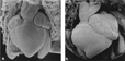
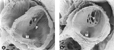
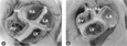
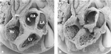
Frontal views of the unopened hearts demonstrate the striking similarities between our chicken hearts and human hearts with left juxtaposition of the atrial appendages (Fig. 1). After opening the cardiac chambers, the usual viscero-atrial arrangement was found to be present in all hearts. It was always the morphologically right atrial appendage, therefore, that was malpositioned with respect to the great arteries. The systemic venous connections were always normal, but totally anomalous pulmonary venous connection was noted in one case (Fig. 2). The atrioventricular connections were concordant in nine hearts, with a univentricular connection to the left ventricle, due to absence of the right atrioventricular connection (Figs 3–5), being found in the other heart. The ventriculo-arterial connections, in contrast, were abnormal in every case. In eight hearts, there was double outlet from the right ventricle (Figs 3a; 4c; 6). The remaining two cases had a single outlet from the right ventricle, one case via a common arterial trunk, and the other via a solitary arterial trunk (Figs 3b; 4d). Consequently, the blood from the left ventricle exited through the interventricular communication in every heart examined (Figs 4c; 6b). The atrial septum, which in the chick is normally formed only by the primary septum, was normally developed in nine cases. The remaining case had a small atrioventricular septal defect (Fig. 7).

Figure 1. Ventral views of hearts and great arteries of 10-day-old chick embryos. External aspects of a normal heart (a) and of a heart with left juxtaposition of the atrial appendages (b).

Figure 2. Hearts with left juxtaposition of the atrial appendages of 8-day-old chick embryos. Internal aspects of the left atrium. There is a normally developed primary septum with normal fenestrations (secondary foramen) in both hearts, but there is absence of pulmonary venous connection to the left atrium in the heart shown in Fig. 2b (×35). F: secondary foramen; M: mitral valve; P: mouth of the pulmonary veins.

Figure 3. Ventral views of opened hearts with left juxtaposition of the atrial appendages of 8-day-old chick embryos. (a) Heart with normally developed atrioventricular valves. (b) Heart with tricuspid atresia, absence of the pulmonary trunk, and totally anomalous pulmonary venous connection. Note the hypoplasia of the right atrium and right ventricle in the heart shown in Fig. 3b (×19). Same abbreviations as before.

Figure 4. Hearts with left juxtaposition of the atrial appendages of 8-day-old chick embryos. The plane of the cardiac valves is viewed from the atrial cavities (a,b), and viewed from the cardiac apex (c,d). Note the abnormal left-sided position of both atrial cavities with respect to the origin of the great arterial trunks. Note also the absence of the tricuspid valve in the heart shown in Fig. 4b,d. (×35). Ao: aortic valve; IA: interatrial septum; M: mitral valve; P: pulmonary valve; S: mouth of systemic venous sinus; T: tricuspid valve; [starf ]: the borders of the ventricular septal defect.

Figure 5. Tricuspid atresia. Higher magnification view of the right atrium as shown in Fig. 4b. Note the presence of a slight depression ([starf ]) at the normal side of the orifice of the tricuspid valve (×65). F: secondary foramen; IA: interatrial septum; LA: left atrium; S: mouth of systemic venous sinus.

Figure 6. Same heart with atrial juxtaposition as shown in Fig. 1b. The internal aspects of the cardiac chambers, and of the aorta and the brachiocephalic arteries, have been exposed by stepwise dissection. Note the anterior position of the aorta, the left-sided position of the atrioventricular valves with respect to the great arterial trunks, the ventriculo-arterial connection of double-outlet right ventricle, and the only outlet of the left ventricle via an interventricular communication (×19). Ao: aorta; LA: left atrium; LV: left ventricle; RA: right atrium; RV: right ventricle; S: mouth of systemic venous sinus.

Figure 7. Atrioventricular septal defect. (a) The plane of the cardiac valve is viewed from the cardiac apex (×50). (b) Internal aspect of the left atrium. There is an almost normally developed primary septum with normal fenestrations (secondary foramen). (×35). AV: opening of the common atrioventricular valve; F: secondary foramen; L: left portion of the atrioventricular canal; O: ventricular outlet portion; R: right portion of the atrioventricular canal; [starf ]: the crest of the interventricular septum.
Anomalies of the atrioventricular valves were found in five hearts, including one case of tricuspid atresia (Figs 4b,d; 5), three cases of tricuspid stenosis, and one case of common atrioventricular junction and valve (Fig. 7a,b).
At the ventriculo-arterial junction, we found two arterial valves in eight hearts only. A single arterial valve was found in the two hearts with single outlet from the right ventricle. The position of the aortic valve with respect to the pulmonary valve was abnormal in all of the eight hearts with two arterial valves. The aortic valve was directly anterior in three, anterior and to the right in four, and side-by-side and to the right of the pulmonary valve in one of the cases.
The positional relationships between the atrioventricular valves and the arterial valves were also abnormal in every heart. This anomaly was not simply the abnormal position of the aorta typically found in “classical” cases of hearts with double-outlet right ventricle. Malpositions of the valves reflected also the positional anomaly of the atrial appendages, the two atrioventricular valves being located almost completely to the left of the arterial valves (Figs. 4a–d; 6b). Malposition of the morphologically right atrium with respect to the great arteries was, therefore, not confined to its appendage, but also involved the body of the chamber and the systemic venous sinus.
Examination of the great arteries displayed a frequent association with anomalies of the arterial trunks themselves, such abnormalities being found in seven cases. Most of lesions, five in all, involved the pulmonary circulation, including absence of one or of both of the branches of the pulmonary trunk in four cases, and absence of the trunk and its intrapericardial branches in the one heart with solitary arterial trunk (Fig. 3b). Absence of the right brachiocephalic artery was found four times, and anomalies of the aortic arch were found twice, the latter comprising interruption of the aortic arch, and presence of a left aortic arch instead of the normal right aortic arch of chicks.
Discussion
The term “juxtaposition of the atrial appendages” describes the situation in which the two atrial appendages do not embrace the arterial trunks, but lie side-by-side, either to the left or to the right of the arterial pedicle.4, 5, 6, 10 Hearts with such a side-by-side arrangement of the atrial appendages can then be divided as left- and right juxtapositions, respectively, taking the position of the two juxtaposed atrial appendages relative to the great arterial trunks to be the defining feature.3 With such left juxtapositions, it is usually the morphologically right atrial appendage that is malpositioned with respect to the great arteries, whereas in right juxtapositions it is typically the morphologically left appendage that is juxtaposed. Both types of juxtaposition are frequently associated with severe cardiovascular malformations.4–6
This classical classification has recently been challenged by the finding that the identities of the juxtaposed appendages are reversed in the extremely rare settings of atrial juxtaposition with mirror-imaged viscero-atrial arrangement.4, 10 Based on this finding, a new classification was proposed, which used the morphological identity of the malpositioned appendage as the defining feature.4, 10 This classification based on morphology then distinguishes between juxtaposition of the morphologically right atrial appendage, corresponding to the majority of cases previously classified as left juxtaposition, and juxtaposition of the morphologically left atrial appendage, which corresponds to the majority of cases previously classified as right juxtaposition. It was also suggested that each of these syndromes had a distinct pattern of associated cardiovascular defects.4, 10 Juxtaposition of the morphologically right atrial appendage, for example, was reported to be associated with hypoplasia of the right ventricle and abnormal infundibular morphology, whereas juxtaposition of the left atrial appendage was reported to be associated with hypoplasia of the left ventricle, but normal infundibular structures. This concept itself, however, is challenged by other data showing that the patterns of cardiovascular lesions associated with juxtaposed atrial appendages do not strictly depend on the morphological identity of the malpositioned atrial appendage, but are related to the specific type of atrioventricular connection.11, 12
In the present study, we describe the pathomorphological features of chicken hearts with left juxtaposition of the atrial appendages induced by the administration of the drug suramin to embryos during the early phase of organogenesis. In all theses hearts, it was the morphologically right atrial appendage that was juxtaposed. Our cases of juxtaposition, therefore, can also be classified as juxtaposition of the morphologically right atrial appendage. For simplicity of description, nonetheless, we have retained the classical terminology.
The pattern of cardiovascular malformations found in our chicken hearts was very similar to that described for the corresponding human cases.4–6 Thus, we found frequent associations with stenosis or atresia of the tricuspid valve, abnormal relationships between the great arteries, typically an anterior aorta, abnormal ventriculo-arterial connections, and anomalies of the aortic and pulmonary arterial pathways.
On the other hand, however, we have to state that there also seem to be some differences from the human situation. In our chicken hearts with left juxtaposition, double-outlet from the right ventricle was the predominant form of ventriculo-arterial connection, whereas discordant ventriculo-arterial connections were not found. In the human situation, double-outlet right ventricle has been seen, but discordant ventriculo-arterial connections predominate.4–6 A second difference seems to concern the position of the atrial chambers. In our chicken hearts, the malposition with respect to the great arteries was not confined to the appendage of the right atrium but regularly included almost its entire chamber so that these heart defects would be best classified as complete malposition of the morphologically right atrium to the left of the great arterial trunks. This situation seems to differ from that found in the human, since several authors have stated that, in the human cases, the positional anomaly is confined only to the appendage of the right atrium, with the body of the chamber and the systemic venous sinus being located in the usual position behind and to the right of the roots of the arterial trunks.13–16 When reviewing the literature, however, we also found descriptions of atrial anatomy in the setting of human hearts with left juxtaposition that suggest that the malposition is not always confined to the right atrial appendage. Allwork and coauthors,17 for example, noted that “[mldr ] the right atrium, instead of forming the right border of the heart, was immediately anterior to the left.” Moreover, we have become aware of at least three case reports on human hearts with left juxtaposition that have described or illustrated a left-sided position of the tricuspid valve with respect to the origin of the great arteries.3, 18, 19 Interestingly, these three human hearts not only showed the same kind of malposition of the right atrial chamber as did our chicken hearts with left juxtaposition, but additionally had the same ventriculo-arterial connection, namely double-outlet from the right ventricle. Fragoyannis and Nickerson20 have also described a heart with left juxtaposition in which the right atrial chamber was positioned entirely to the left of the great arteries. In this heart, a solitary ventricle of undetermined morphology gave rise to the pulmonary trunk, and the tricuspid and aortic valves were atretic.
We draw the following conclusions. First, in human beings, the diagnostic group of congenital cardiac malformations with left juxtaposition of the atrial appendages seems to encompass a spectrum of cardiac malformations ranging from hearts with malposition of only the right atrial appendage to hearts with malposition of the entire right atrium. They might, therefore, be more heterogeneous than previously thought. Second, our chicken hearts with left juxtaposition do not reflect the entire spectrum of the human entity, but correspond to malposition of the entire morphologically right atrium. The fact that hearts of this subpopulation, chicken as well as human, usually show the ventriculo-arterial connection of double-outlet from the right ventricle raises the question of whether the type of ventriculo-arterial connection found in human hearts with left juxtaposition might depend on the degree of malposition of the right atrial chamber. We speculate that the occurrence of discordant ventriculo-arterial connections is limited only to those hearts with normal position of the body of the right atrial chamber and the systemic venous sinus. Unfortunately, information on the position of the tricuspid valve with respect to the roots of the great arteries is not given in the majority of the papers previously published.
The apparent restriction of the present series of chicken hearts with left juxtaposition to one subpopulation of the human spectrum might be explained in part by the process of selection of the specimens from the pool of hearts from suramin-treated chicks. Among this pool, there were not only specimens with left juxtaposition of the atrial appendages but also specimens in which the right atrial appendage was behind the great arteries.2 At the time-point of examination of our specimens, the basic features of the four-chambered heart are present but the atrial appendages usually are not very well developed. It is conceivable that, at this developmental stage, recognition of two poorly developed atrial appendages to the left of the great arteries is only possible in hearts with malposition of the entire right atrium. The milder forms of atrial malposition, which might have a potential for later manifestations of juxtaposed appendages, could therefore escape diagnosis.
Our animal model for juxtaposition of the atrial appendages is not the first for this type of human congenital cardiac malformation. A review of the literature showed that similar cardiac phenotypes have previously been induced in rat and chick embryos by the administration of trypan blue.21–25 Surprisingly, the authors of the papers did not realize that these abnormal cardiac phenotypes had their counterparts in human cardiovascular pathology. The main interest in this model, therefore, focussed on the pathogenesis of the induced abnormal ventriculo-arterial connections.23, 24 Trypan blue shares some chemical similarities with suramin. We have found that both agents cause not only a similar spectrum of cardiac malformations, but also a similar spectrum of non-cardiac malformations.2 With respect to juxtaposition of the atrial appendages, however, there also seem to be some differences. In our suramin-induced chicken model, we observed only left juxtaposition, and double- outlet right ventricle was the only type of abnormal ventriculo-arterial connections found. In the trypan blue-induced rat models, however, both left and right juxtapositions were found, and discordant ventriculo-arterial connections occurred in addition to double-outlet right ventricles.23 The occurrence of left and right juxtapositions in the rat models might be explained by the fact that the administration of trypan blue to pregnant rats was performed during a period when the left and right identities of the embryonic body halves were being established. The rat embryos, therefore, might have been sensitive to possible interferences of trypan blue with the establishment of left-right body asymmetry. In contrast, the administration of suramin to our chick embryos was carried out one day after the normal establishment of the bodily asymmetry, which precluded any possible interference of suramin with these processes. The occurrence of discordant ventriculo-arterial connections in the rat model might be explained by a particular potential of the mammalian embryonic heart to develop such connections or by the possibility that, in the cases with discordant ventriculo-arterial connections, the atrial malposition might have been confined only to the atrial appendages.
Another cardiac phenotype with left juxtaposition of the atrial appendages has recently been described, namely in Pitx2 knockout mice,26 suggesting that mutations of the Pitx2 gene might be involved in the aetiology of this kind of cardiac malformation. We do not, however, regard the Pitx2 knockout mouse as a proper model for left juxtaposition of the atrial appendages. First, a cardiac phenotype with left juxtaposition of the atrial appendages was reported only in one study of these knockout mice that, additionally, did not contain any data on the incidence of the anomaly.26 Other investigators have not found atrial juxtaposition within the spectrum of abnormal cardiac phenotypes found in Pitx2 knockout mice.27, 28 Second, juxtaposition of the atrial appendages has not been found in abnormal hearts of human patients with the Axenfeld-Rieger anomaly,29 which can be associated with mutations in the human Pitx2 gene. Third, the hearts of Pitx2 knockout mice with left juxtaposition are all characterized by complete absence of the interatrial septum and the presence of a common atrioventricular junction and valve.26 Such features are found in only a small minority of human hearts with juxtaposition of the atrial appendages,4 as well as in a minority of animal hearts with suramin- and trypan blue-induced juxtapositions.
The fact that hearts with the main characteristics of left juxtaposition of the atrial appendages as seen in man can be induced in avian and mammalian embryos by the administration of suramin or the chemically related dye trypan blue might have some important implications for the understanding of the aetiology and morphogenesis of these congenital cardiac malformations. Finding the molecular target(s) of suramin and trypan blue within the developing heart, for example, might facilitate the identification of genetic defects possibly underlying some of the human congenital cardiac malformations with juxtaposition of the atrial appendages. Furthermore, the chemical similarities between suramin and trypan blue, which are both polysulfonated agents, might facilitate the identification of exogenous factors with the potential of inducing such malformations. In this respect, we should not forget that suramin is used for the chemotherapy of frequent infectious diseases, such as African trypanosomiasis and onchocerciasis. This fact poses the question as to whether suramin might induce in such patients the birth of children with juxtaposition of the atrial appendages. In view of the use of suramin mainly in developing countries, however, the answer to this question will not be easy.
Various hypotheses have been published to explain the morphogenesis of human hearts with left juxtaposition of the atrial appendages. Due to the lack of embryological data, none of these hypotheses has thus far been confirmed. With our chicken model for left juxtaposition, we now possess a tool that might clarify the morphogenesis. Further study of our chicken model might not only help to clarify the basic developmental errors underlying the human hearts with left juxtaposition, but also facilitate the unravelling of some of the morphogenetic mechanisms underlying normal cardiac development. This is because the abnormal positional relationship between the atriums and great arteries present in left juxtaposition resembles the normal situation of the embryonic heart loop at the beginning of the late phase of cardiac looping. Left juxtaposition of the atrial appendages has, therefore, been interpreted by many authors as the result of a developmental arrest of the process of cardiac looping.4, 18–20, 30–32 If this hypothesis is true, our chicken model might also help unravel the morphogenetic mechanisms underlying this late phase of normal cardiac looping, characterised by a shift of the proximal portion of the outflow tract of the embryonic heart from a right lateral position with respect to the atriums towards its final position ventral to the right atrium.33
Acknowledgements
We thank Mrs. Kirsten Falk-Stietenroth and Mr. Hannes Sydow for technical and photographic assistance, Mrs. Cyrilla Maelicke for correcting the English manuscript, and Prof. Gerd Steding for critical reading of the manuscript.
References
Männer J, Sydow H-G, Heinicke F, Hesse H. Teratogene Wirkungen von Suramin bei Hühnerembryonen. Ann Anat 2002; 184 (Suppl.): 107.Google Scholar
Männer J, Seidl W, Heinicke F, Hesse H. Teratogenic effects of suramin on the chick embryo. Anat Embryol 2003; 206: 229–237.Google Scholar
Dixon ASJ. Juxtaposition of the atrial appendages: two cases of an unusual congenital cardiac deformity. Brit Heart J 1954; 16: 153–164.Google Scholar
Van Praagh S, O'Sullivan, Brili S, Van Praagh R. Juxtaposition of the morphologically right atrial appendage in solitus and inversus atria: A study of 35 postmortem cases. Am Heart J 1996; 132: 382–390.Google Scholar
Maître Azcárate MJ, Quero Jiménez M, Cabrera Duro A, Berrazueta JR, Otero Coto E, Raposo Sonnenfeld I. Juxtaposition der Herzohren. Herz 1980; 5: 339–348.Google Scholar
Anjos RT, Ho SY, Anderson RH. Surgical implications of juxtaposition of the atrial appendages. A review of forty-nine autopsied hearts. J Thorac Cardiovasc Surg 1990; 99: 897–904.Google Scholar
Hamburger V, Hamilton HL. A series of normal stages in the development of the chick embryo. J Morphol 1951; 88: 49–92.Google Scholar
Männer J, Seidl W, Steding G. Experimental study on the significance of abnormal cardiac looping for the development of cardiovascular anomalies in neural crest-ablated chick embryos. Anat Embryol 1996; 194: 289–300.Google Scholar
Ho SY, Baker EJ, Rigby ML, Anderson RH. Basic principles of diagnosis. In: Ho SY, Baker EJ, Rigby ML, Anderson RH (eds). Color atlas of congenital heart disease. Morphologic and clinical correlations. Mosby-Wolfe, 1995, pp 25–34.
Van Praagh S, O'Sullivan JJ, Brili S, Van Praagh R. Juxtaposition of the morphologically left atrial appendage in solitus and inversus atria: A study of 18 postmortem cases. Am Heart J 1996; 132: 391–402.Google Scholar
Anderson RH, Smith A, Wilkinson JL. Right juxtaposition of the auricular appendages. Eur J Cardiol 1976; 4: 495–503.Google Scholar
Lai WW, Ravishankar C, Gross RP, et al. Juxtaposition of the atrial appendages: a clinical series of 22 patients. Pediatr Cardiol 2001; 22: 121–127.Google Scholar
Birmingham A. Extreme anomaly of the heart and great vessels. J Anat Physiol 1893; 27: 139–150.Google Scholar
Wagner HR, Alday LE, Vlad P. Juxtaposition of the atrial appendages. A report of six necropsied cases. Circulation 1970; 42: 157–163.Google Scholar
Mendelsohn G, Hutchins GM. Juxtaposition of atrial appendages. Arch Pathol Lab Med 1977; 101: 490–492.Google Scholar
Muñoz Castellanos L, de la Cueva R, Zavaleta D, Kuri Nivón M. Yuxtaposicion de las Orejuelas (Juxtaposition of the atrial appendages). Arch Inst Mex 1989; 59: 375–382.Google Scholar
Allwork SP, Urban AE, Anderson RH. Left juxtaposition of the auricles with l-position of the aorta. Report of 6 cases. Br Heart J 1977; 39: 299–308.Google Scholar
Kettler L. Ein besonders gearteter Fall von Transposition der großen Gefäße. Virchow Arch Path Anat 1932; 287: 10–28.Google Scholar
Fragoyannis S, Nickerson D. An unusual congenital heart anomaly. Tricuspid atresia, aortic atresia and juxtaposition of atrial appendages. Am J Cardiol 1960; 6: 678–681.Google Scholar
Fox MH, Goss CM. Experimental production of a syndrome of congenital cardiovascular defects in rats. Anat Rec 1956; 124: 189–208.Google Scholar
Fox MH, Goss CM. Experimentally produced malformations of the heart and great vessels in rat fetuses. Atrial and caval abnormalities. Anat Rec 1957; 129: 309–332.Google Scholar
Fox MH, Goss CM. Experimentally produced malformations of the heart and great vessels in rat fetuses. Transposition complexes and aortic arch abnormalities. Am J Anat 1958; 102: 65–91.Google Scholar
Monie IW, Takacs E, Warkany J. Transposition of the great vessels and other cardiovascular abnormalities in rat fetuses induced by trypan blue. Anat Rec 1966; 156: 175–190.Google Scholar
Stéphan F, Mick G. Action du bleu Trypan sur le Coeur et les Arcs Aortiques de l'Embryon de Poulet. C R Ass Anat 1963; 121: 408–417.Google Scholar
Kitamura K, Miura H, Miyagawa-Tomita S, et al. Mouse Pitx2 deficiency leads to anomalies of the ventral body wall, heart, extra- and periocular mesoderm and right pulmonary isomerism. Development 1999; 126: 5749–5758.Google Scholar
Lu MF, Pressmann C, Dyer R, Johnson RL, Martin JF. Function of Rieger syndrome gene in left-right asymmetry and craniofacial development. Nature 1999; 401: 276–278.Google Scholar
Lin CR, Kioussi C, O'Connell S, et al. Pitx2 regulates lung asymmetry, cardiac positioning and pituitary and tooth morphogenesis. Nature 1999; 401: 279–282.Google Scholar
Grosso S, Farnetani MA, Berardi R, et al. Familial Axenfeld-Rieger anomaly, cardiac malformations, and sensineural hearing loss: A provisionally unique genetic syndrome? Am J Med Genet 2002; 111: 182–186.Google Scholar
Ngai SK. Congenital anomaly of the heart: report of a case, with embryological discussion. Am J Pathol 1935; 11: 309–321.Google Scholar
Bredt H. Formdeutung und Entstehung des mißgebildeten menschlichen Herzens. I–V. Virchow Arch Path Anat 1936; 296: 114–157.Google Scholar
Smyth NPD. Lateroposition of the atrial appendages. A case of levoposition of the appendages. Arch Pathol 1955; 60: 259–266.Google Scholar
Männer J. Cardiac looping in the chick embryo: a morphological review with special reference to terminological and biomechanical aspects of the looping process. Anat Rec 2000; 259: 248–256.Google Scholar
Ventral views of hearts and great arteries of 10-day-old chick embryos. External aspects of a normal heart (a) and of a heart with left juxtaposition of the atrial appendages (b).
Hearts with left juxtaposition of the atrial appendages of 8-day-old chick embryos. Internal aspects of the left atrium. There is a normally developed primary septum with normal fenestrations (secondary foramen) in both hearts, but there is absence of pulmonary venous connection to the left atrium in the heart shown in Fig. 2b (×35). F: secondary foramen; M: mitral valve; P: mouth of the pulmonary veins.
Ventral views of opened hearts with left juxtaposition of the atrial appendages of 8-day-old chick embryos. (a) Heart with normally developed atrioventricular valves. (b) Heart with tricuspid atresia, absence of the pulmonary trunk, and totally anomalous pulmonary venous connection. Note the hypoplasia of the right atrium and right ventricle in the heart shown in Fig. 3b (×19). Same abbreviations as before.

Hearts with left juxtaposition of the atrial appendages of 8-day-old chick embryos. The plane of the cardiac valves is viewed from the atrial cavities (a,b), and viewed from the cardiac apex (c,d). Note the abnormal left-sided position of both atrial cavities with respect to the origin of the great arterial trunks. Note also the absence of the tricuspid valve in the heart shown in Fig. 4b,d. (×35). Ao: aortic valve; IA: interatrial septum; M: mitral valve; P: pulmonary valve; S: mouth of systemic venous sinus; T: tricuspid valve; [starf ]: the borders of the ventricular septal defect.

Tricuspid atresia. Higher magnification view of the right atrium as shown in Fig. 4b. Note the presence of a slight depression ([starf ]) at the normal side of the orifice of the tricuspid valve (×65). F: secondary foramen; IA: interatrial septum; LA: left atrium; S: mouth of systemic venous sinus.
Same heart with atrial juxtaposition as shown in Fig. 1b. The internal aspects of the cardiac chambers, and of the aorta and the brachiocephalic arteries, have been exposed by stepwise dissection. Note the anterior position of the aorta, the left-sided position of the atrioventricular valves with respect to the great arterial trunks, the ventriculo-arterial connection of double-outlet right ventricle, and the only outlet of the left ventricle via an interventricular communication (×19). Ao: aorta; LA: left atrium; LV: left ventricle; RA: right atrium; RV: right ventricle; S: mouth of systemic venous sinus.
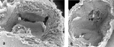
Atrioventricular septal defect. (a) The plane of the cardiac valve is viewed from the cardiac apex (×50). (b) Internal aspect of the left atrium. There is an almost normally developed primary septum with normal fenestrations (secondary foramen). (×35). AV: opening of the common atrioventricular valve; F: secondary foramen; L: left portion of the atrioventricular canal; O: ventricular outlet portion; R: right portion of the atrioventricular canal; [starf ]: the crest of the interventricular septum.
 You have
Access
- 6
- Cited by