Pulmonary atresia with ventricular septal defect is a complex congenital cardiac anomaly. The pulmonary circulation is usually supplied by a patent ductus arteriosus or a major aortopulmonary collateral artery in patients with pulmonary atresia with ventricular septal defect. Rarely, the source of the pulmonary blood flow is from the coronary arteries. These are called coronary artery–pulmonary artery fistulas. Reference Yadav, Bhargava, Buxi and Sirvi1 We present coronary artery–pulmonary artery fistula in two patients with pulmonary atresia with ventricular septal defect. In the first patient who was operated 10 years ago, the main pulmonary artery was supplied by a fistula from the left main coronary artery. In the second patient, the right pulmonary artery was supplied by a fistula from the left main coronary artery and advanced to the right lung posteriorly to the aorta. The left pulmonary artery originated from the patent ductus arteriosus. The difference in our cases is that the coronary artery pulmonary artery fistulas behave like major aortopulmonary collateral arteries originating from the coronary arteries. These fistulas were the main source of pulmonary blood flow. A limited number of case reports on pulmonary atresia with ventricular septal defect currently exist in the literature.
Case reports
Patient 1
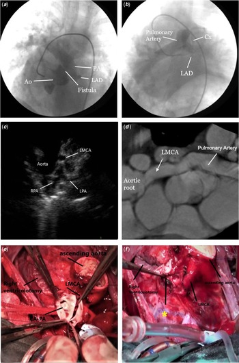
A 4-year-old boy was diagnosed with pulmonary atresia with ventricular septal defect and admitted to our hospital. On cardiac examination, there was a single S2 sound with a continuous 3/6 murmur radiating to the left midsternal border. Transthoracic echocardiography revealed a large ventricular septal defect and pulmonary atresia. A fistula was present between the left main coronary artery and the main pulmonary artery (Fig 1). A diagnostic catheter angiography confirmed transthoracic echocardiography findings. Major aortopulmonary collateral arteries that supplied blood from the descending aorta to both lungs were identified.
( a ) Cardiac catheterisation image of the main pulmonary artery (PA) originating from the LMCA. ( b ) The pulmonary artery with contrast during coronary angiography. ( c ) Transthoracic echocardiography image of the main pulmonary artery originating from the LMCA. ( d ) CT image of the main pulmonary artery originating from the LMCA. ( e ) Coronary artery–pulmonary artery (CA–PA) fistula. ( f ) Pulmonary artery stump (marked with *) after fistula closure. LMCA = left main coronary artery.

During the operation, cardiopulmonary bypass was initiated with aortic and bicaval cannulation. After isolation of the left and right pulmonary arteries from the heart, the aortic cross-clamp was placed and the aortotomy was performed. The left coronary ostium was observed to be greatly dilated. The left main coronary artery branched from the posterior of the aorta and bifurcated into the left anterior descending and circumflex arteries. After the pulmonary artery was incised longitudinally, the fistula from the left main coronary artery to the pulmonary artery was identified (Fig 1). The fistula between the left main coronary artery and main pulmonary artery was transected and the left main coronary artery roof was closed with a pericardial patch. The ventricular septal defect was closed transatrially and transventricularly with a polytetrafluoroethylene (Goretex) patch. Due to high right heart pressures, a 4–5-mm window was opened in the middle of the patch. The right ventricular outflow tract and the transected pulmonary artery were reconstructed with a 19 no Labcor stentless valved pulmonary conduit (Labcor, Belo Horizonte, Brazil).
The patient experienced pulmonary haemorrhage in the intensive care and was referred to cardiac catheterisation. Major aortopulmonary collateral arteries were occluded with coils. Following the procedure, oxygen saturation remained at 88% and the mean pulmonary artery pressure decreased from 56 mmHg to 51 mmHg. Due to difficulty with weaning from mechanical ventilation, a tracheostomy was performed on the 42nd post-operative day. The patient was eventually weaned from mechanical ventilation and was discharged on the third post-operative month. During transthoracic echocardiography examination at follow-up, a two-way shunt was observed in the fenestrated interventricular patch.
The patient was admitted 8 years later with symptoms of NYHA class III and ascites. Transthoracic echocardiography examination showed a one-way shunt from left to right resulting in high pulmonary artery pressures and closure of the ventricular septal defect was decided. The ventricular septal defect was closed with a 16-mm muscular ventricular septal defect occluder. After the procedure, the right ventricular pressure was 50 mmHg with improved right ventricular systolic functions and a mild-to-moderate stenosis in the pulmonary conduit. The patient is currently alive 10 years after the first operation at NYHA class I.
Patient 2
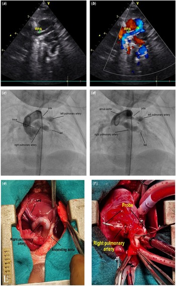
A 40-day-old patient weighing 3350 g with a pulmonary atresia with ventricular septal defect was admitted to our hospital. The patient was tachycardic, with feeding difficulties. Oxygen saturation was 90%. Cardiac examination revealed a single S2 sound with a continuous 3/6 murmur radiating to the left midsternal border. Transthoracic echocardiography showed a wide subaortic ventricular septal defect and a 60% dextroposed overriding aorta. Cardiac catheterisation was performed for further examination. The right pulmonary artery originated from the left main coronary artery and continued to the right posterior to the ascending aorta. The left pulmonary artery originated from the patent ductus arteriosus. A dilated left anterior descending was seen distal to the left main coronary artery (Fig 2). No major aortopulmonary collateral arteries were identified in this patient.
( a , b ) Transthoracic echocardiography image of the right pulmonary artery originating from the LMCA. ( c , d ) Cardiac catheterisation image of the right pulmonary artery (PA) originating from the LMCA. The LAD is seen distal to the right PA. The left PA originates from the PDA. ( e ) The right pulmonary artery originating from the LMCA (intraoperative view). ( f ) A probe is advanced from the LMCA to the right pulmonary artery. LMCA = left main coronary artery, LAD = left anterior descending, PDA = patent ductus arteriosus.

Patency of the patent ductus arteriosus was maintained with PgE1 infusion until the operation. In this case, coronary artery–-pulmonary artery fistula causing a high blood flow to the pulmonary vascular bed and a dilated left anterior descending was seen after the origin of the right pulmonary artery from left main coronary artery (Fig 2). The right pulmonary artery was isolated and cardiopulmonary bypass was initiated with aortic and bicaval cannulation. The right pulmonary artery was separated from the left main coronary artery, and the left main coronary artery was repaired with sutures. The left pulmonary artery was transected from the patent ductus arteriosus,, and patent ductus arteriosus was closed. The right and left pulmonary arteries were anastomosed posteriorly and augmented with fresh pericardium anteriorly. A modified Blalock–Taussing shunt with a 4-mm Polytetrafluoroethylene graft was created between the right subclavian artery and right pulmonary artery. The patient, who had an ICU stay of 2 days, was discharged on the ninth post-operative day.
Discussion
Coronary artery to pulmonary artery fistula is a rare condition in pulmonary atresia with ventricular septal defect patients. The prevalence of coronary artery–pulmonary artery fistula has been reported as 7–11% in retrospective case series. Reference Sathanandam, Loomba, Ilbawi and Van Bergen2 Collison et al reported four cases of coronary artery–pulmonary artery fistula (8%) among 50 pulmonary atresia with ventricular septal defect patients. Reference Collison, Dagar, Kaushal, Radhakrishanan, Shrivastava and Iyer3 In another review by Amin et al 9 (10%), coronary artery–pulmonary artery fistula patients were detected in 87 pulmonary atresia with ventricular septal defect patients. Reference Amin, McElhinney, Reddy, Moore, Hanley and Teitel4 Similar rates were reported in other studies. In our study, among 78 pulmonary atresia with ventricular septal defect patients, 2 (2.5%) patients had coronary artery–pulmonary artery fistula.
Coronary artery–pulmonary artery fistula originates most frequently from the left coronary artery than the right coronary artery. In the left coronary system, the most common origin of coronary artery–pulmonary artery fistulas is the circumflex coronary artery. Reference Sathanandam, Loomba, Ilbawi and Van Bergen2 In the study by Yadav et al with two patients, the pulmonary arteries of both patients originated from the left main coronary artery. Reference Yadav, Bhargava, Buxi and Sirvi1 In our study, coronary artery–pulmonary artery fistula originated from the left main coronary artery in both of our patients and these fistulas were the main source of pulmonary blood flow. Small coronary pulmonary fistulas between coronaries and pulmonary arteries are common in patients with tetralogy of Fallot and patients with pulmonary atresia with ventricular septal defect. However, in these two cases, this coronary–pulmonary connection is the main source of pulmonary blood flow, and coronary artery–pulmonary artery fistulas act as major aortopulmonary collateral arteries.
Another important detail in this pathology is the presence of major aortopulmonary collateral arteries. Major aortopulmonary collateral arteries were present in 90% of the previously reported cases. The remaining 10% did not have major aortopulmonary collateral artery and fistulas were the sole blood supply of the pulmonary circulation. Reference Sathanandam, Loomba, Ilbawi and Van Bergen2 There is a risk of coronary steal in pulmonary atresia with ventricular septal defect patients with coronary artery–pulmonary artery fistula, but this has not been reported in the literature. Reference Sathanandam, Loomba, Ilbawi and Van Bergen2 We have observed an increased blood flow through the fistulas to the lungs in our patients without any Electrocardiogram changes.
Different surgical approaches were utilised in these patients according to the experience of the surgeons and pulmonary artery development. In the review by Amin et al, all cases had additional major aortopulmonary collateral arteries, seven patients from the left and two from the right coronary system. One-stage unifocalisation was applied to all their patients. Reference Amin, McElhinney, Reddy, Moore, Hanley and Teitel4 Collison et al performed two-stage repairs on all four patients. Unifocalisations were followed by complete repairs. Reference Collison, Dagar, Kaushal, Radhakrishanan, Shrivastava and Iyer3 In our first patient, major aortopulmonary collateral arteries were closed with coils after the operation. In our second patient without major aortopulmonary collateral arteries, unifocalisation of the right and left pulmonary arteries and a m-BT shunt were carried out. Ventricular septal defect closure was necessary in the next operation. Both of our patients are currently alive.
Conclusion
Coronary artery–pulmonary artery fistula is a very rare anomaly. The necessary surgical intervention is unifocalisation of the pulmonary arteries, supply of pulmonary flow, and the closure of fistulas. After the unifocalisation of the pulmonary arteries, to continue with a shunt or total correction is decided depending on the development of the pulmonary arteries.
Financial support
This research received no specific grant from any funding agency, commercial or not-for-profit sectors.
Conflicts of interest
None.
Ethical standards
The authors assert that all procedures contributing to this work comply with the ethical standards of the relevant national guidelines on human experimentation and with the Helsinki Declaration of 1975, as revised in 2008, and has been approved by the institutional committees.