INTRODUCTION
Trichomonas vaginalis is the causal agent of trichomonosis, the primary non-viral sexually transmitted infection. Men and women with this condition are at higher risk for HIV seroconversion (Laga et al. Reference Laga, Manoka, Kivuvu, Malele, Tuliza, Nzila, Goeman, Behets, Batter and Alary1993; Cohen, Reference Cohen2000; McClelland et al. Reference McClelland, Sangare, Hassan, Lavreys, Mandaliya, Kiarie, Ndinya-Achola, Jaoko and Baeten2007; Van Der Pol et al. Reference Van Der Pol, Kwok, Pierre-Louis, Rinaldi, Salata, Chen, Van De Wijgert, Mmiro, Mugerwa, Chipato and Morrison2008; Kissinger et al. Reference Kissinger, Secor, Leichliter, Clark, Schmidt, Curtin and Martin2008, Reference Kissinger, Amedee, Clark, Dumestre, Theall, Myers, Hagensee, Farley and Martin2009). Women with trichomonosis will have adverse pregnancy outcomes (Cotch et al. Reference Cotch, Pastorek, Nugent, Hillier, Gibbs, Martin, Eschenbach, Edelman, Carey, Regan, Krohn, Klebanoff, Rao and Rhoads1997) and elevated susceptibility to cervical cancer (Viikki et al. Reference Viikki, Pukkala, Nieminen and Hakama2000).
T. vaginalis possesses numerous cysteine proteases (CPs). There is evidence to indicate the CPs are promiscuous in activity and appear to be involved in cytoadherence (Arroyo and Alderete, Reference Arroyo and Alderete1989; Mendoza-Lopez et al. Reference Mendoza-Lopez, Becerril-Garcia, Fattel-Facenda, Avila-Gonzalez, Ruiz-Tachiquin, Ortega-Lopez and Arroyo2000), haemolysis (Dailey et al. Reference Dailey, Chang and Alderete1990), cytotoxicity (Alvarez-Sanchez et al. Reference Alvarez-Sanchez, Avila-Gonzalez, Becerril-Garcia, Fattel-Facenda, Ortega-Lopez and Arroyo2000, Reference Alvarez-Sanchez, Solano-Gonzalez, Yañez-Gomez and Arroyo2007), disruption of the host cell membrane skeleton (Fiori et al. Reference Fiori, Rappelli, Addis, Mannu and Cappuccinelli1997), and degradation of human immunoglobulins (Provenzano and Alderete, Reference Provenzano and Alderete1995; Min et al. Reference Min, Hyun, Ryu, Ahn and Cho1998). In addition to the CPs, a metallo-peptidase has been identified that could be involved in the processing of hydrogenosomal proteins (Brown et al. Reference Brown, Goldstone, Bastida-Corcuera, Delgadillo-Correa, McArthur and Johnson2007). The genomic sequence of T. vaginalis indicates that there are 33 subtilisin-like serine proteases, but the proteases are annotated as putative based on sequence similarity (Carlton et al. Reference Carlton, Hirt, Silva, Delcher, Schatz, Zhao, Wortman, Bidwell, Alsmark, Besteiro, Sicheritz-Ponten, Noel, Dacks, Foster, Simillion, Van De, Miranda-Saavedra, Barton, Westrop, Muller, Dessi, Fiori, Ren, Paulsen, Zhang, Bastida-Corcuera, Simoes-Barbosa, Brown, Hayes, Mukherjee, Okumura, Schneider, Smith, Vanacova, Villalvazo, Haas, Pertea, Feldblyum, Utterback, Shu, Osoegawa, De Jong, Hrdy, Horvathova, Zubacova, Dolezal, Malik, Logsdon, Henze, Gupta, Wang, Dunne, Upcroft, Upcroft, White, Salzberg, Tang, Chiu, Lee, Embley, Coombs, Mottram, Tachezy, Fraser-Liggett and Johnson2007). To our knowledge, there are no experimental reports on subtilisin-like serine proteases for T. vaginalis.
In an ongoing project in our laboratory a putative subtilisin-like gene (referred to as sub1) was identified in a T. vaginalis genomic clone as an open reading frame adjacent to an actin gene. We have attempted to characterize the sub1 gene and the encoded SUB1 protein regarding its expression and localization. As such, this work may be the first experimental characterization of a putative subtilisin-like serine protease in T. vaginalis. This 906-aa protein possesses a signal peptide and a pro-peptide at the amino terminal end. The protein, termed SUB1, is detected on the parasite surface by a specific antibody, and this is consistent with a putative transmembrane domain. Interestingly, the sub1 gene is modulated in expression by distinct mechanisms involving iron and contact with host epithelial cells. The implications of our findings are discussed.
MATERIALS AND METHODS
Parasites and cell cultures
The T. vaginalis isolate CNCD147 was used in this study (Alvarez-Sanchez et al. Reference Alvarez-Sanchez, Avila-Gonzalez, Becerril-Garcia, Fattel-Facenda, Ortega-Lopez and Arroyo2000). Parasites were grown in Diamond's trypticase-yeast extract-maltose (TYM) medium (Diamond, Reference Diamond1957) supplemented with 10% (v/v) heat-inactivated horse serum. The iron concentration in TYM is ~20 μm (Alvarez-Sanchez et al. Reference Alvarez-Sanchez, Solano-Gonzalez, Yañez-Gomez and Arroyo2007). Iron-depleted and iron-rich trichomonads were obtained as described (Lehker et al. Reference Lehker, Arroyo and Alderete1991). Parasites were grown in culture medium supplemented with 250 μm ferrous ammonium sulfate (high-iron or iron-rich) (Sigma Chemical Co., St Louis, MO, USA) or with 150 μm of the iron chelator 2,2-dipyridil (low-iron or iron-depleted) (Sigma Chemical Co). Parasites in the logarithmic-phase of growth were used in all assays.
Cloning of the sub1 gene
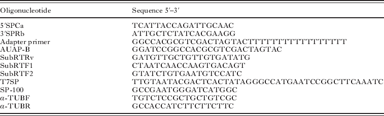
An incomplete open reading frame (ORF) of 2326-bp with similarity to subtilisin-like serine proteases (termed sub1) was identified in a clone isolated in our laboratory from a genomic EcoRI library of T. vaginalis isolate CNCD147. To obtain the missing 3′ end of the sub1gene, a BLAST search was made on the TIGR database of T. vaginalis (www.tigr.org) using the 2326-bp fragment as query. This search retrieved the contig 83372 (TIGR Assembly 83372). The 3′ end of sub1 gene was amplified from genomic DNA using the sense primer 5′SPCa and the antisense primer 3′SPRb (Table 1). The PCR product was cloned in the PCR Blunt TOPO vector (Invitrogen, Carlsbad, CA, USA), and the selected clones were sequenced. The complete sub1 gene sequence is accessed in the GeneBank database under Accession number FJ384963.
Oligonucleotides used in this work

Reverse transcriptase coupled PCR (RT-PCR)
RT-PCR was used to determine the expression of sub1 in T. vaginalis. Total RNA was prepared from parasites grown in TYM, high-iron or low-iron medium using the TRIZOL reagent (Invitrogen) according to the manufacturer's instructions. cDNA was synthesized with the Superscript III RNase H- kit (Invitrogen) and adapter primer (Table 1) using 3 μg of total RNA that was quantified in an Ultrospec 3100 pro spectrometer (Amersham Pharmacia Biotech, Uppsala, Sweden). For semi-quantitative RT-PCR a fragment of 284-bp of the sub1 cDNA was amplified by PCR using the primers SubRTF1 and SubRTRv. As an internal reference the α-tubulin fragment of 370-bp was amplified using the primers α-TUBF and α-TUBR (Table 1). PCR reactions without the cDNA synthesis step were used as controls to detect contaminant genomic DNA. PCR conditions were as follows. Fifty μl reactions were set up with 25 pmol of each primer, 1X PCR buffer, 10 mm each dNTP, 25 mm MgCl2, 2·5 units of Taq DNA polymerase (recombinant, Invitrogen) and 0·5 μl of total cDNA of each condition as template. The amplification reactions consisted of 19, 23 or 27 cycles of 92°C denaturing for 1 min, 55°C annealing for 1 min, and 72°C extension for 3 min, all followed by a 5 min extension at 72°C. Ten μl of each PCR reaction were loaded in agarose gels and stained with ethidium bromide. Gels were analysed with Quantity One software (Bio-Rad Laboratories).
Rapid amplification of 3′ cDNA ends (3′ RACE)
The 3′ end of the sub1 mRNA was determined by a 3′ RACE assay. cDNA was synthesized using 3 μg of total RNA, Superscript III RNase H- kit (Invitrogen) and adapter primer (Table 1). The sub1 cDNA end was amplified by PCR from the cDNA pool using the SubRTF2 primer and the AUAP-B adapter primer (Table 1). A second (semi-nested) PCR amplification was performed on the initial PCR product using the SubRTF1 and the AUAP-B adapter primer. The PCR product was cloned into TOPO vector (Invitrogen, Carlsbad, CA) and sequenced in our institutional sequencing facility.
Ribonuclease protection assay (RPA)
Since we identified 2 possible Inr promoter sequences at the 5′ end of the sub1 gene, an RPA was performed to determine the transcription start point of this gene. The RNA antisense probe was synthesized by in vitro transcription using the MAXIscript T7 kit (AMBION Inc., Austin, TX), [α-32P]UTP (Amersham Biosciences UK Ltd) and 1 μg of a PCR product containing the 2 putative sub1 Inr sequences as shown in Fig. 2A. The 533-bp PCR product used as template was obtained from the sub1 clone with primers T7SP and SP-100 (Table 1). The RPA III kit (AMBION Inc. Austin, TX) was used following the manufacturer′s instructions. RPA reactions were performed essentially as described (Lopez-Villaseñor et al. Reference Lopez-Villaseñor, Contreras, Lopez-Griego, Alvarez-Sanchez and Hernandez2004) using 10–20 μg of total RNA and ~60 000 cpm of gel purified probe. After hybridization and digestion, the protected fragments were separated on a denaturing gel comprised of 5% acrylamide-bisacrylamide (19:1) and 8 m urea. The T. vaginalis rRNA gene transcription start point (Lopez-Villaseñor et al. Reference Lopez-Villaseñor, Contreras, Lopez-Griego, Alvarez-Sanchez and Hernandez2004) was used as an internal control in a parallel reaction.
Antibody production and Western blot assay
A mouse antiserum to SUB1 was raised against a synthetic peptide (484-NSETAERKCDFSS-496). This sequence was found to be specific of SUB1 from the T. vaginalis genome database. The peptide was covalently linked as an 8-branched multiple antigen peptide (MAP-8), and 300 μg were mixed with Freund's complete adjuvant (Sigma Immuno Chemicals) and used to immunize mice. The animals were given boosted injections once with 150 μg of synthetic peptide homogenized with Freund's incomplete adjuvant (Sigma Immuno Chemicals) and 5 times with 75 μg of synthetic peptide dissolved in water at 1 to 2-week intervals. This antiserum was used in Western blots and immunofluorescence assays. Pre-immune normal mouse serum was obtained before the immunization schedule began.
For Western blots, 1·5×107T. vaginalis organisms were washed, centrifuged, and lysed with 100 μl of 10% SDS in 400 μl of freshly prepared electrophoresis dissolving buffer (0·6 m Tris, pH 6·8, 2% SDS, 10% glycerol, 0·1% bromophenol blue) with 5% β-mercaptoethanol. Samples were heated at 75°C for 10 min and stored on ice before loading. Total T. vaginalis proteins were separated by PAGE-SDS and electrophoresed at 30 mA (constant). After electrophoresis, the proteins were transferred onto nitrocellulose membranes, blocked with 3% skim milk in PBS for 18 h at 4°C, and incubated with either pre-immune normal mouse serum or mouse anti-SUB1 serum (1:250) for 1 h at 25°C. Blots were then washed 5 times with PBS-0·05% Tween-20 and incubated with peroxidase-conjugated anti-mouse IgG secondary antibody (1:50 000) (Pierce Biotechnology, Rockford, IL, USA) for 1 h at 25 °C prior to developing with hydrogen peroxide and luminol (Supersignal West Pico, Pierce Biotechnology, Rockford, IL, USA). All antibodies were diluted in 1% skim milk-3%-bovine serum albumin in PBS-0·05% Tween-20.
Immunofluorescence assay
Immunofluorescence was performed using established protocols (Allan, Reference Allan2000). Briefly, parasites were washed twice with PBS before fixation in 4% paraformaldehyde for 30 min at RT and washed again with PBS. Permeabilized parasites were treated with 0·1% Triton X-100 in PBS for 7 min at RT after the fixation step. Parasites were then washed once with PBS and blocked with 2% fetal bovine serum in PBS for 1 h at RT, and washed with PBS. Next, the parasites were suspended in 100 μl of the anti-SUB1 serum diluted 1:100 in PBS and incubated 1 h at RT. Parasites were washed once with PBS, suspended in 200 μl of FITC-conjugated anti-mouse IgG (GE Healthcare, Buckingham, UK) (1:100 in PBS) and incubated for 30 min at RT. Controls were performed with non-permeabilized and permeabilized parasites handled identically, except using the pre-immune normal mouse serum and the secondary antibody. The parasites were then examined with a confocal laser scanning microscope (Olympus FV1000).
Bioinformatic analyses of SUB1 and subtilisin-like serine protease genes of Trichomonas vaginalis
The deduced amino acid sequence of SUB1 was analysed with several programs: the signalP 3.0 software (www.cbs.dtu.dk/services/SignalP/) to detect putative signal peptides (Nielsen et al. Reference Nielsen, Engelbrecht, Brunak and Von1997; Bendtsen et al. Reference Bendtsen, Nielsen, Von and Brunak2004), TMHMM 2.0 software (www.cbs.dtu.dk/services/TMHMM-2.0/) to detect transmembrane spanning regions and Phobious software (http://phobius.sbc.su.se/) for a combined transmembrane topology and signal peptide prediction (Kall et al. Reference Kall, Krogh and Sonnerhammer2004, Reference Kall, Krogh and Sonnerhammer2007). The hydropathy plot (window=7) (Kyte and Doolittle, Reference Kyte and Doolittle1982) and gene maps were constructed with the Vector NTI software (Invitrogen). Subtilisin sequences obtained from the MEROPS database (merops.sanger.ac.uk/) were used for amino acid alignment and for the construction of a phylogenetic tree using the CLC sequence viewer software (Neighbor-joining algorithm).
The theoretical model of SUB1 was constructed using the PHYRE software (Protein Homology/analogY Recognition Engine, http://www.sbg.bio.ic.ac.uk/phyre/) (Kelley et al. Reference Kelley, MacCallum and Sternberg2000; Bennett-Lovsey et al. Reference Bennett-Lovsey, Herbert, Sternberg and Kelley2008). The model obtained with PHYRE was tested for errors in the three-dimensional structure with the ProSA software (http://prosa.services.came.sbg.ac.at/prosa.php) (Sippl, Reference Sippl1993; Wiederstein and Sippl, Reference Wiederstein and Sippl2007) and scanned for calcium-binding sites with the WebFEATURE software (http://feature.stanford.edu/webfeature/) (Liang et al. Reference Liang, Banatao, Klein, Brutlag and Altman2003). The protein 3D structures and the electrostatic surface potential were generated with the PyMOL molecular graphics system v0.99 (http://pymol.org/). The assignment of S1, S2, S3 and S4 subsites in the theoretical model of SUB1 was based in the localization of the active site in subtilisin like serine proteases (Siezen and Leunissen, Reference Siezen and Leunissen1997).
The deduced amino acid sequence of the 33 subtilisin-like serine protease genes annotated in the T. vaginalis database (http://trichdb.org/trichdb/) were analysed with the programs signalP 3.0 (www.cbs.dtu.dk/services/SignalP/) (Nielsen et al. Reference Nielsen, Engelbrecht, Brunak and Von1997; Bendtsen et al. Reference Bendtsen, Nielsen, Von and Brunak2004), TMHMM 2.0 (www.cbs.dtu.dk/services/TMHMM-2.0/), Phobious (http://phobius.sbc.su.se/) (Kall et al. Reference Kall, Krogh and Sonnerhammer2004, Reference Kall, Krogh and Sonnerhammer2007), Pfam (pfam.sanger.ac.uk/) (Finn et al. Reference Finn, Tate, Mistry, Coggill, Sammut, Hotz, Ceric, Forslund, Eddy, Sonnhammer and Bateman2008), and Interpro (www.ebi.ac.uk/Tools/InterProScan/) (Hunter et al. Reference Hunter, Apweiler, Attwood, Bairoch, Bateman, Binns, Bork, Das, Daugherty, Duquenne, Finn, Gough, Haft, Hulo, Kahn, Kelly, Laugraud, Letunic, Lonsdale, Lopez, Madera, Maslen, McAnulla, McDowall, Mistry, Mitchell, Mulder, Natale, Orengo, Quinn, Selengut, Sigrist, Thimma, Thomas, Valentin, Wilson, Wu and Yeats2009) to identify functional domains.
RESULTS
The sub1 gene encodes a novel subtilisin-like serine protease in Trichomonas vaginalis
The T. vaginalis genome sequence indicates there are 33 genes encoding subtilisin-like serine proteases. However, these genes are annotated as putative based on sequence similarity and nothing is known about their expression, protein localization and cellular function. Here, we identified a subtilisin-like gene, called sub1, in a genomic clone of the T. vaginalis CNCD147 isolate. This sequence has been accessed in the GenBank database under Accession number FJ384963. The equivalent sequence from the G3 isolate is annotated in the T. vaginalis genome database as TVAG_090450 (Carlton et al. Reference Carlton, Hirt, Silva, Delcher, Schatz, Zhao, Wortman, Bidwell, Alsmark, Besteiro, Sicheritz-Ponten, Noel, Dacks, Foster, Simillion, Van De, Miranda-Saavedra, Barton, Westrop, Muller, Dessi, Fiori, Ren, Paulsen, Zhang, Bastida-Corcuera, Simoes-Barbosa, Brown, Hayes, Mukherjee, Okumura, Schneider, Smith, Vanacova, Villalvazo, Haas, Pertea, Feldblyum, Utterback, Shu, Osoegawa, De Jong, Hrdy, Horvathova, Zubacova, Dolezal, Malik, Logsdon, Henze, Gupta, Wang, Dunne, Upcroft, Upcroft, White, Salzberg, Tang, Chiu, Lee, Embley, Coombs, Mottram, Tachezy, Fraser-Liggett and Johnson2007). The sub1 complete ORF (2,721-bp) does not contain intervening sequences, and encodes for a predicted 101-kDa protein. A BLAST search with the complete deduced amino acid sequence shows that SUB1 has 26–34% similarity with bacterial subtilisins.
In order to determine the gene copy number of sub1 in the T. vaginalis genome, we performed Southern analysis using restriction enzymes that restrict both inside and outside of the coding region. The hybridization pattern expected for a single copy gene was obtained (data not shown), although additional weak hybridization bands were detected that could correspond to other subtilisin-like serine protease related genes described in the T. vaginalis genome (Carlton et al. Reference Carlton, Hirt, Silva, Delcher, Schatz, Zhao, Wortman, Bidwell, Alsmark, Besteiro, Sicheritz-Ponten, Noel, Dacks, Foster, Simillion, Van De, Miranda-Saavedra, Barton, Westrop, Muller, Dessi, Fiori, Ren, Paulsen, Zhang, Bastida-Corcuera, Simoes-Barbosa, Brown, Hayes, Mukherjee, Okumura, Schneider, Smith, Vanacova, Villalvazo, Haas, Pertea, Feldblyum, Utterback, Shu, Osoegawa, De Jong, Hrdy, Horvathova, Zubacova, Dolezal, Malik, Logsdon, Henze, Gupta, Wang, Dunne, Upcroft, Upcroft, White, Salzberg, Tang, Chiu, Lee, Embley, Coombs, Mottram, Tachezy, Fraser-Liggett and Johnson2007; Hirt et al. Reference Hirt, Noel, Sicheritz-Ponten, Tachezy and Fiori2007).
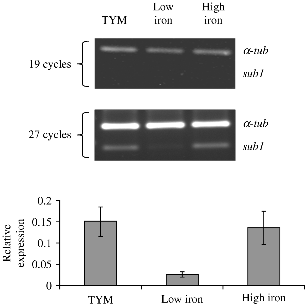
As an initial approach to determine the expression of sub1, Northern blot analyses were performed using up to 40 μg of total T. vaginalis RNA. Unexpectedly, no hybridization bands were detected indicating either a very low or absent expression (data not shown). It is important to mention that the RNA used for these analyses was judged to be of high quality by the presence of intact ribosomal bands on gel-electrophoresis and the readily detectable hybridization band with a control probe for a specific mRNA. As an alternative approach, we evaluated the presence of sub1 mRNA by reverse transcriptase-(RT) PCR analysis using T. vaginalis cDNA and SubRTF1 and SubRTRv oligonucleotides. An amplicon of the expected size was detected after 27 cycles (see below, Fig. 3), suggesting a low abundance of sub1 mRNA in this trichomonad isolate.
The features identified in the genomic and deduced amino acid sequences of sub1 are illustrated in Fig. 1. Two potential initiator (Inr) promoter elements (Liston and Johnson, Reference Liston and Johnson1999) were identified in the 5′ region separated by 142-bp as shown in Fig. 1A: CTAAATT (position 507, distal) and TTACTTC (position 656, proximal). Further, 2 in-frame ATGs were found that could be involved in translation initiation (Fig. 1A, positions 526 and 673). To determine the functional transcription initiator in the sub1 gene, we performed a Ribonuclease Protection Assay (RPA). In the case that this gene is transcribed from the distal initiator a 408-bp protected fragment is predicted, whereas a 259-bp protected fragment would be produced if transcription is initiated at the proximal initiator (Fig. 2A), as annotated for the corresponding TVAG_090450 sequence accessed in the T. vaginalis genome database (Carlton et al. Reference Carlton, Hirt, Silva, Delcher, Schatz, Zhao, Wortman, Bidwell, Alsmark, Besteiro, Sicheritz-Ponten, Noel, Dacks, Foster, Simillion, Van De, Miranda-Saavedra, Barton, Westrop, Muller, Dessi, Fiori, Ren, Paulsen, Zhang, Bastida-Corcuera, Simoes-Barbosa, Brown, Hayes, Mukherjee, Okumura, Schneider, Smith, Vanacova, Villalvazo, Haas, Pertea, Feldblyum, Utterback, Shu, Osoegawa, De Jong, Hrdy, Horvathova, Zubacova, Dolezal, Malik, Logsdon, Henze, Gupta, Wang, Dunne, Upcroft, Upcroft, White, Salzberg, Tang, Chiu, Lee, Embley, Coombs, Mottram, Tachezy, Fraser-Liggett and Johnson2007). The result shows a ~408-bp protected fragment, indicating that transcription of the sub1 gene initiates at the distal Inr promoter sequence (CTAAATT, Fig. 2B). This result also evidences a low level of expression of the sub1 transcript. A RPA assay for the rRNA transcription initiation site (Lopez-Villaseñor et al. Reference Lopez-Villaseñor, Contreras, Lopez-Griego, Alvarez-Sanchez and Hernandez2004) was done in parallel as a control for this experiment (Fig. 2C).
Features of sub1 gene and protein sequences. (A) Partial DNA sequence of sub1 gene. Only the 5′ and 3′ regions are shown (2500 bp of the internal coding region have been omitted). The non-coding sequence is shown in lower case and the coding sequence in upper case. In the 5′ region the two potential transcription initiator elements are in bold (positions 507 and 656). The distal transcription initiator element experimentally confirmed in this work including the putative transcription start site is shown with an arrow. 5′ and 3′ UTR regions are in italics. The two possible AUG codons for translation initiation are underlined. In the 3′ UTR region two contiguous UAAA polyadenylation signals are in bold. T-rich elements and possible binding sites for Myb1 and Myb2 transcription factors are boxed. (B) Deduced amino acid sequence of SUB1. The putative signal peptide is underlined. The predicted transmembrane domain is underlined with a dotted line. The catalytic triad residues D163, H208 and S504 are boxed. Panel C: Hydropathy plot for SUB1. Two hydrophobic regions are found in the N- and C- terminal regions (arrows).

Identification of the transcription start site for sub1. (A) Schematic representation of the sub1 gene indicating the two potential Inr elements (arrows). The relative position of the antisense RNA probe used in the ribonuclease protection assay (RPA) is shown with a solid line. The sizes of the two possible protected fragments are shown as shadowed boxes. (B) RPA of the sub1 transcript. Lanes 1 and 2 (duplicates): Total T. vaginalis RNA+sub1 antisense probe+RNase treatment (protected probe). Lane 3: sub1 antisense probe (non digested probe). Lane 4: total yeast RNA+sub1 antisense probe+RNase reatment (non protected probe). Over-exposure of the autoradiograph was necessary for the detection of protected bands (lanes 1 and 2). (C) RPA of the rRNA transcript used as a positive control. Lane 1: rRNA antisense probe+total yeast RNA+RNase treatment (non protected probe); Lane 2: Total T. vaginalis RNA+rRNA antisense probe+RNase treatment (protected probe). Lane 3: rRNA antisense probe (non-digested probe).

Regarding the 3′ region of the sub1 gene, it was found that the stop codon of sub1 is UAG and is not coupled to the UAAA proposed polyadenylation signal as has been described for several T. vaginalis mRNAs (Espinosa et al. Reference Espinosa, Hernandez, Lopez-Griego and Lopez-Villaseñor2002). The 3′-UTR region of sub1 mRNA was determined by a 3′-RACE assay. This region is 56-nt long with 2 UAAA proposed polyadenylation signals at 31-nt and 38-nt downstream of the stop codon (Fig. 1A). These elements are consistent with previously proposed polyadenylation motifs for T. vaginalis (Espinosa et al. Reference Espinosa, Hernandez, Lopez-Griego and Lopez-Villaseñor2002).
The deduced amino acid (aa) sequence of SUB1 shows several sequence motifs conserved in other subtilisin-like serine proteases, particularly within a region of 342-aa (aa 163 to 504). This region contains the highly conserved residues of the catalytic triad (D163, H208 and S504) and the oxyanion hole (N309) within locally conserved residues (Fig. 1B). The primary sequence alignment of the catalytic domains of SUB1 and equivalent sequences from other subtilisins show similarity among these sequences (data not shown). The sub1 coding sequence here analysed obtained from the CNCD147 isolate was compared with the TVAG_090450 sequence accessed in the T. vaginalis genome database (Carlton et al. Reference Carlton, Hirt, Silva, Delcher, Schatz, Zhao, Wortman, Bidwell, Alsmark, Besteiro, Sicheritz-Ponten, Noel, Dacks, Foster, Simillion, Van De, Miranda-Saavedra, Barton, Westrop, Muller, Dessi, Fiori, Ren, Paulsen, Zhang, Bastida-Corcuera, Simoes-Barbosa, Brown, Hayes, Mukherjee, Okumura, Schneider, Smith, Vanacova, Villalvazo, Haas, Pertea, Feldblyum, Utterback, Shu, Osoegawa, De Jong, Hrdy, Horvathova, Zubacova, Dolezal, Malik, Logsdon, Henze, Gupta, Wang, Dunne, Upcroft, Upcroft, White, Salzberg, Tang, Chiu, Lee, Embley, Coombs, Mottram, Tachezy, Fraser-Liggett and Johnson2007). Eight nucleotide differences were found that represent 5 changes at the amino acid level (4 non-conserved and 1 conserved). None of these changes are within the domains of the catalytic triad.
A hydropathy analysis of the SUB1 deduced amino acid sequence identified 2 hydrophobic peaks, one in the amino terminal region and the second near the carboxy-terminal region (Fig. 1C), suggesting the occurrence of a signal peptide in the first 17-aa and a transmembrane domain of 23-aa towards the carboxy-terminal end (position 862–884) (Fig. 1B). A further analysis of the amino-terminal region suggests a potential pro-peptide of about 114 residues (positions 18–131); the assignment of this region was based on the position of the aspartate residue of the catalytic triad usually found at position 32 of the mature subtilisins.
sub1 expression is modulated by the availability of iron
Of interest was to analyse whether the expression of sub1 gene responded to iron availability. Parasites were grown in TYM, high-iron and low-iron medium as described in the Materials and Methods section. Based on semi-quantitative RT-PCR and as shown in Fig. 3, a decreased level of sub1 transcript was evident in trichomonads grown in low-iron medium as compared with the level observed in trichomonads grown in TYM and in high-iron medium, with α-tubulin as an internal reference. These data suggest that the level of expression of sub1 mRNA may be modulated by the external availability of iron in the growth medium. Nevertheless, further experiments are necessary to determine whether iron directly regulates sub1 gene expression.
The expression of sub1 gene is modulated by iron. RT-PCR amplification products of α-tubulin (α-tub) and sub1 using cDNA from parasites grown in TYM medium, iron-depleted medium (low iron) and iron-rich medium (high iron). Amplification products obtained after 19 and 27 cycles from 1 representative experiment are shown. Graph values represent the average of 3 independent experiments and vertical lines indicate the standard error.

Identification and cellular localization of SUB1
We performed an immunoblot analysis of total trichomonad proteins using the SUB1 peptide antiserum. Not unexpectedly, a single band of ~82-kDa was detected (Fig. 4A, lane 1). No reactivity was evident using normal mouse serum as a control (Fig. 4A, lane 2).
Western blot of total Trichomonas vaginalis proteins and immunolocalization of SUB1. (A) Western blot of total T. vaginalis proteins probed with anti-SUB1 antibody (lane 1) and pre-immune serum (lane 2). Total T. vaginalis proteins stained with Coomassie blue (lane 3). (B) Immunofluorescence assay using the anti-SUB1 antibody in non-permeabilized and permeabilized parasites. Parasites were observed with 100X magnification objective. DIC: Differential interference contrast microscopy.

We next performed indirect immunofluorescence using the SUB1 peptide antiserum with non-permeabilized and permeabilized parasites (Fig. 4B). Fluorescence was detected on the trichomonad surface using non-permeabilized parasites. Although fluorescence was distributed across the surface, the increased intensity observed toward a pole may represent capping. In the fluorescence pattern for permeabilized parasites (middle panel), a punctuated labelling suggestive of vesicles was observed in the cytoplasm. Fluorescence was also observed on the surface of permeabilized parasites. No signal was observed when the pre-imune normal mouse serum was used in permeabilized (lower panel) or non-permeabilized parasites (data not shown).
Bioinformatic analysis of SUB1
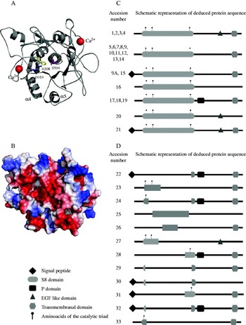
A phylogenetic tree was constructed with the amino acid sequences of reported subtilisins from animals, plants, fungi, bacteria and protozoa. The cladogram was generated with a Neighbor-joining algorithm, with bootstrap values obtained from 1000 replicates. T. vaginalis SUB1 was found to be more related to subtilisin-like serine proteases from plants than to proteases from bacteria or protozoa (Fig. 5). Additionally, a theoretical model for SUB1 was constructed using the Protein Homology/analogY Recognition Engine (PHYRE). The model so generated is consistent with the typical structure of subtilisins (Fig. 6A) (Siezen and Leunissen, Reference Siezen and Leunissen1997).
Phylogenetic tree constructed with subtilisins of different organisms. The cladogram was generated by use of the Neighbor-joining algorithm. Bootstrap values from 1000 replicates are shown. The amino acid sequences were obtained of the MEROPS database: B. licheniformis (MER000309), B. pumillus (MER001755), B. anthracis (MER020846), T. vulgaris (MER000333), O. iheyensis (MER021983), P. falciparum (MER004690), P. chabaudi (MER049747), A. glutinosa (MER002779), A. thaliana (MER015439), O. sativa (MER019141), L. longiflorum (MER000363), M. truncatula (MER063464), P. trichocarpa (MER134612), S. lycopersicum (MER002760), R. communis (MER177246), S. tuberosum (MER050332), V. vinifera (MER165001), L. casei (MER070961), H. sapiens (MER022416), D. ordii (MER158080), C. porcellus (MER103883), B. floridae (MER160186), B. rerio (MER171403), M. mulatta (MER161661), M. domestica (MER077833), M. musculus (MER022415), O. latipes (MER110645), X.laevis (MER172800), C. melo (MER001561), A. benhamiae (MER054530), C. immitis (MER142327), M. canis (MER005078), T. rubrum (MER029350), E. coli (MER037258), A. healyi (MER006074), A. castellanii (MER125416), C. botulinum (MER092670), D. discoideum (MER026564), A. pernix (MER005707), T. onnurineus (MER145074), B. divergens (MER087792), T. gondii (MER028365), B. sp (MER016985), B. amyloliquiefaciens (MER000311).

Theoretical model of SUB1 and structural analysis of the 33 subtilisin-like serine proteases annotated in the Trichomonas vaginalis genome. (A) Theoretical 3D model for SUB1. Residues of the catalytic triad: Asp 163 (blue), His 208 (yellow) and Ser 504 (purple) are shown in sticks. The α4 and α5 helixes, important for the interaction with the pro-peptide, are also shown. The putative calcium-binding sites associated with protein stability are shown as red spheres. (B) Theoretical model of SUB1 surface and electrostatic potential. S1, S2, S3 and S4 subsites are indicated. Negatively charged regions are shown in red, positively charged regions are blue. (C) Schematic representation of the elements identified by bioinformatics in the SUB1 protein (9A) and in the subtilisin-like serine proteases annotated in the T. vaginalis TrichDB database. These sequences possess the complete catalytic triad characteristic of subtilisin-like serine proteases. Accession numbers are: 1: TVAG_124770; 2: TVAG_213290; 3: TVAG_227060; 4: TVAG_379760; 5: TVAG_003700; 6: TVAG_035840; 7: TVAG_045540; 8: TVAG_062550; 9: TVAG_090450; 9A: GenBank FJ384963 (SUB1 protease); 10: TVAG_109360; 11: TVAG_162950; 12: TVAG_230700; 13: TVAG_418990; 14: TVAG_376790; 15: TVAG_054370; 16: TVAG_079860; 17: TVAG_083790; 18: TVAG_103310; 19: TVAG_161600; 20: TVAG_238680; 21: TVAG_482670. For comparison, drawings are not to scale. (D) Genes annotated as subtilisin-like serine proteases in the T. vaginalis TrichDB database. These translated proteins lack some residues of the catalytic triad and have an incomplete S8 domain. The Accession numbers are: 22: TVAG_028690; 23: TVAG_035830; 24: TVAG_042330; 25: TVAG_090240; 26: TVAG_142720; 27: TVAG_151300; 28: TVAG_184220; 29: TVAG_270490; 30: TVAG_276930; 31: TVAG_282560; 32: TVAG_321410; 33: TVAG_465250. For comparison, drawings are not to scale.

DISCUSSION
Protozoan parasites are rich in proteases, many of which are involved in important processes like cellular metabolism, host cell rupture and invasion, excystation, cytoadherence, maturation and activation of other proteins (Klemba and Goldberg, Reference Klemba and Goldberg2002). The study of proteases in protozoan parasites has been focused in the cysteine-type proteases, while serine proteases have remained poorly studied. Nevertheless, experimental data suggest that serine-type proteases may play important roles in the life cycle of parasites (Dejkriengkraikhul and Wilairat, Reference Dejkriengkraikhul and Wilairat1983; Leher et al. Reference Leher, Silvany, Alizadeh, Huang and Niederkorn1998; Conseil et al. Reference Conseil, Soete and Dubremetz1999).
One of the largest families of serine proteases is the subtilisins (Clan SB, family S8A) or subtilisin-like serine proteases. Subtilisins are an ancient group of calcium-dependent enzymes with very similar three-dimensional structures that are widely dispersed throughout evolution (Subbian et al. Reference Subbian, Yabuta and Shinde2004). These enzymes are functionally diverse but in most cases are secreted to function either within the secretory transport system or extracellularly. This family of proteases is characterized by a highly similar arrangement in the primary sequence of the amino acids that form the catalytic triad: aspartate (D), histidine (H) and serine (S) residues. These amino acids are grouped in the domains DSG, HGTH and GTS. In most cases subtilisins are synthesized as pre-pro-enzymes that are subsequently translocated through a cell membrane or the endoplasmic reticulum via the pre-peptide (or signal peptide). These enzymes are activated by cleavage of the pro-peptide which also functions as an intramolecular chaperone that aids in the correct folding of the mature enzyme (Inouye, Reference Inouye1991; Siezen and Leunissen, Reference Siezen and Leunissen1997).
T. vaginalis is a parasite with numerous proteases, most of which appear to be of the cysteine type. Three cysteine proteases (CP65, CP39 and CP30) have been shown to be involved in adherence and cytotoxicity of the parasite to the vaginal epithelial cells (VECs) (Alvarez-Sanchez et al. Reference Alvarez-Sanchez, Avila-Gonzalez, Becerril-Garcia, Fattel-Facenda, Ortega-Lopez and Arroyo2000; Hernandez-Gutierrez et al. Reference Hernandez-Gutierrez, Ortega-Lopez and Arroyo2003, Reference Hernandez-Gutierrez, Avila-Gonzalez, Ortega-Lopez, Cruz-Talonia, Gomez-Gutierrez and Arroyo2004; Mendoza-Lopez et al. Reference Mendoza-Lopez, Becerril-Garcia, Fattel-Facenda, Avila-Gonzalez, Ruiz-Tachiquin, Ortega-Lopez and Arroyo2000). The numerous CPs are found in vaginal secretions of patients (Hernandez-Gutierrez et al. Reference Hernandez-Gutierrez, Avila-Gonzalez, Ortega-Lopez, Cruz-Talonia, Gomez-Gutierrez and Arroyo2004). T. vaginalis CPs appear to induce apoptosis in VECs (Sommer et al. Reference Sommer, Costello, Hayes, Beach, Gilbert, Lucas and Singh2005) and have been shown to degrade all human immunogloblulin subtypes (Provenzano and Alderete, Reference Provenzano and Alderete1995; Min et al. Reference Min, Hyun, Ryu, Ahn and Cho1998). In contrast, only 1 metallo-protease has been characterized to date (Brown et al. Reference Brown, Goldstone, Bastida-Corcuera, Delgadillo-Correa, McArthur and Johnson2007). To our knowledge, serine proteases have only been proposed by bioinformatic analyses of the sequenced T. vaginalis genome (Carlton et al. Reference Carlton, Hirt, Silva, Delcher, Schatz, Zhao, Wortman, Bidwell, Alsmark, Besteiro, Sicheritz-Ponten, Noel, Dacks, Foster, Simillion, Van De, Miranda-Saavedra, Barton, Westrop, Muller, Dessi, Fiori, Ren, Paulsen, Zhang, Bastida-Corcuera, Simoes-Barbosa, Brown, Hayes, Mukherjee, Okumura, Schneider, Smith, Vanacova, Villalvazo, Haas, Pertea, Feldblyum, Utterback, Shu, Osoegawa, De Jong, Hrdy, Horvathova, Zubacova, Dolezal, Malik, Logsdon, Henze, Gupta, Wang, Dunne, Upcroft, Upcroft, White, Salzberg, Tang, Chiu, Lee, Embley, Coombs, Mottram, Tachezy, Fraser-Liggett and Johnson2007). This work represents the first experimental characterization of a putative subtilisin-like serine protease in T. vaginalis.
The sub1 gene appears to be single copy by hybridization assays (data not shown), albeit additional weak hybridization bands were detected that could correspond to other subtilisin-like serine protease-related genes described in the T. vaginalis genome (Carlton et al. Reference Carlton, Hirt, Silva, Delcher, Schatz, Zhao, Wortman, Bidwell, Alsmark, Besteiro, Sicheritz-Ponten, Noel, Dacks, Foster, Simillion, Van De, Miranda-Saavedra, Barton, Westrop, Muller, Dessi, Fiori, Ren, Paulsen, Zhang, Bastida-Corcuera, Simoes-Barbosa, Brown, Hayes, Mukherjee, Okumura, Schneider, Smith, Vanacova, Villalvazo, Haas, Pertea, Feldblyum, Utterback, Shu, Osoegawa, De Jong, Hrdy, Horvathova, Zubacova, Dolezal, Malik, Logsdon, Henze, Gupta, Wang, Dunne, Upcroft, Upcroft, White, Salzberg, Tang, Chiu, Lee, Embley, Coombs, Mottram, Tachezy, Fraser-Liggett and Johnson2007; Hirt et al. Reference Hirt, Noel, Sicheritz-Ponten, Tachezy and Fiori2007). One main difference between the genome data (TVAG_090450) and our data (GenBank FJ384963) is the localization of the transcription start point for the sub1 gene. In the 5′ region of sub1 we identified 2 potential transcription initiator promoter elements (Inr) (Liston and Johnson, Reference Liston and Johnson1999) and 2 possible in-frame ATGs for initiation of translation that are separated by 144-nt (49 aa). The correct mapping of the sub1 transcription start point is important since transcription from the distal initiator would produce a translated product containing a putative signal peptide, as predicted from hidrophobicity and bioinformatics analyses. Alternatively, transcription of sub1 from the proximal initiator would yield a protein without a putative signal peptide. In the genome database the annotated ORF (TVAG_090450) corresponding to the sub1 gene starts at the proximal ATG. In our experimental conditions we found that sub1 transcription starts at the distal initiator element, although a very low level of expression from the proximal initiator cannot be ruled out. Therefore, the translated SUB1 protein would have 17-aa at the amino-terminal end that could function as a pre-peptide or signal peptide (positions 1–17).
In studies done with AhSUB, a secreted subtilisin of Acanthamoeba healyi, the signal peptide is necessary for the proper intracellular trafficking and secretion of the protease (Moon et al. Reference Moon, Lee, Chung and Kong2006). In subtilisins of apicomplexan parasites such as PfSUB-1 of Plasmodium falciparum and TgSUB1 of Toxoplasma gondii, the signal peptide is probably cleaved in the endoplasmic reticulum during the secretory transport (Sajid et al. Reference Sajid, Withers-Martinez and Blackman2000; Miller et al. Reference Miller, Binder, Blackman, Carruthers and Kim2001). The putative signal peptide here identified in T. vaginalis SUB1 could direct the protein to the parasite's membrane through the endoplasmic reticulum. This is in good agreement with the finding of SUB1 on the trichomonad surface of non-permeabilized parasites. Although speculative, the localization of SUB1 on the parasite surface would allow SUB1 to participate, along with other proteases, in promoting the unmasking of adhesins thus facilitating vaginal epithelial cell attachment. This model has been hypothesized before (Arroyo and Alderete, Reference Arroyo and Alderete1989) and is supported in recent work (Garcia et al. Reference Garcia, Benchimol and Alderete2005; Alvarez-Sanchez et al. Reference Alvarez-Sanchez, Carvajal-Gamez, Solano-Gonzalez, Martinez-Benitez, Garcia, Alderete and Arroyo2008). Indeed, subtilsin-like serine proteases have been proposed as processing proteases for proteins involved in the interaction with host cells in protozoan parasites (Miller et al. Reference Miller, Binder, Blackman, Carruthers and Kim2001, Reference Miller, Thathy, Ajioka, Blackman and Kim2003; Withers-Martinez et al. Reference Withers-Martinez, Jean and Blackman2004). Interestingly, we have found that the expression of sub1 mRNA is increased upon parasites in contact with epithelial cells (data not shown), as has been described for α-enolase (Mundodi et al. Reference Mundodi, Kucknoor and Alderete2008) and for adhesins (Garcia et al. Reference Garcia, Chang, Benchimol, Klumpp, Lehker and Alderete2003). These observations are consistent with the occurrence of cell surface proteolytic activity for unmasking of adhesins and adherence of T. vaginalis to the host cells (Alderete and Neale, Reference Alderete and Neale1989; Arroyo and Alderete, Reference Arroyo and Alderete1989). If this was the case for SUB1, the diversification of proteolytic activities would give the parasite the ability to degrade a broad spectrum of substrates under different conditions.
In mature subtilisins, the aspartate residue of the catalytic triad is usually found in position 32. Consequently, we propose a putative pro-region or pro-peptide of 114 residues (positions 18–131) in SUB1. Many of the reported subtilisins are synthesized as pre-pro-enzymes. The pro-region is essential for protease activation given that the pro-peptide acts as an intramolecular chaperone that allows correct folding of the protein (Inouye, Reference Inouye1991; Chang et al. Reference Chang, Kadokura, Yoda and Yamasaki1996). Another important feature found in the trichomonad SUB1 deduced sequence is the presence of a potential transmembrane helix close to the C terminal and would indicate that SUB1 is a Type I transmembrane protease.
It has been reported that iron is an essential nutrient for T. vaginalis and modulates its metabolism and growth (Wilson and Britigan, Reference Wilson and Britigan1998). Iron positively regulates the synthesis of adhesins (Lehker et al. Reference Lehker, Arroyo and Alderete1991) and resistance to lysis by complement (Alderete et al. Reference Alderete, Provenzano and Lehker1995) due to the upregulation of the expression of adhesins and of proteinases involved in complement (C3b) degradation. During the menstrual cycle the iron concentration in the vaginal cavity is constantly changing, providing a variable environment for this parasite. An iron-responsive promoter has been described for the ap65-1 gene (Ong et al. Reference Ong, Huang, Liu and Tai2004, Reference Ong, Hsu, Liu, Chu and Tai2006, Reference Ong, Hsu, Liu, Chu and Tai2007). Two Myb proteins are involved in iron regulation: Myb1 binds to the sequences ANAACGAT and ATCG (Ong et al. Reference Ong, Hsu, Liu, Chu and Tai2006) and Myb2 binds to the sequences CGATA and tAtCGTc (Ong et al. Reference Ong, Hsu, Liu, Chu and Tai2007). Three T-rich (ATTTT, ATTATT and ATTTT) sequences are also important in iron regulation (Ong et al. Reference Ong, Huang, Liu and Tai2004). Since our data show that the sub1 gene is modulated by external availability of iron in the growth medium, we compared the ap65-1 and the sub1 upstream regions. In the 5′region of sub1 gene we found the motif −369ATCG−366 that could be a binding site for Myb1, −258ACGATA−253 and −466TATCGAC−460 that could be binding sites for Myb2 and three T-rich sequences (−170ATTATT−165, −20ATTTTT−15 and −12ATTTTT−7). Nevertheless, further experiments are necessary to determine if iron directly regulates sub1 gene expression.
Bioinformatic analyses of the SUB1 deduced amino acid sequence suggest that it has all the elements necessary to be an active subtilisin. At a primary sequence level, the catalytic core of SUB1 shows significant similarity with known subtilisins: SUB1 possesses the 3 amino acid residues conserved among known subtilisins (the catalytic triad residues Asp163, His208, and Ser504), and an asparagine (Asn309) residue necessary to form the oxyanion hole (Siezen and Leunissen, Reference Siezen and Leunissen1997). A theoretical model of SUB1 was constructed with PHYRE 2.0 based on the crystallographic structure (at 1·30 Å) of the alkaline serine protease kp-43 of Bacillus sp (Nonaka et al. Reference Nonaka, Fujihashi, Kita, Saeki, Ito, Horikoshi and Miki2004). This program was capable of modelling the catalytic conserved core, with a precision value of 100%. The theoretical model of SUB1 was then analysed with ProSA-web software for recognition of errors in 3-dimensional structures of proteins and the obtained z-score value (−5·43) suggests that the model is a good approximation of the real SUB1 structure. In this model the residues of the catalytic triad are in the correct spatial conformation to carry out the peptidase activity. It also possesses the α4 and α5 helices identified in subtilisin E (SbtE) of Bacillus subtilis (Subbian et al. Reference Subbian, Yabuta and Shinde2004) that interact with the pro-peptide, and 2 calcium-binding sites important for protein stability (Alexander et al. Reference Alexander, Ruan and Bryan2001; Subbian et al. Reference Subbian, Yabuta and Shinde2004). However, the assignment of SUB1 as an active subtilisin protease is still putative and needs additional experimental corroboration.
A phylogenetic tree was constructed with subtilisin sequences from several organisms. In this tree subtilisins are mainly distributed in 5 groups: Plants, animals, fungi, protozoa and bacteria. It has been reported that subtilisins may have different functions in each of these groups of organisms. In animals proprotein convertases process many precursor substrates including neuropeptides, peptide hormones, receptors, enzymes and adhesion molecules (Rholam and Fahy, Reference Rholam and Fahy2009). In plants, subtilisins have been involved in cellular signalling and processing of enzymes. In Arabidopsis thaliana subtilisin SDD1 (localized at the plasma membrane of developing stomata mother cells) generates signals that move to neighbouring cells (van der Hoorn, Reference van der Hoorn2008). In pathogenic fungi, subtilisins are involved in the degradation of host tissues (St Leger et al. Reference St Leger, Joshi and Roberts1997; Bagga et al. Reference Bagga, Hu, Screen and St Leger2004) while in protozoan parasites it has been reported that subtilisins are involved as virulence factors and in the processing proteins (Hong et al. Reference Hong, Kong, Ock, Kim and Chung2000; Harris et al. Reference Harris, Yeoh, Dluzewski, O′Donell, Whiters-Martinez, Hackett, Bannister, Mitchell and Blackman2005; Carruthers, Reference Carruthers2006). Bacterial subtilisins are mainly involved in nutrition, although they have also been reported as virulence factors (Brown et al. Reference Brown, Dave, Gey van Pittius, Stevens, Ehlers and Beyers2000; Anderson et al. Reference Anderson, Wetherell, Winter, Olmsted, Cleary and Matsuka2002). It is interesting to note that in the phylogenetic tree depicted in Fig. 5, the branch corresponding to T. vaginalis SUB1 is found in the same clade as plants. Consequently, it is possible that SUB1 may be involved in cellular roles similar to those of plant subtilisins, for example, the processing of surface proteins or cellular signalling.
In an attempt to propose a possible amino acid preference for T. vaginalis SUB1, we identified the amino acid preference of plant subtilisins in the MEROPS database. S1 site preference is for polar uncharged residues such as glutamine (31·25%) and serine (12·25%), followed by hydrophobic residues (leucine and alanine, 12·25% respectively) this is consistent with the reported properties for the S1 site (Graycar et al. Reference Graycar, Ballinger, Wells, Barrett, Rawlings and Woessner2004). Moreover, the surface electrostatic potential for the S1 site of SUB1 has the same negative electrostatic potential that S1 sites of plant subtilisins. Whether the amino acid preference of SUB1 is the same as plant subtilisins remains to be determined.
In total, 33 subtilisin-like serine protease genes belonging to the S8 family have been annotated in the T. vaginalis genome (Carlton et al. Reference Carlton, Hirt, Silva, Delcher, Schatz, Zhao, Wortman, Bidwell, Alsmark, Besteiro, Sicheritz-Ponten, Noel, Dacks, Foster, Simillion, Van De, Miranda-Saavedra, Barton, Westrop, Muller, Dessi, Fiori, Ren, Paulsen, Zhang, Bastida-Corcuera, Simoes-Barbosa, Brown, Hayes, Mukherjee, Okumura, Schneider, Smith, Vanacova, Villalvazo, Haas, Pertea, Feldblyum, Utterback, Shu, Osoegawa, De Jong, Hrdy, Horvathova, Zubacova, Dolezal, Malik, Logsdon, Henze, Gupta, Wang, Dunne, Upcroft, Upcroft, White, Salzberg, Tang, Chiu, Lee, Embley, Coombs, Mottram, Tachezy, Fraser-Liggett and Johnson2007) and the general structural organization of surface proteases has been proposed (Hirt et al. Reference Hirt, Noel, Sicheritz-Ponten, Tachezy and Fiori2007). A detailed in silico analysis of the subtilisin-like serine proteases was carried out for all 33 proteins with several programs, as described in the Materials and Methods section. Only 21 out of the 33 annotated genes predict proteins with the 3 conserved residues of the catalytic triad and the complete S8 domain characteristic of subtilisin-like serine proteases. From these 21 annotated subtilisins, 6 have the EGF (Epidermal Growth Factor) motif particularly found in the extracellular domain of membrane-bound proteins, most of them have a predicted transmembranal domain that may anchor proteins to a membrane and only 2 have a predicted signal peptide for intracellular trafficking. The annotated sequence in the T. vaginalis genome database (TVAG_090450) corresponding to the sub1 gene has been assigned starting at the proximal transcription initiation that would render a protein without a signal peptide. Nevertheless, our experimental data show that sub1 is transcribed from the distal promoter and therefore possesses a putative signal peptide that could direct the protease towards the cytoplasmic membrane.
Most of the remaining 12 genes annotated as subtilisin-like serine proteases have a single residue of the catalytic triad and were probably classified as such because of a small region of similarity with subtilisin-like serine proteases. The expression, localization and characterization of the other 20 subtilisin-like serine protease genes annotated in the T. vaginalis genome remain to be determined.
ACKNOWLEDGEMENTS
The authors thank Diego González-Halphen for helpful suggestions throughout the development of this work. Technical assistance from Lorena López Griego, Leticia Ávila and Juliana Herrera López is acknowledged. Finally, we thank Vasanthakrishna Mundodi of the Alderete laboratory for performing some of the RT-PCR reactions.
FINANCIAL SUPPORT
This work was supported by the Consejo Nacional de Ciencia y Tecnología, CONACYT (grants number P45037-Q and number 79983) and by the PAPIIT, UNAM (grant number IN214006), México. P. A. H. R. was recipient of a scholarship from the DGEP, UNAM and CONACYT, México.