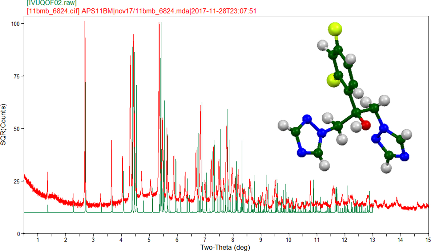
Fluconazole (CAS Registry number 86386-73-4) is used to treat a number of fungal infections, particularly candidiasis, and has many polymorphs. There are five low-precision experimental patterns of fluconazole in the Powder Diffraction File (00-057-1444, 00-058-1926, 00-059-1308, 00-062-1568, and 00-062-1569); one of them, 00-059-1308, corresponds to polymorph V, but does not contain all of the peaks observed here. In this study a commercial sample of fluconazole crystallized in the orthorhombic space group Pbca (#61) with a = 12.97949(12), b = 6.13624(7), c = 35.1757(5) Å, V = 2801.57(8) Å3, and Z = 8 at 295 K. A reduced cell search in the Cambridge Structural Database (Groom et al., Reference Groom, Bruno, Lightfoot and Ward2016) yielded a previously reported crystal structure of polymorph V (Karanam et al., Reference Karanam, Dev and Choudury2012; Refcode IVUQOF02) collected at 100 K. In this work, the sample was ordered from Sigma-Aldrich (Product # F8929-100 mg) and analyzed as received. Other commercial samples evaluated as part of this study were found to have consisted of mixtures of polymorphs. The room-temperature crystal structure was refined using synchrotron (λ = 0.412708 Å) powder diffraction data, density functional theory, and Rietveld refinement techniques. The diffraction data were collected on beamline 11-BM at the Advanced Photon Source, Argonne National Laboratory (see crystallographic details in the supplementary material). Figure 1 shows the powder X-ray diffraction pattern of the compound. The thermal expansion between 100 and 295 K is anisotropic; the expansion is 0.40, 1.86, and 0.98% along a, b, and c, respectively. The cell volume is 3.27% larger at room temperature. The hydroxyl group forms a strong hydrogen bond to a ring nitrogen atom along the a-axis, but the crystal packing is dominated by ring–ring interactions. The pattern and room-temperature crystal structure have been submitted to ICDD for inclusion in the Powder Diffraction File.
(Color online) Observed (295 K, red) and calculated (100 K, green) powder X-ray diffraction patterns of fluconazole polymorph V. Note the shift to higher 2θ (smaller d-spacing) for the 100 K diffraction pattern when compared to the 295 K diffraction pattern.

SUPPLEMENTARY MATERIAL
The supplementary material for this article can be found at https://doi.org/10.1017/S0885715618000659.
ACKNOWLEDGEMENTS
The use of the Advanced Photon Source at Argonne National Laboratory was supported by the U.S. Department of Energy, Office of Science, Office of Basic Energy Sciences, under Contract No. DE-AC02-06CH11357. This work was partially supported by the International Centre for Diffraction Data. We thank Lynn Ribaud and Saul Lapidus for their assistance in data collection.