Introduction
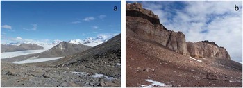
The geological evolutionary history and geographical isolation of the Antarctic continent have produced a unique environment inhabited by species adapted to extreme conditions especially in the interior (Hodgson et al. Reference Hodgson, Convey, Verleyen, Vyverman, McInnes, Sands, Fernández-Carazo, Wilmotte, de Wever, Peeters, Tavernier and Willems2010). Terrestrial ecosystems in Antarctica are restricted to a few ice-free areas (0.34% of the continent), most of which are small and isolated from one another (Convey Reference Convey2011). These areas span from the deserts, nunataks, cliffs and boulder/scree fields of the continental interior to the moist cryptogam-dominated fellfields of Maritime Antarctic (Convey Reference Convey2011). The largest ice-free area in Antarctica, the McMurdo Dry Valleys (Fig. 1), is among the most remote and harsh environments for terrestrial life anywhere on Earth (Doran et al. Reference Doran, Priscu, Lyons, Walsh, Fountain, McKnight, Moorhead, Virginia, Wall, Clow, Fritsen, McKay and Parsons2002a).
General landscape of a. Miers Valley and b. University Valley in the McMurdo Dry Valleys.

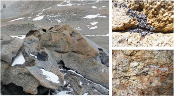
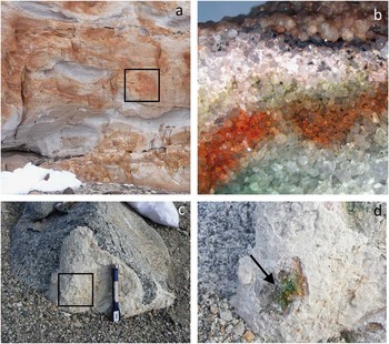
Antarctica is the only continent dominated by microorganisms that exclusively drive the biogeochemical transformations of carbon (Hopkins et al. Reference Hopkins, Sparrow, Gregorich, Elberling, Novis, Fraser, Scrimgeour, Dennis, Meier-Augenstein and Greenfield2009, Namsaraev et al. Reference Namsaraev, Mano, Fernández and Wilmotte2010). The presence and abundance of microbial communities in the rocks of Antarctica’s terrestrial areas indicate their ability to colonize the lithic substrate. Microbial life in Antarctica retreats to niches that formed as a result of biotic–abiotic relationships on, under or within rocks (Pointing & Belnap Reference Pointing and Belnap2012). Green algal-fungal symbiosis gives rise to diverse lichen species as the most visible components of lithic microbial ecosystems, especially when they grow on rock surfaces (Fig. 2a). These epilithic forms may scarcely cover rocks or, in areas where conditions are less extreme such as Maritime Antarctic, rocks may be heavily colonized. Under harsher environmental conditions, such as those of the Dry Valleys, crustose and foliose lichens preferentially occupy cavities or protected areas (Fig. 2b) and chasmoendolithic forms can be observed in weathered rocks (Fig. 2c). Some lichen symbiotic associations hide within the rock interior (Friedmann Reference Friedmann1982) only to be seen after the rock has been fractured (Fig. 3a & b). Cyanobacteria dominated communities also inhabit these rocks (Fig. 3c & d) (Friedmann & Ocampo-Friedmann 1988, De los Rios et al. Reference De los Rios, Grube, Sancho and Ascaso2007, Büdel et al. Reference Büdel, Bendix, Bicker and Green2008). Here we review current knowledge of the lithic microbial ecosystems of Antarctica’s Dry Valleys with special attention paid to their microhabitats and specific adaptations to these habitats.
a. Granite boulder showing epilithic lichen colonization and snow deposits, which could favour lichen community development. b. Umbilicaria aprina Nyl. thalli occupying protected areas of the rock surface. c. Lecideoid lichen colonizing weathered granite.

a. Sandstones from University Valley showing a lack of epilithic colonization. b. Fractured sandstone showing bands of microbial colonization in the area indicated with a square in a. c. Granite boulder from the Miers Valley showing a lack of epilithic colonization. d. Fractured granite boulder showing signs of endolithic cyanobacterial colonization corresponding to the area indicated with a square in c.

The lithobiontic ecological niche
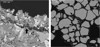
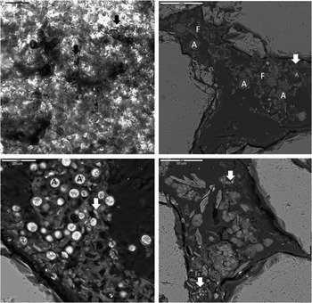
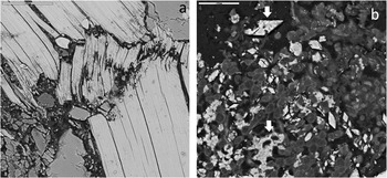
Microorganisms and lichen associations in Antarctica appear both on rock surfaces (epilithic forms) and hidden inside rocks (endolithic forms). Antarctic endolithic microorganisms adapt to different ecological niches within the lithic substrate. They can be found living beneath rock surfaces in interstitial habitats of cracks and fissures of weathered siliceous rocks (chasmoendolithic, arrow in Fig. 4a) but also in pore spaces between mineral grains of porous rocks (cryptoendolithic, arrows in Fig. 4b) (Golubic et al. Reference Golubic, Friedmann and Schneider1981). The type of colonization is determined mainly by the micromorphological, structural, physical and mineralogical properties of the rock substrate.
a. SEM-BSE image of chasmoendolithic colonization of granite rock. Arrow shows the fungal colonized fissure. b. SEM-BSE image showing cryptoendolithic colonization in pores of sandstones (arrows).

The development of the ‘SEM-BSE technique’ (scanning electron microscope in backscattered electron mode; Wierzchos & Ascaso Reference Wierzchos and Ascaso1994), which enabled the study of the lithobiontic community within its rock microhabitat, has permitted the characterization of the different ecological niches and the spatial organization of lithic microbial ecosystems (Wierzchos & Ascaso Reference Wierzchos and Ascaso2001, Ascaso & Wierzchos Reference Ascaso and Wierzchos2003, De los Ríos et al. Reference De los Ríos, Wierzchos, Sancho and Ascaso2003, Reference De los Ríos, Wierzchos, Sancho, Green and Ascaso2005a, Reference De los Ríos, Sancho, Grube, Wierzchos and Ascaso2005b, Wierzchos et al. Reference Wierzchos, Ascaso, Sancho and Green2003, Reference Wierzchos, Sancho and Ascaso2005). Chasmoendolithic and cryptoendolithic communities share some features. In both, two main classes of community are defined: eukaryotic communities dominated by lichens and algal components, and prokaryotic cyanobacteria-dominated communities (Friedmann & Ocampo-Friedmann 1988, De la Torre et al. Reference De la Torre, Goebel, Friedmann and Pace2003). Both classes can be found in close proximity but they occupy different zones within the rock: fissures or cracks for chasmoendolithic communities (Broady Reference Broady1981, De los Ríos et al. Reference De los Ríos, Sancho, Grube, Wierzchos and Ascaso2005b, Reference De los Rios, Grube, Sancho and Ascaso2007) and colonization of pore spaces between mineral grains in the case of cryptoendolithic communities (Friedmann Reference Friedmann1982). Chasmoendolithic growth forms have been reported in granite, marble, silicified sandstone, gypsum crusts and anorthosite (Broady Reference Broady1981, Friedmann Reference Friedmann1982, Ascaso & Wierzchos Reference Ascaso and Wierzchos2003, De los Ríos et al. Reference De los Ríos, Wierzchos, Sancho, Green and Ascaso2005a, Reference De los Rios, Grube, Sancho and Ascaso2007, Guglielmin et al. Reference Guglielmin, Cannone, Strini and Lewkowicz2005, Büdel et al. Reference Büdel, Bendix, Bicker and Green2008), while cryptoendolithic microorganisms have been detected mainly in porous sandstones (Friedmann Reference Friedmann1977, 1982, Friedmann & Ocampo-Friedmann 1988, Siebert et al. Reference Siebert, Hirsch, Hoffmann, Gliesche, Peissl and Jendrach1996, Wierzchos & Ascaso Reference Wierzchos and Ascaso2001, De la Torre et al. Reference De la Torre, Goebel, Friedmann and Pace2003, Blackhurst et al. Reference Blackhurst, Genge, Kearsley and Grady2005, Selbmann et al. Reference Selbmann, de Hoog, Mazzaglia, Friedmann and Onofri2005, Wierzchos et al. Reference Wierzchos, Sancho and Ascaso2005, Pointing et al. Reference Pointing, Chan, Lacap, Lau, Jurgens and Farrell2009).
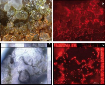
The first endolithic Antarctic communities described were cryptoendolithic microorganisms inhabiting sandstones (Friedmann & Ocampo-Friedmann Reference Friedmann and Ocampo-Friedmann1976, Friedmann Reference Friedmann1982, Friedmann & Ocampo-Friedmann 1988). Shortly afterwards, Ascaso & Wierzchos (Reference Ascaso and Wierzchos2002) and Wierzchos & Ascaso (Reference Wierzchos and Ascaso2002) reported the presence of microbial fossils within these rocks. Cryptoendolithic microbial colonization normally occurs as different coloured bands running parallel to the rock surface (Friedmann Reference Friedmann1982). These bands are composed of different groups of microorganisms at varying depths. Layers of photosynthetic microorganisms (Fig. 5) alternate with layers dominated by fungi (Fig. 6a & b). Typically from the surface to greater rock depths, the pattern consists of a black zone under the crust followed by a white zone, a green and sometimes a blue-green zone (Fig. 3b) (Friedmann Reference Friedmann1982, Ruisi et al. Reference Ruisi, Barreca, Selbmann, Zucconi and Onofri2007). Although the sequence of black, white and green is generally constant, any one or two of these zones can be absent (Friedmann Reference Friedmann1982). The first black layer exclusively contains melanized rock fungi (Fig. 6a), the white layer harbours cryptoendolithic lichen associations (Figs 5b & 6b); and the green layer (Fig. 6c) contains several species of non-lichenized chlorophycean algae, such as Hemichloris antarctica Tschermak-Woess & Friedmann, coexisting with cyanobacteria of the genera Choroococcidiopsis and Gloeocapsa (Friedmann & Ocampo-Friedmann 1988). Finally, a layer of cyanobacterial colonization may be observed in some rock samples (Figs 5d & 6d). In addition, melanized rock fungi can appear in the different layers (arrows in Fig. 6b, c & d).
a. Closer view of the cryptoendolithic microbial colonization of sandstone. b. Same area as in a. visualized by epifluorescence microscopy showing autofluorescence emitted by algal cells (red signal). c. Microscopy image of a sandstone area colonized by cyanobacteria. d. Same area as in c. visualized by epifluorescence microscopy showing autofluorescence emitted by phototrophic microorganisms (red signal).

Cryptoendolithic colonization of Antarctic sandstones. Arrows indicate the presence of black fungi. a. Microscopy image of an area colonized by black fungi on sandstone. b. SEM-BSE image of the white band occupied by algal (A) and fungal (F) associations. c. SEM-BSE image of free-living green algae colonizing a sandstone pore. d. SEM-BSE image of a sandstone pore colonized by cyanobacteria (C).

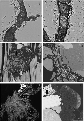
The fissures and cracks appearing in weathered siliceous rocks can also feature extensive chasmoendolithic microbial growth in the Dry Valleys (Broady Reference Broady1981, Ascaso & Wierzchos Reference Ascaso and Wierzchos2003, De los Ríos et al. Reference De los Ríos, Wierzchos, Sancho, Green and Ascaso2005a, Reference De los Ríos, Sancho, Grube, Wierzchos and Ascaso2005b, Reference De los Rios, Grube, Sancho and Ascaso2007). Rock freeze-fracturing could favour the formation of chasmoendolithic microhabitats (Cowan & Tow Reference Cowan and Tow2004). As with the cryptoendolithic communities, some zonation is observed for chasmoendoliths. Certain fissures are occupied by cyanobacterial colonies frequently associated with heterotrophic bacteria (Fig. 7a), while others harbour algal-fungal lichen associations (Fig. 7b). Free-living fungi may also occur (Fig. 7c), generally close to areas colonized by lichen associations. Fissures only occupied by non-lichenized algae have also been detected in some areas (Fig. 7d). Lichen-dominated chasmoendolithic growth to some extent follows the lichen thallus structure. In fissure zones close to the surface, algal and fungal symbionts intermix to form something similar to an algal layer (Fig. 7b), while only mycobiont cells inhabit deeper areas, as in the medulla of a lichen thallus (Fig. 7e). Chasmoendolithic microbial communities in the Dry Valleys are generally observed in areas lacking epilithic growth but may also be detected under epilithic lichen thalli (arrow in Fig. 7f).
Chasmoendolithic colonization of Antarctic granites. a. SEM-BSE image of fissures colonized by cyanobacteria. b. SEM-BSE image of fissures colonized by lichen algal-fungal associations. c. SEM-BSE image of fissures colonized by free-living fungi (arrow). d. SEM-BSE image of fissures colonized by free-living algae. e. Confocal scanning light microscopy image of glutaraldehyde-fixed endolithic lichen showing colonization of a rock fissure. f. Epilithic lichen colonizing granite. Arrow indicates the separation of mica layers on the rock surface and endolithic colonization through these layers.

The zonation shown by cryptoendolithic microbial communities has been correlated with the steep light gradient that occurs in the top few millimetres of the rock (Nienow et al. Reference Nienow, McKay and Friedmann1988). The occurrence of cyanobacteria in the lowermost zone may reflect their ability to grow at lower light levels and in microaerophilic conditions, whereas green algae in the outer zones may require higher light and oxygen levels (Broady Reference Broady1981). Chasmoendolithic colonization by algae and cyanobacteria is also determined by the existence of sufficient light for a community to become established. Most extensive colonization by cyanobacteria has been described in close-to-surface areas of a fissure (De los Ríos et al. Reference De los Rios, Grube, Sancho and Ascaso2007). The algae of chasmoendolithic lichens also show a preference for zones that are closer to the surface and are frequently observed under the more translucent minerals (De los Ríos et al. Reference De los Ríos, Wierzchos, Sancho, Green and Ascaso2005a).
Besides epilithic and endolithic colonization of Antarctica’s terrestrial environments, hypolithic colonization also exists as microbial assemblages present on the undersides of translucent rocks (Cowan Reference Cowan2009, Pointing et al. Reference Pointing, Chan, Lacap, Lau, Jurgens and Farrell2009, Cowan et al. Reference Cowan, Pointing, Stevens, Cary, Stomeo and Tuffin2011). While this zone acts as a refuge for microorganisms from extreme environmental conditions (Cowan et al. Reference Cowan, Khan, Pointing and Cary2010) and a high diversity of microbial communities have been reported (Khan et al. Reference Khan, Tuffin, Stafford, Cary, Lacap, Pointing and Cowan2011, Chan et al. Reference Chan, Lacap, Lau, Ha, Warren-Rhodes, Cockell, Cowan, McKay and Pointing2012, Reference Chan, van Nostrand, Zhou, Pointing and Farrell2013), their description is outside the scope of this review.
Features of the Antarctic Dry Valley microhabitat
Microhabitat conditions are an important determining factor for the establishment of lithic microbial ecosystems (Friedmann Reference Friedmann1980) and are generally less extreme than external environmental conditions. We review the main features of the Antarctic lithic microhabitat below.
Water availability
Water is essential for biological processes and is, hence, a requirement for microbial life. In Antarctic lithic microbial ecosystems, water availability varies according to geographical and local topographical factors. In the Maritime Antarctic conditions are milder (Green et al. Reference Green, Schroeter and Sancho2007) and liquid water is available for microbial and lichen growth. However, liquid water availability is one of the most limiting factors for the distribution and abundance of terrestrial organisms in the Dry Valleys (Doran et al. Reference Doran, Priscu, Lyons, Walsh, Fountain, McKnight, Moorhead, Virginia, Wall, Clow, Fritsen, McKay and Parsons2002a, Ruisi et al. Reference Ruisi, Barreca, Selbmann, Zucconi and Onofri2007). These differences in water availability are reflected in lichen growth rates, which are close to two orders of magnitude lower in the Dry Valleys than on the Antarctic Peninsula (Sancho et al. Reference Sancho, de la Torre, Horneck, Ascaso, de los Ríos, Pintado, Wierzchos and Schuster2007). The presence of liquid water in the Dry Valleys is limited to transient meltwater from ice or snow driven by solar heating when rock temperatures are near or above 0°C (summer) (Friedmann Reference Friedmann1978, Broady Reference Broady1981, Doran et al. Reference Doran, McKay, Clow, Dana, Fountain, Nylen and Lyons2002b). Humidity within the rock can be two-fold that detected in the atmospheric air (Green et al. Reference Green, Schroeter and Sancho2007). Water can enter the endolithic habitat via percolation into subsurface pore spaces or through fissures or weathered areas (Friedmann Reference Friedmann1978, McKay & Friedmann Reference McKay and Friedmann1985) where evaporation is limited due to reduced gas exchange between the air and subsurface, especially when rock varnish is present (Friedmann & Ocampo-Friedmann Reference Friedmann and Ocampo-Friedmann1984). Hence, the existence of points of snow or ice accumulation has a positive effect on the development of lithobiontic communities (Broady Reference Broady1981, Winkler et al. Reference Winkler, Kappen and Schulz2000) (Fig. 2a). In addition, the physical structure and water-adsorbing capacity of the lithic substrate helps to retain moisture following precipitation events (Kappen et al. Reference Kappen, Friedmann and Garty1981, Friedmann et al. Reference Friedmann, McKay and Nienow1987, Friedmann & Ocampo-Friedmann 1988). These properties confer advantages on the endolithic microhabitat over other habitats in the Dry Valleys, where water is inherently scarce (Omelon Reference Omelon2008). For example, Büdel et al. (Reference Büdel, Bendix, Bicker and Green2008) observed that chasmoendolithic cyanobacteria could use available moisture from dew/rime deposition on rocks.
Temperature
Low temperatures and temperature fluctuations are among the most decisive factors for the existence of lithobiontic microbial communities in Antarctica because they can arrest metabolic activity (Friedmann & Weed Reference Friedmann and Weed1987). Specific climatic conditions, i.e. sunny days with a gentle breeze, generate frequent temperature oscillations (within few minutes), while low-frequency oscillations occur on a longer timescale (McKay & Friedmann Reference McKay and Friedmann1985). Rapid temperature fluctuations due to gusty winds are common in the Dry Valleys (Friedmann Reference Friedmann1982). These conditions give rise to freeze-thaw cycles that can severely impact microorganisms that do not have specific anti-freeze mechanisms (Nienow et al. Reference Nienow, McKay and Friedmann1988). Depending on topographical factors, such as aspect and altitude, mean annual air temperatures in the McMurdo Dry Valleys range from -15 to -30°C, with differences between the absolute maximum and minimum of c. 50°C (Doran et al. Reference Doran, Priscu, Lyons, Walsh, Fountain, McKnight, Moorhead, Virginia, Wall, Clow, Fritsen, McKay and Parsons2002a, Reference Doran, McKay, Clow, Dana, Fountain, Nylen and Lyons2002b). Field microclimate measurements have revealed that the rock surface and shallow subsurface are considerably warmer, by up to 20°C higher, than the ambient air during sunny periods at high altitude. These conditions are attributed to solar radiative heating of the rock surface, and the heat capacity and transparency of the rock (Friedmann Reference Friedmann1977, Kappen et al. Reference Kappen, Friedmann and Garty1981, McKay & Friedmann Reference McKay and Friedmann1985, Green et al. Reference Green, Schroeter and Sancho2007). Furthermore, the heat absorption capacity of the rock dampens rapid temperature oscillations, especially important when temperatures are c. 0°C (McKay & Friedmann Reference McKay and Friedmann1985).
Light
Light is a vital factor for the development and functioning of lithic microbial ecosystems because it drives autotrophic processes. In the Dry Valleys, mean annual solar flux ranges from 73–117 W m-2 and photosynthetic active radiation (PAR) ranges from 170–238 µM s-1 in relation to topographical effects (Doran et al. Reference Doran, McKay, Clow, Dana, Fountain, Nylen and Lyons2002b). Rock surfaces and narrow or deep valleys that face north receive more sunlight and are, therefore, more frequently colonized by endolithic microbial communities than south facing surfaces (Friedmann Reference Friedmann1977, McKay & Friedmann Reference McKay and Friedmann1985).
Light attenuation patterns have been linked to the limited distribution depth of algae and cyanobacteria within colonized rocks (Matthes et al. Reference Matthes, Turner and Larson2001). Given the reduced light that reaches microbial ecosystems in the endolithic habitat, the amount of insolation on rock surfaces becomes a more relevant factor for the distribution of endolithic communities. Using mathematical methods, Nienow et al. (Reference Nienow, McKay and Friedmann1988) showed that within the endolithic sandstone habitat light was diminished by up to 90% per millimetre of depth and that, in addition to the physicochemical features of the rock, light attenuation depended on the presence of water in the rock and/or its microbial colonization. Greater light penetration was observed through a hydrated rock but light penetration was markedly reduced if the rock was colonized by epilithic and endolithic microorganisms.
Microbial photosynthetic activity is dependent on a sufficient amount of PAR reaching the microbial community. However, an excess of radiation can also be detrimental; this is especially true in Antarctica where ultra violet (UV-A and UV-B) transmission to the lower atmosphere and surface is substantially higher in spring than elsewhere in the world as a result of a depleted ozone layer (Madronich et al. Reference Madronich, McKenzie, Björn and Caldwell1998). Incoming solar UV radiation is harmful to microorganisms and is, therefore, an important factor controlling the surface development of biological colonizers (Caldwell et al. Reference Caldwell, Björn, Bornman, Flint, Kulandaivelu, Teramura and Tevini1998, Hughes et al. Reference Hughes, Lawley and Newsham2003, Tosi et al. Reference Tosi, Onofri, Brusoni, Zucconi and Vishniac2005, Hughes Reference Hughes2006). To protect themselves against excessive radiation and UV-B rays, Antarctic microbiota synthesize UV-photoprotective pigments (such as scytonemin or mycosporine-like amino acids) and photoprotective-quenching pigments (e.g. carotenoids), and also have special DNA repair mechanisms (Ruisi et al. Reference Ruisi, Barreca, Selbmann, Zucconi and Onofri2007, Selbmann et al. Reference Selbmann, Isola, Zucconi and Onofri2011, Singh et al. Reference Singh, Dubey and Singh2011). A number of Antarctic endolithic communities take advantage of geoprotective screens, such as iron oxide (haematite), goethite or calcium sulphate (gypsum), that protect them from potential lethal radiation (Villar et al. Reference Villar, Edwards and Seaward2005).
Other factors
Although temperature, radiation and water availability critically influence the composition and survival of microbial communities, other factors such as wind, nutrient availability, salinity and the physicochemical properties of local colonized rock (which can vary at the microscale level) are also important.
Wind has both direct and indirect effects on the distribution of lithobiontic communities. While summer temperatures are strongly controlled by coastal winds, warming as they move inland and cooling with inland elevation, sporadic katabatic windstorms have a pronounced effect on non-summer temperatures (Doran et al. Reference Doran, McKay, Clow, Dana, Fountain, Nylen and Lyons2002b). Broady (Reference Broady1981) noted that snowdrift from the continental ice plateau is a source of moisture upon melting, promoting the survival of microbial colonizers. However, high winds enhance evaporation, making lithobiontic colonization especially difficult at or close to the rock surface (Ruisi et al. Reference Ruisi, Barreca, Selbmann, Zucconi and Onofri2007). In addition, erosion due to a sandblasting effect from wind-driven particles can prevent microbial colonization, as shown by Broady (Reference Broady1981) in the Vestfold Hills area. Finally, interactions between topography and air masses can greatly influence climate in microhabitats (Doran et al. Reference Doran, McKay, Clow, Dana, Fountain, Nylen and Lyons2002b) and subsequently the possibility of lithobiontic colonization.
Nutrients are scarce in Antarctic lithic habitats. Birds and other animals are a source of nutrients, especially nitrogen and phosphorous, in coastal areas (Green et al. Reference Green, Schroeter and Sancho2007) but this supply is minimal in other regions. In the endolithic habitat, nutrients may be obtained from in situ-weathered bedrock or allochthonous sediments, such as dust (Johnston &Vestal Reference Johnston and Vestal1989, Omelon et al. Reference Omelon, Polllard and Ferris2007, Walker & Pace Reference Walker and Pace2007), and thereby depend on the local physicochemical properties of the colonized rocks. However, the nutrient requirements of slow-growing communities cannot be too great and there seem to be no signs of nutrient limitation for cryptoendolithic photosynthetic metabolism (Johnston & Vestal Reference Johnston and Vestal1991).
In Antarctica, high evaporation rates often lead to high concentrations of salts on certain rocks and soil surfaces (Nishiyama Reference Nishiyama1977). Furthermore, strong winds can pick up salt spray from the sea and saline lakes and deposit this salt on downwind land areas (Broady Reference Broady1981). Salinity may be a stress factor for microbial colonization and determine community structure (Broady Reference Broady1981, Ruisi et al. Reference Ruisi, Barreca, Selbmann, Zucconi and Onofri2007).
Diversity of lithic microbiota
Epilithic communities
The colonization of rock surface in the Dry Valleys is dominated by lichens. Although lichens are among the most diverse organisms inhabiting Antarctica, the magnitude of this diversity is still unclear mainly due to difficulties in identification related to the presence of numerous sterile forms and extensively modified thalli, including those of endolithic growth forms (Kappen Reference Kappen2004, Pérez-Ortega et al. Reference Pérez-Ortega, Ortiz-Alvarez, Green and de los Ríos2012). Øvstedal & Lewis-Smith (Reference Øvstedal and Smith2011) have estimated the number of lichen species in Antarctica and South Georgia at c. 500. Several taxonomic (Hertel Reference Hertel1987, Reference Hertel1988, Ruprecht et al. Reference Ruprecht, Lumbsch., Brunauer, Green and Türk2010) and floristic (Castello & Nimis Reference Castello and Nimis1995, Øvstedal & Lewis-Smith Reference Øvstedal and Smith2001, Reference Øvstedal and Smith2009, Reference Øvstedal and Smith2011, Castello Reference Castello2010, Seppelt et al. Reference Seppelt, Türk, Green, Moser, Pannewitz, Sancho and Schroeter2010, Green et al. Reference Green, Sancho, Türk, Seppelt and Hogg2011a) studies exist, but DNA-based diversity estimates including modified forms have only been undertaken recently for three McMurdo ice-free valleys: Garwood, Marshall and Miers (Pérez-Ortega et al. Reference Pérez-Ortega, Ortiz-Alvarez, Green and de los Ríos2012). Higher lichen diversity estimates are obtained when morphological and molecular characters are used for identification. In Continental Antarctica, a much lower diversity of lichens has been reported than in the Antarctic Peninsula region (Green et al. Reference Green, Schroeter and Sancho2007, Armstrong & Bradwell Reference Armstrong and Bradwell2010). Lichen communities, composed of foliose and crustaceous lichens, are practically the only types of epilithic community in Continental Antarctica. A clear diversity gradient exists in the Antarctic Peninsula, with a strong decline in species richness, albeit in steps, from 62°S to around 70°S (Peat et al. Reference Peat, Clarke and Convey2007). However, despite a lack of evidence for a major drop in lichen diversity with increasing latitude in Victoria Land and the Transantarctic Mountains, lichen diversity hotspots have been discovered in these areas (Green et al. Reference Green, Schroeter and Sancho2007, 2011a, Peat et al. Reference Peat, Clarke and Convey2007, Seppelt et al. Reference Seppelt, Türk, Green, Moser, Pannewitz, Sancho and Schroeter2010). These observations suggest that the distribution of lichens in Continental Antarctica could be more highly influenced by microclimatic features, such as the concurrent presence of warmth, water, light and shelter in a microhabitat, and historical events than by direct climate effects (Schroeter et al. Reference Schroeter, Green, Pannewitz, Schlensog and Sancho2010, Green et al. Reference Green, Brabyn, Beard and Sancho2011b).
To examine the diversity of lithic microbial ecosystems dominated by lichens, as well as knowledge of mycobiont diversity (determining lichen nomenclature), the diversity of the photobionts forming lichen associations needs to be known. A good understanding of the diversity of photobionts and specificity of their associations acquires special relevance in extreme environments, such as Antarctica, where the availability of algae could be a limiting factor for lichen-dominated microbial communities (De los Ríos et al. Reference De los Ríos, Wierzchos, Sancho, Green and Ascaso2005a). In a recent study, Pérez-Ortega et al. (Reference Pérez-Ortega, Ortiz-Alvarez, Green and de los Ríos2012) found that photobiont diversity in three of the McMurdo Dry Valleys was not much lower than in non-Antarctic regions. Five putative species of lichen photobionts were associated with 27 species of lichen-forming fungi in the three valleys examined. There was a general trend of low selectivity but a number of species showed high selectivity. Other studies have also shown low selectivity of the photobiont for the mycobiont in Maritime Antarctica (Romeike et al. Reference Romeike, Friedl, Helms and Ott2002, Wirzt et al. Reference Wirtz, Lumbsch, Green, Türk, Pintado, Sancho and Schroeter2003) and in lecideoid lichens in Continental and Maritime Antarctic (Ruprecht et al. Reference Ruprecht, Brunauer and Printzen2012).
Endolithic communities
Before the introduction of molecular biology methods, the diversity of lithobiontic communities was described based on cultures and microscopic observations of morphological and ultrastructural features. Using these methods, Friedman & Ocampo-Friedmann (Reference Friedmann, Hua and Ocampo-Friedmann1988) distinguished and characterized the diversity of the different classes of cryptoendolithic community inhabiting the Ross Desert: cyanobacteria-dominated communities and two types of eukaryotic communities, lichen-dominated and Hemichloris communities.
Cyanobacteria of the genera Gloeocapsa found under quartz-like rocks in southern Victoria Land were the first primary producers described in Antarctica’s cold desert ecosystems (Friedmann & Ocampo-Friedmann Reference Friedmann and Ocampo-Friedmann1976). Since then, different cyanobacteria-dominated communities have been described within cryptoendolithic and chasmoendolithic ecological niches. In total, 17 cryptoendolithic cyanobacteria from the McMurdo Dry Valleys were described in cyanobacteria-dominated communities from Beacon Supergroup sandstone (Friedman & Ocampo-Friedmann Reference Friedmann, Hua and Ocampo-Friedmann1988). Depending on the dominating cyanobacterium, three communities were defined: Gloeocapsa, Hormathonema-Gloeocapsa and Chroococcidiopsis. In Taylor Valley, cyanobacteria-dominated communities were composed of the cyanobacterium Chroococcidiopsis sp., with the occasional presence of Cyanothece cf. aeruginosa (Nägeli) Komárek and Nostoc sp. (Büdel et al. Reference Büdel, Bendix, Bicker and Green2008). In the Granite Harbour area, two different chasmoendolithic cyanobacteria-dominated communities have been described (De los Ríos et al. Reference De los Rios, Grube, Sancho and Ascaso2007). One of them, loosely adhered to the substrate, formed by a cyanobacteria closely related to Acaryochloris marina Miyashita et Chihara (the only organism known to contain chlorophyll d). The second community, more closely associated with the rock minerals, harbours Gloeocapsa-like cells.
The non-lichenized algae most commonly detected in the cryptoendolithic niches of the McMurdo Dry Valleys belong to the genus Hemichloris (Friedmann & Ocampo-Friedmann Reference Friedmann and Ocampo-Friedmann1984, Tschermak-Woess & Friedmann Reference Tschermak-Woess and Friedmann1984). In addition, several black fungi have been isolated from cryptoendolithic lichen-dominated communities living within sandstones in the McMurdo Dry Valleys (Friedmann & Ocampo-Friedmann Reference Friedmann and Ocampo-Friedmann1976, Friedmann Reference Friedmann1982, Nienow & Friedmann Reference Nienow and Friedmann1993, Onofri et al. Reference Onofri, Seltimann, de Hoog, Grube, Barreca, Ruisi and Zucconi2007a, Selbmann et al. Reference Selbmann, de Hoog, Zucconi, Isola, Ruisi, van den Ende, Ruibal, de Leo, Urzi and Onofri2008). These fungi form a heterogeneous, taxonomic, phylogenetic group characterized by thick, heavily-pigmented cell walls, apparent lack of reproductive structures and slow growth. Members of this group show different growth styles, such as the formation of daughter cells by yeast-like multilateral or polar budding (black yeasts) or the formation of aggregates of thick-walled, melanized cells, which enlarge and reproduce by isodiametric division (meristematic black fungi) (Ruibal et al. Reference Ruibal, Gueidan, Selbmann, Gorbushina, Crous, Groenewald, Muggia, Grube, Isola, Schoch, Staley and Lutzoni2009). The biodiversity of Antarctic black fungi has only recently been addressed and although sampling is still limited, several new genera have been described: Friedmanniomyces, Cryomyces, Acidomyces, Recurvomyces and Elasticomyces within the class Dothideomycetes (Selbmann et al. Reference Selbmann, de Hoog, Mazzaglia, Friedmann and Onofri2005, Reference Selbmann, de Hoog, Zucconi, Isola, Ruisi, van den Ende, Ruibal, de Leo, Urzi and Onofri2008). With the exception of the genus Cryomyces considered to be Dothideomycetes incertae sedis (Ruibal et al. Reference Ruibal, Gueidan, Selbmann, Gorbushina, Crous, Groenewald, Muggia, Grube, Isola, Schoch, Staley and Lutzoni2009), all these genera have been ascribed to the order Capnodiales (Selbmann et al. Reference Selbmann, de Hoog, Zucconi, Isola, Ruisi, van den Ende, Ruibal, de Leo, Urzi and Onofri2008, Ruibal et al. Reference Ruibal, Gueidan, Selbmann, Gorbushina, Crous, Groenewald, Muggia, Grube, Isola, Schoch, Staley and Lutzoni2009). Among the filamentous hyphomycetes, the other ecological group of rock-inhabiting fungi represented in Antarctica, a strain of Verticillium sp. has been identified in a microbial endolithic community inhabiting gypsum crusts at Two Step Cliffs on Alexander Island (Hughes et al. Reference Hughes, Lawley and Newsham2003).
De la Torre et al. (Reference De la Torre, Goebel, Friedmann and Pace2003) conducted the first rRNA-based molecular survey of the two main classes of endolithic communities (lichen-dominated and cyanobacteria-dominated) described by Friedmann et al. (1998) in Beacon sandstone. In both classes, a dominant fungal and algal phylotype was detected. The fungal phylotype was closely related to the epilithic Buellia spp. and the algal phylotype was Trebouxia jamesii (Hildreth & Ahmadjian) Gärtner. The most commonly detected cyanobacterium was closely related to Plectonema spp. In cyanobacteria-dominated communities, many sequences of the Thermus-Deinococcus bacterial phylogenetic group and a representative of the α-proteobacteria have also been detected. Pointing et al. (Reference Pointing, Chan, Lacap, Lau, Jurgens and Farrell2009) reported that endolithic communities in the McKelvey Valley were dominated by different cyanobacteria and showed a great complexity including 16 phyla. Chrococcidiopsis and Nostocales phylotypes were the most common endoliths. An additional source of bacterial phototrophy was indicated by the presence of Chloroflexi. A lower phylogenetic diversity was detected for chlorophytes with two closely related groups within the Trebouxiophyaceae and a single Brateococcus phylotype.
Physiological peculiarities of microbiota in Antarctica
Antarctic microorganisms generally have specific physiological adaptations in order to cope with the harsh environmental conditions, such as metabolic activity at subzero temperatures, the ability to persist in metabolically inactive states under unfavourable conditions and the presence of cell protection strategies (e.g. photoprotective mechanisms and extracellular polymeric substance (EPS) production). These mechanisms are probably not driven by the extreme climate conditions per se, but by the extreme range of conditions encountered (Friedmann Reference Friedmann1993, Yergeau et al. Reference Yergeau, Bokhorst, Huiskes, Boschker, Aerts and Kowalchuk2007, Omelon Reference Omelon2008). In endolithic communities, physiological adaptations act in combination with the inherent protective properties of the host rock microenvironment, leading to successful colonization.
Microbial growth and metabolism are clearly limited at low temperature. Cold-adapted microorganisms include both psychrophilic and psychrotrophic organisms. In the lithic habitat, psychrotrophic microorganisms are probably more common than psychrophilic forms. Psychrophiles include organisms whose optimum growth temperatures are <15°C and upper limits are <20°C, while the optimum and maximum growth temperatures of psychrotrophs are >15°C (although they are still capable of growing at or close to zero) (Morita Reference Morita1975). Maximum photosynthetic rates of 10°C suggest a psychrophilic temperature response for photosynthesis in cryptoendolithic lichen communities, which contrasts with psychrotrophic cryptoendolithic cyanobacterial communities that achieve maximum photosynthetic rates at 20–30°C (Johnston & Vestal Reference Johnston and Vestal1991). Only a few Antarctic cyanobacteria can be considered true psychrophiles and these have been mostly associated with the polar freshwater ecosystem (Pandey et al. Reference Pandey, Shukla, Shukla, Giri, Singh, Singh and Kashyap2004). Siebert et al. (Reference Siebert, Hirsch, Hoffmann, Gliesche, Peissl and Jendrach1996) observed psychrophilic and psychrotrophic cryptoendolithic bacteria in cultures of sandstone samples obtained from Linneus Terrace. Antarctic black fungi have also been described as psychrotolerant. These fungi are able to grow at c. 0°C but optimal growth is observed at much higher temperatures than those to which they are generally exposed. This enables them to tolerate daily and seasonal temperature fluctuations (Selbmann et al. Reference Selbmann, de Hoog, Mazzaglia, Friedmann and Onofri2005).
Effective strategies against freezing temperatures and desiccation (Selbmann et al. Reference Selbmann, de Hoog, Mazzaglia, Friedmann and Onofri2005, Reference Selbmann, Isola, Zucconi and Onofri2011, Kranner et al. Reference Kranner, Beckett, Hochman and Nash2008, Billi Reference Billi2009) enable microorganisms to persist in Antarctic terrestrial environments but the in situ primary production is very limited, including in the abundant cryptoendolithic microbial communities (Johnston & Vestal Reference Johnston and Vestal1991, Hopkins et al. Reference Hopkins, Sparrow, Gregorich, Elberling, Novis, Fraser, Scrimgeour, Dennis, Meier-Augenstein and Greenfield2009). Most microorganisms exist in a metabolic dry and/or frozen state for most of the year and are only active during the short summer period, when temperatures rise and water from snow or ice melt is available (Friedmann Reference Friedmann1982, Selbmann et al. Reference Selbmann, de Hoog, Mazzaglia, Friedmann and Onofri2005, Schroeter et al. Reference Schroeter, Green, Pannewitz, Schlensog and Sancho2011, Sterflinger et al. Reference Sterflinger, Tesei and Zakharova2012).
Lichens are able to survive under extreme environmental conditions that are adverse to most other organisms owing to their anhydrobiotic behaviour. Lichens are even able to recover their metabolic activity after exposure to space conditions (Sancho et al. Reference Sancho, de la Torre, Horneck, Ascaso, de los Ríos, Pintado, Wierzchos and Schuster2007, Raggio et al. Reference Raggio, Pintado, Ascaso, de la Torre, de los Ríos, Wierzchos, Horneck and Sancho2011). Some lichens show positive net photosynthesis at subzero temperatures of -5–-20°C (Lange Reference Lange1965, Kappen & Redon Reference Kappen and Redon1987, Schroeter et al. Reference Schroeter, Green, Kappen and Seppelt1994, Bártak et al. Reference Barták, Vaczi, Hájek and Smykla2007). However, Antarctic lichens seem to feature lower net rates of photosynthesis than lichens in other parts of the world (Sancho et al. Reference Sancho, Valladares, Schroeter and Kappen2000, Green et al. Reference Green, Schroeter and Sancho2007). Low molecular weight antioxidants, such as glutathione, and reactive oxygen species (ROS)-processing enzymes and peroxidases have been described as key components in the desiccated state and during subsequent rehydration to avoid the effects of oxidative stress (Kranner et al. Reference Kranner, Beckett, Hochman and Nash2008). In response to high light intensities, photoprotective mechanisms via non-photochemical energy dissipation, synthesis of antioxidants and production of light-screening compounds by mycobiont hyphae forming the upper cortex have also been described in Antarctic lichens (Kappen et al. Reference Kappen, Schroeter, Green and Seppelt1998, Edwards et al. Reference Edwards, Wynn-Williams and Villar2004, Barták et al. Reference Barták, Hájek and Očenášová2012). Photobionts and mycobionts feature different mechanisms and those facilitating tolerance are more effective in the whole lichen than in the separate symbionts (Kranner et al. Reference Kranner, Cram, Zorn, Wornik, Yoshimura, Stabentheiner and Pfeifhofer2005). Additionally, Friedmann & Sun (Reference Friedmann and Sun2005) observed that the contribution of community adaptation to heat was greater than the mechanisms functioning at the organism level.
Meristematic black fungi that make up some cryptoendolithic communities are not only able to tolerate high levels of stress, but are even specialized to do so. The simple morphology, scarcely differentiated structure and reduced life cycle make these fungi particularly suited to live in the harsh environment of the McMurdo Dry Valleys. These fungi can complete their life cycle during the short period of suitable conditions (Selbmann et al. Reference Selbmann, de Hoog, Mazzaglia, Friedmann and Onofri2005). Thick-walled and intensely melanized cells produce UV-B absorbing substances, such as mycosporine-like amino acids (Kogej et al. Reference Kogej, Gorbushina and Gunde-Cimerman2006), conferring these fungi drought- and UV-resistance and thus protecting the cell DNA (Selbmann et al. Reference Selbmann, Isola, Zucconi and Onofri2011).
The extremotolerance exhibited by cyanobacteria found in Antarctic lithobiontic communities, such as Chrococcidiopsis and Nostoc, has also been attributed to protection mechanisms and efficient repair systems (Shirkey et al. Reference Shirkey, McMaster, Smith, Wright, Rodriguez, Jaruga, Birincioglu, Helm and Potts2003, Billi Reference Billi2009, Reference Billi2012). It has been proposed that EPS production is a crucial factor for the stress tolerance of cyanobacteria to photosynthesis during desiccation and freezing-thawing cycles (Grilli Caiola et al. Reference Grilli Caiola, Billi and Friedmann1996, De los Ríos et al. Reference De los Ríos, Wierzchos, Sancho and Ascaso2003, Tamaru et al. Reference Tamaru, Takani, Yoshida and Sakamoto2005). In these anhydrobiotic cyanobacteria genera, EPS could act in synergy with trehalose accumulation in the cytoplasm (Billi Reference Billi2012). The mechanisms causing the death of a few Chroococcidiopsis cells within a dried aggregate are thought to contribute to the survival of the remaining cells, by providing physical protection and/or nutrient supply upon rehydration (Billi Reference Billi2009). Cyanobacterial mechanisms including light-harvesting pigments, light-screening pigments, ROS-quenching compounds (such as carotenoids), membrane fluidity at low temperatures and cold-stable proteins allow for slow yet steady growth in Antarctica (Vincent Reference Vincent2007, Banerjee & Verma Reference Banerjee and Verma2009). Cryptoendolithic cyanobacteria within Antarctic rocks obtain nitrogen through overnight nitrogen fixation using energy generated by daytime photosynthesis or heterotrophic fixation using lichen osmolytes (Banerjee & Verma Reference Banerjee and Verma2009).
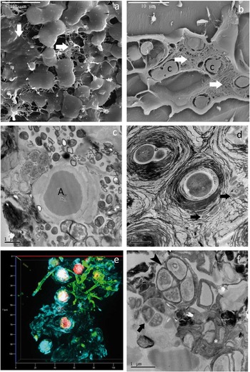
The presence of EPS is a common feature of lithobiontic communities (De los Ríos et al. Reference De los Ríos, Sancho, Grube, Wierzchos and Ascaso2005b). In lithic microbial ecosystems, microorganisms and mineral components are embedded in an organic matrix resulting from the excretion of polymers by resident microorganisms, leading to formation of a protective biofilm (De los Ríos et al. Reference De los Ríos, Wierzchos, Sancho and Ascaso2003). The low-temperature scanning electron microscopy (LTSEM) image in Fig. 8a shows endolithic cyanobacterial cells covered by an EPS matrix. The EPS often confers a compact structure to the community (Fig. 8b) and can play an important role in adapting to Antarctic conditions (De los Ríos et al. Reference De los Ríos, Sancho, Grube, Wierzchos and Ascaso2005b). As with other types of biofilm, EPS could have structural and protective functions in these lithic microbial communities, such as helping to maintain structure, acting as a protective barrier, and facilitating water retention and the sorption of organic compounds and ions (Davey & O’Toole Reference Davey and O’Toole2000, De los Ríos et al. Reference De los Ríos, Wierzchos, Sancho and Ascaso2003, Omelon Reference Omelon2008). At the physiological level, the EPS matrix could facilitate the photosynthesis of endolithic cyanobacteria. Decho et al. (Reference Decho, Kawaguchi, Allison, Louchard, Reid, Stephens, Voss, Wheatcroft and Taylor2003) determined that high EPS concentrations enable sediment-associated, densely-packed photosynthetic cells to trap photons more efficiently. The EPS matrix has also been considered to be important for long-term accumulation of carbon and energy (Wolfaardt et al. Reference Wolfaardt, Lawrence and Korber1999), and may act as a cryoprotectant under cold temperatures (De los Ríos et al. Reference De los Ríos, Wiezchos, Sancho and Ascaso2004).
a. LTSEM image of endolithic cyanobacteria embedded in an extracellular polymeric substance (EPS) matrix (arrows). b. LTSEM image of a cyanobacteria community showing fractured cells surrounded by an EPS matrix. c. TEM image of endolithic microbial cells showing living bacterial cells in close proximity to green alga cells (A). d. TEM image of endolithic Gloeocapsa cyanobacteria cells showing heterotrophic bacteria (arrows) in the cyanobacterial EPS matrix. e. In situ 3D reconstruction of a lithobiontic microbial community visualized by epifluorescence microscopy operated in structural illumination mode. f. TEM image of a group of cyanobacterial cells showing living cells (arrow), cells that have lost their cell contents (arrrowhead) and remnants of cells (asterisk).

Functioning of lithic microbial ecosystems
Microorganisms in rock habitats are integrated within a complex ecological entity, or ecosystem. The dynamics of this integration are driven by multiple relationships involving not only the biological components but also the immediate environment, or lithic substrate, in which they are found. Ecosystem dynamics are driven by independent controls, such as climate, host rock, topography, potential biota and time, and interactive controls which directly affect the activity of the ecosystem and are controlled by feedback (Chapin et al. Reference Chapin, Walker, Hobbs, Hooper, Lawton, Sala and Tilman1997). Dynamics include microclimate, nutrient supply, activity of the lithobiontic community and human activity. In Antarctic environments, the latter is minimal.
Lithobiontic communities are made up of microbial consortia in which several microorganism species make physical, and probably biochemical, contact with one another. In Antarctica, the lithic microbial ecosystem is simple. There are no higher predators, only an autochthonous microflora comprised of cyanobacteria and lichenized algae, playing a small but crucial role in primary production, with mycobionts and non-lichenized fungi as consumers and non-photosynthetic bacteria as decomposers (Friedmann Reference Friedmann1982, De la Torre et al. Reference De la Torre, Goebel, Friedmann and Pace2003, Selbmann et al. Reference Selbmann, de Hoog, Zucconi, Isola, Ruisi, van den Ende, Ruibal, de Leo, Urzi and Onofri2008, Pointing et al. Reference Pointing, Chan, Lacap, Lau, Jurgens and Farrell2009). The presence of heterotrophic microorganisms points to the existence of functional trophic structures. Given that the exogenous input of organic carbon is almost negligible, the nutrient supply for consumers and decomposers is limited to products released by the photosynthetic primary producers, or to the organic matter available after cell lysis or death (Siebert & Hirsch Reference Siebert and Hirsch1988, De los Ríos et al. Reference De los Rios, Grube, Sancho and Ascaso2007, Billi Reference Billi2009). Microscopy has shown close physical associations between the different types of microorganisms (Fig. 8c) indicating possible nutritional relationships (De los Ríos et al. Reference De los Ríos, Wiezchos, Sancho and Ascaso2004, Reference De los Rios, Grube, Sancho and Ascaso2007, Pointing et al. Reference Pointing, Chan, Lacap, Lau, Jurgens and Farrell2009). Heterotrophic bacteria in Antarctic endolithic communities are frequently detected in close association with photosynthetic microorganisms, both cyanobacteria (Fig 8d) and algae (Fig. 8c). Fungal and algal cells forming endolithic lichen associations show close interactions, resulting from symbiont behaviour (Fig. 7b) (De los Ríos et al. Reference De los Ríos, Wierzchos, Sancho, Green and Ascaso2005a), but mycobiont and photobiont cells also establish close relationships with black pigmented free-living fungi in cryptoendolithic communities (Fig. 6b) (Friedmann Reference Friedmann1982, Onofri et al. Reference Onofri, Zucconi, Selbmann, de Hoog, de los Ríos, Ruisi and Grube2007b). Microbial interactions have also been observed at the biochemical level. For example, Micrococcus and Deinococcus isolated from Antarctic rocks of the Dry Valleys showed a preference for sugar alcohols and organic acids, compounds commonly released by lichens, moulds and algae, as the remaining components of cryptoendolithic ecosystems (Siebert & Hirsh 1988). The beneficial use of spent algal medium, or the co-culture with algae, when culturing heterotrophic endolithic bacteria also point to possible nutritional interactions such as the exchange of vitamins and other resources; these interactions could be important for all community populations (Siebert et al. Reference Siebert, Hirsch, Hoffmann, Gliesche, Peissl and Jendrach1996).
Not all the biotic components of lithic microbial ecosystems contribute equally to ecosystem processes. In Antarctic lithobiontic communities, some microorganisms probably only play a potential role within the community (active yet non-productive), others have a functional role and contribute to the production of biomass (active and productive), and the remaining microorganisms only represent particulate organic matter (moribund and dead) (Vestal Reference Vestal1988, De los Rios et al. 2004, Wierzchos et al. Reference Wierzchos, de los Ríos, Sancho and Ascaso2004). Fluorescence microscopy (Fig. 8e) shows the coexistence of cells in different physiological states in Antarctic endolithic communities. Transmission electron microscopy (TEM) reveals living cells with a healthy ultrastructural appearance (arrow in Fig. 8f) associated with cells that have lost their integrity (arrowhead in Fig. 8f) and cellular remains (asterisk in Fig. 8f). The abundant presence of moribund cells is notorious in Antarctic lithobiontic communities; these cells retain their morphological integrity longer than in other environments (Vestal Reference Vestal1988, De los Ríos et al. Reference De los Ríos, Wiezchos, Sancho and Ascaso2004). This physiological heterogeneity and the high proportion of moribund or decayed cells may confer on these isolated communities an ecological advantage. Leakage and lysis products from decaying microorganisms, which break down all their major polymer constituents (except lipids and DNA), support the activity of the survivors (Roszak & Colwell Reference Roszak and Colwell1987, Billi Reference Billi2009). The close relationship observed among microorganisms in different physiological states in lithobiontic communities (Fig. 8f) also includes their contribution to recycling processes. Collectively, these findings suggest that microbial viability in Antarctic lithic ecosystems works more at the community than individual level.
The long-term survival of Antarctic lithobiontic communities has been linked to microbial interactions on the lithic substrate and their effects (Johnston & Vestal Reference Johnston and Vestal1993). An intimate relationship between the rock substrate and microorganisms gives rise to complex mineral–microorganism interactions, which result in physical and chemical alterations to the substrate. Epilithic colonizers and their actions can promote the formation of cracks in the substrate (Fig. 7f); in turn, these cracks provide a microhabitat for chasmoendolithic microorganisms (Ascaso et al. Reference Ascaso, Sancho and Rodriguez-Pascual1990, Sun & Friedmann Reference Sun and Friedmann1999, Ascaso & Wierzchos Reference Ascaso and Wierzchos2002, Wierzchos et al. Reference Wierzchos, Ascaso, Sancho and Green2003, De los Ríos et al. Reference De los Ríos, Sancho, Grube, Wierzchos and Ascaso2005b). Endolithic microorganisms, especially fungi, also interact with subsurface zones altering the rock (Fig. 9a). Mechanical actions are the main force behind these microhabitat modifications, but in most cases chemical actions also play a role. Antarctic endolithic microorganisms show evidence of calcium and potassium biomobilization processes (Ascaso & Wierzchos Reference Ascaso and Wierzchos2003, De los Ríos et al. Reference De los Ríos, Wierzchos, Sancho and Ascaso2003, Reference De los Ríos, Wierzchos, Sancho, Green and Ascaso2005a). In epilithic and endolithic lichens, calcium oxalate accumulation as a result of a biomineralization is also a common phenomenon (Fig. 9b) (Ascaso et al. Reference Ascaso, Sancho and Rodriguez-Pascual1990, Johnston & Vestal Reference Johnston and Vestal1993, Ascaso & Wierzchos Reference Ascaso and Wierzchos2003, De los Ríos et al. Reference De los Ríos, Wierzchos, Sancho, Green and Ascaso2005a, Reference De los Ríos, Sancho, Grube, Wierzchos and Ascaso2005b). The build-up of calcium oxalate may lead to enhanced mineral dissolution, providing nutrients and increasing rock porosity, and ultimately may affect the functioning of the microbial community (Johnston & Vestal Reference Johnston and Vestal1993, Blackhurst et al. Reference Blackhurst, Genge, Kearsley and Grady2005). Calcium oxalate could also protect against solar radiation (Modenesi et al. Reference Modenesi, Piana, Giordani, Tafanelli and Bartoli2000), especially in the presence of EPS. Most microorganism–mineral interactions in a lithic ecosystem occur within the EPS matrix; EPS chemically react with minerals and accelerate dissolution by up to several orders of magnitude. Thereby, EPS participates in the biomobilization of elements (Wierzchos & Ascaso Reference Wierzchos and Ascaso1996), biomineralization phenomena (Banfield et al. Reference Banfield, Barker, Welch and Taunton1999, Barker et al. Reference Barker, Welch, Chu and Banfield1998) and clay mineral transformation processes (Wierzchos & Ascaso Reference Wierzchos and Ascaso1998). This effect is amplified by increases in the amount of reactive surface area produced through the physical action of microbial cells (Barker & Banfield Reference Barker and Banfield1996), such as the exfoliation of biotite and other clays layers (De los Ríos et al. Reference De los Ríos, Wierzchos, Sancho and Ascaso2003, Reference De los Rios, Grube, Sancho and Ascaso2007).
a. SEM-BSE image of fungal endolithic colonization associated with damaged micas. b. Calcium oxalate crystals (arrows) associated with endolithic lichen growth.

Past life and fossilization processes
The 1990s and early 2000s saw the description of indirect biomarkers of the past activity of Antarctic lithobionts (Kappen Reference Kappen1993, Sun & Friedmann Reference Sun and Friedmann1999, Wierzchos & Ascaso Reference Wierzchos and Ascaso2001). Iron leaching bioweathering patterns observed on the surface layers of rocks were first interpreted as signs of previous microbial activity (Friedmann & Weed Reference Friedmann and Weed1987). The biogeophysical weathering of rock surfaces forming characteristic mosaic exfoliation patterns indicating the previous presence and activity of microbial communities was also considered a biomarker (Kappen Reference Kappen1993, Sun & Friedmann Reference Sun and Friedmann1999). The introduction of the SEM-BSE visualization strategy (Wierzchos & Ascaso Reference Wierzchos and Ascaso1994) made it possible to characterize better the mechanical and chemical changes to minerals induced by lithobiontic microorganisms that may give rise to physical or chemical traces of their previous presence along with microbial fossil formation processes (Wierzchos & Ascaso Reference Wierzchos and Ascaso2002, Ascaso & Wierzchos Reference Ascaso and Wierzchos2003, Wierzchos et al. Reference Wierzchos, Sancho and Ascaso2005). Nanocrystals of diagenic iron hydroxide and biogenic clays transformed by the actions of chasmoendolithic microorganisms have been reported as biomarkers of prior chasmoendolithic life in the Granite Harbour area (Wierzchos et al. Reference Wierzchos, Ascaso, Sancho and Green2003). In other regions, physical bioweathering patterns have been proposed. These take the form of exfoliated, undulated biotite sheets and microdivided clay minerals, along with biogeochemical microbial action effects resulting in inorganic deposits such as calcium oxalate and silica (Ascaso & Wierzchos Reference Ascaso and Wierzchos2003).
Furthermore, there is direct evidence of past microbial life in Antarctic deserts in the form of mineralized structures recognizable as microorganism fossils. In extreme regions of Antarctica, microbial fossil traces formed in the past and are currently forming (Friedmann Reference Friedmann1986). The survival of microbial cells in this setting depends upon a precarious balance between biological, geological and climate factors, and any change in the external conditions upsetting this balance will lead to their death. This may then be followed by trace microfossil formation (Friedmann Reference Friedmann1993, Wierzchos & Ascaso Reference Wierzchos and Ascaso2001, Wierzchos et al. Reference Wierzchos, Ascaso, Sancho and Green2003). Progressive cell damage stages can be observed in Antarctic endolithic colonizers (De los Ríos et al. Reference De los Ríos, Wiezchos, Sancho and Ascaso2004, Wierzchos et al. Reference Wierzchos, Sancho and Ascaso2005). These stages are the steps that precede the start of the fossilization process. Depending on the environmental conditions prevailing at the time of death, fossilization may preserve the remains of this organic material (Schopf Reference Schopf1993, Westall & Folk Reference Westall and Folk2003) or lead to complete cell mineralization (Wierzchos & Ascaso Reference Wierzchos and Ascaso2002). The relicts of cell structures in these mineralized cells can be observed through the combined use of SEM-BSE and energy dispersive x-ray spectroscopy (EDS) (Wierzchos & Ascaso Reference Wierzchos and Ascaso2001). Careful interpretation of morphological features, such as preserved cell walls, cytoplasm elements and occasionally chloroplast membranes, will lead to the identification of mineralized cells as microfossils (Wierzchos & Ascaso Reference Wierzchos and Ascaso2002, Ascaso & Wierzchos Reference Ascaso and Wierzchos2003, Wierzchos et al. Reference Wierzchos, Sancho and Ascaso2005). Presumably, in the presence of liquid water, organic matter is silicified generating coloured areas on the surface of the rock and colonized area (Friedmann & Weed Reference Friedmann and Weed1987). Although a limited quantity of liquid water is sufficient, its presence is a prerequisite for the fossilization of microorganisms. Wierzchos & Ascaso (Reference Wierzchos and Ascaso2002) proposed a scenario for microbial fossil formation in rocks of the Mount Fleming area. The first stage of microbial cell biomineralization occurs when microorganisms are biologically active and/or after their decay; pores containing microbial fossils become infiltrated with jarosite and gypsum. Wierzchos et al. (Reference Wierzchos, Sancho and Ascaso2005) later proposed that an organic matter template may guide the diffusion of chemical elements and give rise to the characteristic distribution pattern observed inside the fossilized cells. Mineralization leading to good preservation could be the result of the extensive biomobilization of elements occurring when the microorganisms are still biologically active or after their decay, and seems to be favoured by the extraordinary slow growth rate and the long lifespan of Antarctic cryptoendoliths (Wierzchos & Ascaso Reference Wierzchos and Ascaso2002). Apart from silicon, which mineralizes the core of algal cells, the presence of elements such as aluminium and iron has been detected in mineralized algae; aluminium usually occupies a more peripheral location in the cytoplasm whereas iron appears on the wall or close to the wall inside cells (Ascaso & Wierzchos Reference Ascaso and Wierzchos2002). It has not been clearly established if it is the microorganisms that induce the mineral precipitation associated with microbial fossilization processes. Although Wierzchos et al. (Reference Wierzchos, Sancho and Ascaso2005) consider that the organic template is the main factor provoking the initial diffusion of mineral elements into decaying cells, they propose that inorganic processes occurring after cell death are also key factors for microbial fossil formation.
Biogeography
Owing to the geographical and environmental features of Antarctica, its lithic microbial ecosystems are probably not consistent with the ‘global ubiquity hypothesis’ (Finlay Reference Finlay2002). This hypothesis proposes that microorganisms are ubiquitous and that it is the environment that selects out resistant and/or tolerant organisms. The existence of highly-specialized communities characterized by a low biodiversity of taxa tolerant to extreme cold and dry conditions, along with some extent of endemism in areas of the Dry Valleys, points to the uniqueness in Continental Antarctica of its microbial assemblages (de la Torre et al. Reference De la Torre, Goebel, Friedmann and Pace2003, Selbmann et al. Reference Selbmann, de Hoog, Mazzaglia, Friedmann and Onofri2005, Convey & Stevens Reference Convey and Stevens2007, Peat et al. Reference Peat, Clarke and Convey2007, Ruisi et al. Reference Ruisi, Barreca, Selbmann, Zucconi and Onofri2007, Convey et al. Reference Convey, Gibson, Hillenbrand, Hodgson, Pugh, Smellie and Stevens2008, De Wever et al. Reference De Wever, Leliaert, Verleyen, Vanormelingen, van der Gucht, Hodgson, Sabbe and Vyverman2009, Pointing et al. Reference Pointing, Chan, Lacap, Lau, Jurgens and Farrell2009, Hodgson et al. Reference Hodgson, Convey, Verleyen, Vyverman, McInnes, Sands, Fernández-Carazo, Wilmotte, de Wever, Peeters, Tavernier and Willems2010).
Isolation could be a structuring factor for the biogeography of lithobiontic microorganisms in Antarctica. In addition to the harsh environmental conditions, the geographical, oceanic and atmospheric isolation of Antarctica’s terrestrial ecosystems will have hindered the dispersal, survival and establishment of lithobiontic microbial communities and promote dependence on stochastic processes (Convey Reference Convey2011). The presence of good colonizers, defined as those adapted for efficient dispersal with phenotypic flexibility and an ability to reproduce freely under suitable conditions (Block Reference Block1994), is an insufficient explanation for the current distribution of lithobiontic microorganisms, and their arrival would have had to coincide with the appropriate conditions. The dispersal of local species may be more efficient than long-distance dispersal determining the considerable influence of intense environmental selection and microbial adaptive strategies, especially for subsequent development and maintenance (Franzmann Reference Franzmann.1996, De Wever et al. Reference De Wever, Leliaert, Verleyen, Vanormelingen, van der Gucht, Hodgson, Sabbe and Vyverman2009, Hodgson et al. Reference Hodgson, Convey, Verleyen, Vyverman, McInnes, Sands, Fernández-Carazo, Wilmotte, de Wever, Peeters, Tavernier and Willems2010). In lithic microbial ecosystems, the physicochemical features of the lithic substrate, along with the contribution of lithobiont activity in the formation of proper microenvironments for settling, growth and development, may condition colonization success (De los Ríos et al. Reference De los Ríos, Wierzchos and Ascaso2002, Pearce et al. Reference Pearce, Bridge, Hughes, Sattler, Psenner and Russell2009). Hence, the establishment and development of the lithobiontic community could depend on the success of processes that operate over short timescales, such as dispersal, colonization and extinction, but this success will be, in turn, strongly affected by regional changes in geophysical and climatic conditions (Selbmann et al. Reference Selbmann, de Hoog, Mazzaglia, Friedmann and Onofri2005, Vyverman et al. Reference Vyverman, Verleyen, Wilmotte, Hodgson, Willems, Peeters, van de Vijver, de Wever, Leliaert and Sabbe2010, Green et al. Reference Green, Sancho, Türk, Seppelt and Hogg2011a, Reference Green, Sancho, Pintado and Schroeter2011c).
Final remarks
Rocks as habitats across the planet have been scarcely explored, including those in extreme environments such as Antarctica’s ice-free areas where lithic microbial ecosystems constitute most of the terrestrial life. Although our knowledge of these microbial ecosystems has expanded since early work on the lithobiontic communities of Antarctica’s Dry Valleys led by Professor E.I. Friedmann, there is still much to be learnt. If we are to fill this void, novel ice-free areas of Antarctica will need to be explored in surveys using both conventional and molecular biology techniques.
Acknowledgements
The authors thank all those who kindly provided the Antarctic rock samples used in our investigations, especially Prof Sancho and Dr E.I. Friedmann, and A. Burton for reviewing the English. We also thank Drs Alan Green and Chris McKay for facilitating the collection of samples from the McMurdo Dry Valleys, and the investigators of the Antarctic New Zealand Program for their excellent scientific and logistic organization of the Dry Valley expeditions. We are very grateful to D.W.H. Walton for the invitation to write this synthesis and two anonymous reviewers for their constructive comments. This work was supported by grant CTM2012-38222-CO2-02 and CGL2010-16004 from the Spanish Ministry of Economy and Competitiveness.