Introduction
Cartilage has proved to be a promising graft material for closing perforations in the tympanic membrane.Reference Dornhoffer1 Cartilage is the preferred graft material for tympanic membrane reconstruction in our otological practice. We use tragal cartilage for both tympanic membrane and ossicular reconstruction.
The rigidity of the cartilage prevents resorption, re-perforation and retraction, even in the context of continuous eustachian tube dysfunction.Reference Dornhoffer1 However, this rigidity can counteract sound conduction properties. For optimal acoustic transfer behaviour, the cartilage should be cut as thinly as possible. Zahnert et al.Reference Zahnert, Huttenbrink, Murbe and Bornitz2 showed that a tympanic membrane with a large perforation that was repaired using a thick cartilage fragment had poor conductivity in response to low-frequency sounds, which could be improved by reducing the cartilage thickness. Further investigation by Zahnert et al.Reference Zahnert, Huttenbrink, Murbe and Bornitz2 showed that, compared with a normal tympanic membrane, 500-µm thick cartilage maintained mechanical stability and reduced sound energy loss during sound transmission. The average thickness of the tragal cartilage reported in the literature is 1 mm.
OverboschReference Overbosch3 was the first to describe the microslice technique to improve the acoustic properties of a reconstructed tympanic membrane. He cut the cartilage using a dermatome into plates with thicknesses of 0.1–0.2 mm. Such thinned cartilage plates are used for the reconstruction of total tympanic membrane defects, and to protect total or partial ossicular replacement prosthesis implants underneath the tympanic membrane.
In order to prevent thick cartilage interfering with sound conduction, we prefer to slice the tragal cartilage. Different techniques to split the thickness of tragal cartilage have been reported. In our technique of cartilage-perichondrium composite graft tympanoplasty, we split the tragal cartilage with a precise mathematical Kurz® Precise Cartilage Splitter to attain cartilage of a definite thickness.Reference Khan and Parab4 This cartilage splitter consists of metallic distance plates of 0.1, 0.2 and 0.3 mm thickness, which give the expected respective thickness of slices.Reference Khan and Parab4 However, tragal cartilage is not of uniform thickness in every patient; hence, it is difficult to attain perfect slices of 0.1, 0.2, 0.3 or 0.5 mm thickness.
We have noted that there are sex- and age-related variations in the thickness of tragal cartilage. This study was undertaken to determine whether these variations in tragal cartilage thickness in our study population (Indian tympanoplasty and mastoidectomy patients) are statistically significant. To our knowledge, no previous study has investigated the intra-operative thickness of tragal cartilage. This is the first study to determine tragal thickness in such a population, and examine sex- and age-related variations in thickness.
Our study was limited to measurements of the thickness of only the harvested tragal cartilage used during tympanoplasty (excluding the top 5 mm, which is retained for cosmesis purposes). In addition, it did not include the lower or inferior parts of the tragus.
Materials and methods
All the patients who underwent tympanoplasty and mastoidectomy from July 2012 to August 2012 at our institution in Pune, India were included in the study. A total of 61 tragal cartilages were included in the study for thickness measurements. The study population comprised 29 males and 32 females, ranging from 8 to 56 years of age.
Procedure
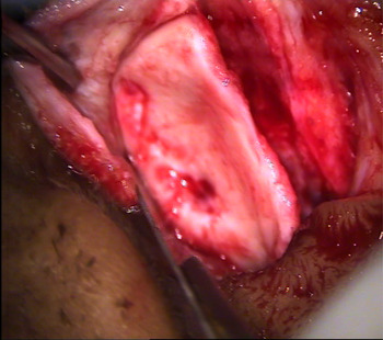
Tragal cartilage (a larger graft than would be routinely used) was harvested using the same endaural incision as reported in our previous study (Figure 1).Reference Khan and Parab4 For each patient, a representative, arbitrary whole-length bar of tragal cartilage was excised and given to the assistant for measurement of thickness. To avoid inter-observer variations, all measurements were repeated by the second author (SP).
Tragal cartilage harvesting through endaural incision.

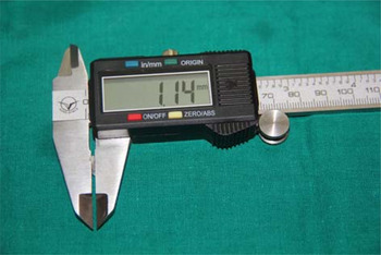
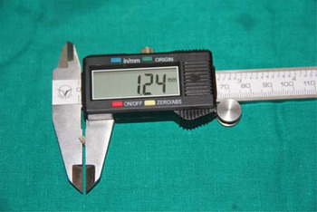
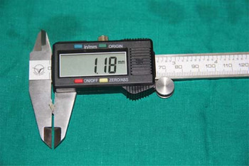
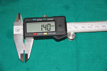
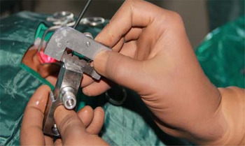
Digital callipers were used to determine cartilage thickness (Figure 2). To avoid discrepancy and minimise error, four readings were taken: the first reading was of the superior part of the tragus, the second reading was of the middle, the third reading was of the inferior end and the fourth reading was of the overall thickness of the whole length of the tragus. Hence, the measurements comprised three readings of the horizontal thickness and one reading of the vertical thickness (Figures 3–5).
Digital callipers with tragal cartilage.

Second reading for tragal cartilage thickness, at middle part of tragus.

Third reading for tragal cartilage thickness, at inferior end of tragus.

Fourth reading for tragal cartilage thickness, along whole length of tragus.

Patients' details (e.g. name, age, sex, address and mobile number) were also recorded. The data collected were then grouped and analysed statistically (using the SPSS® statistical software package) to determine age- and sex-related average thicknesses of the tragal cartilage.
Results
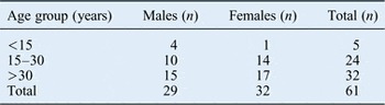
A total of 61 tragal cartilages were measured for thickness. The patients consisted of 29 males and 32 females, ranging from 8 to 56 years of age. The age and sex distribution of the study population is summarised in Table I. The average thicknesses of tragal cartilage in males and females are tabulated in Table II.
Distribution of patients according to age and sex

Average thickness of tragal cartilage according to sex

SD = standard deviation
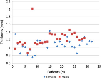
The total average tragal cartilage thickness was 1.228 ± 0.204 mm in males and 1.090 ± 0.162 mm in females. Statistical analysis revealed that thickness was significantly lower in females compared with males (p < 0.005). A scatter diagram (Figure 6) summarises the tragal cartilage thickness data for the males and the females of our study population.
Scatter diagram of tragal cartilage thickness for males and females.

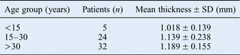
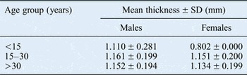
In males, the average tragal cartilage thickness was: 1.110 ± 0.281 mm in those aged less than 15 years, 1.161 ± 0.199 mm in those aged 15 to 30 years, and 1.152 ± 0.194 mm in those aged over 30 years. In females, the average tragal cartilage thickness was: 0.802 ± 0.000 mm in those aged less than 15 years, 1.151 ± 0.200 mm in those aged 15 to 30 years, and 1.134 ± 0.199 mm in those aged over 30 years. Statistical analysis revealed that increased tragal cartilage thickness was marginally significantly associated with increased age. Average tragal cartilage thicknesses for the different age groups, and according to both age and sex, are summarised in Tables III and IV.
Average thickness of tragal cartilage according to age

SD = standard deviation
Average thickness of tragal cartilage according to age and sex

Discussion
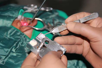
Our technique of cartilage tympanoplasty involves thinning the tragal cartilage with a precise cartilage splitter so as to achieve acoustic benefit (Figure 7).Reference Khan and Parab4 Over the last decade, we have been slicing the tragal cartilage with a cartilage splitter that reportedly gives thicknesses of 0.1, 0.2 and 0.3 mm (Figure 8) following the placement of 1 mm of cartilage graft. The cartilage splitter has a cartilage holder and a blade stabilisation assembly. There are metallic plates of 0.1, 0.2 and 0.3 mm thickness, which are placed within the cartilage-splitting unit (along with the cartilage) to give slices of respective expected thickness. Accordingly, these three metallic plates can be combined in various ways to attain a thickness of up to 0.6 mm (achieved by placing all the plates together).
Tragal cartilage slicing technique, performed using a Kurz® Precise Cartilage Splitter.

Sliced tragal cartilage, in two parts.

The selection of plate (0.1 mm, 0.2 mm or 0.3 mm) is currently considered arbitrary, and depends on the judgement and experience of the surgeons. After slicing the tragal cartilage with the precise cartilage splitter, we get two sliced pieces of tragal cartilage. The thickness of both sliced parts can be estimated only if the total thickness of the tragal cartilage in a particular patient is known. Tragal cartilage thickness may not be uniform in all patients in any given population. We have noted that the tragal cartilage varies in thickness, and in some patients it may be even thinner than 1 mm.
In order to obtain a slice with a definite thickness to be used in tympanoplasty as a graft material, the tragal cartilage thickness of each patient should ideally be known. However, in routine day-to-day practice this is cumbersome, and it would be practically difficult to measure tragal cartilage thickness in every patient intra-operatively before the cartilage is sliced.
Sometimes (in cases of very thin or very thick cartilage), the cartilage is inadequate for the slot in the slicer, and decent slices of the required thickness cannot be obtained. This problem can be minimised if the exact thickness of the tragal cartilage for a particular age group is known. This will aid selection of the most appropriate size of thickness plate to use for slicing the cartilage.
This study was conducted to gather data on tragal cartilage thickness in different age groups (for males and females) in an Indian population. The findings enable us to predict the average tragal cartilage thickness in a particular age group so that a slice of the most appropriate thickness can be selected for use in tympanoplasty. Total average tragal cartilage thickness was 1.228 ± 0.204 mm in the males and 1.090 ± 0.162 mm in the females. The average thicknesses were 1.018 ± 0.139 mm in those aged less than 15 years, 1.139 ± 0.238 mm in those aged 15–30, and 1.189 ± 0.155 in those aged over 30 years.
In a cadaveric study by Hizalan et al.Reference Hizalan, Dokuzlar, Kafa and Sendemir5 it was indicated that, on average, at most 2.19 ± 0.53 cm2 of cartilage can be used as tragal reconstruction material. In cases where the cartilage harvested from the tragus is insufficient, 1.36 ± 0.67 cm2 of cartilage on the root of the external meatus was ready to be utilised as reserve material. The greatest thickness was 2.20 mm, on the inferior part of the tragus. The thinnest part was the superior medial part of the tragus and measured 0.7 mm. In another cadaveric study, conducted on 40 tragi, it was concluded by Ishida et al. that the average thickness of tragal cartilage is 1.6 mm.Reference Ishida, Pereira and Andrews6
To our knowledge, the current study is the first study to measure the intra-operative thickness of tragal cartilage and determine tragal thickness in a given study population, assessing sex- and age-related variations in thickness. Determining the thickness of the tragal cartilage is important for the following reasons. Firstly, it can provide information regarding age-related variations in thickness for a given population. Secondly, it can aid selection of the most appropriate plate size to use in a particular population or age group. Thirdly, in paediatric tympanoplasty, knowledge of cartilage thickness will inform as to whether a full thickness cartilage graft is needed or whether slicing should be carried out.
The simplest way of slicing tragal cartilage that is more than 1 mm thick would be to split it without using a thickness plate, in order to yield two slices, one of which should have a thickness of 1 mm. The 1 mm slice can then be sliced with 0.1, 0.2 and 0.3 mm plates to yield respective thickness slices. However, this same logic cannot be applied if the tragal cartilage is less than 1 mm.
Based on our study findings, we propose that metallic plates of specific thicknesses are used (utilising the Kurz Cartilage Splitter) in the three age groups, to achieve tragal cartilage thicknesses of 0.5 mm or less for acoustic benefit. In those aged less than 15 years, a metallic plate of 0.3 mm thickness should be used; in those aged 15–30 years, the plate should be 0.2–0.3 mm thick; and in those aged over 30 years, the plate should be 0.2 mm thick.
• Sliced tragal cartilage-perichondrial grafts are used for tympanic membrane and middle-ear reconstructions
• Tragal cartilage is sliced with a precise cartilage splitter to achieve a cartilage thickness of less than 0.5 mm for acoustic benefit
• Tragal cartilage thickness in a given population may not be uniform
• Knowledge of total tragal cartilage thickness is useful to achieve tragal cartilage of a particular thickness
• This study was conducted to gather data on tragal cartilage thicknesses in different age groups in an Indian population
• This is the first study to measure intra-operative tragal cartilage thickness, and examine sex- and age-related variations
In terms of limitations of the study, we acknowledge that the fact the cartilage was a compressible material may have led to errors in measurement.
Conclusion
Our study, which has a level of evidence 2c, revealed a total average tragal cartilage thickness of 1.228 ± 0.204 mm in the males and 1.090 ± 0.162 mm in the females. Overall thickness was 1.018 ± 0.139 mm in those aged less than 15 years, 1.139 ± 0.238 mm in those aged 15–30 years, and 1.189 ± 0.155 mm in those aged over 30 years.
Knowledge of the average tragal cartilage thickness for a particular age group aids pre-operative planning by informing the selection of the most appropriate plate thickness for cartilage tympanoplasty.
Acknowledgement
We wish to acknowledge Mrs Swati Raje, Assistant Professor in Biostatistics (Department of Preventive and Social Medicine, Maharashtra Institute of Medical Education and Research Medical College, Pune, India), for the statistical analysis of the data.