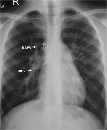
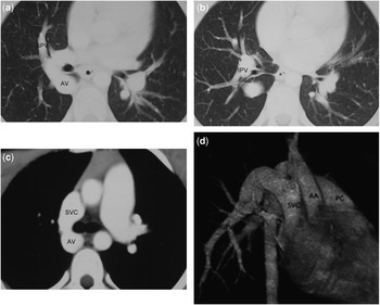
An 11-year-old boy was admitted for investigation of an asymptomatic cardiac murmur. On chest X-ray (Fig 1), a prominent azygos vein was noted in proximity to the right upper and right lower pulmonary veins. Transthoracic echocardiography showed mild dilatation of the right atrium and the right ventricle, with increased superior caval vein flow and a small atrial defect in the oval fossa. Only two pulmonary veins could be seen connected to the left atrium, and partial anomalous pulmonary venous return was suspected. Multidetector computed tomography on a 64-detector-row scanner (Aquillon; Toshiba Medical Systems, Tokyo, Japan; Fig 2a–c) confirmed that the right superior caval vein and inferior pulmonary veins drained to the azygos vein and hence to the superior caval vein. A three-dimensional reconstruction of the volume-rendered images is shown in Figure 2d (AV = azygos vein; RSPV = right superior pulmonary veins; RLPV = right lower pulmonary veins; SCV = superior caval vein; SPV = superior pulmonary vein; IPV = inferior pulmonary vein; AA = ascending aorta; PC = pulmonary trunk). These findings were confirmed at operation and the malformation was repaired. Secundum septum was totally resected. Baffle repair of the defect using pericardial patch was performed, directing anomalous pulmonary return to the left atrium through the surgically created atrial septal defect. The post-operative course was uneventful and a subsequent echocardiographic examination showed the complete restoration of the circulation. Although echocardiography is usually sufficient to delineate pulmonary venous drainage, this case illustrates the value of computed tomographic angiography for imaging rare and atypical morphology.
Figure 1

Figure 2