Magnetic resonance imaging and magnetic resonance angiography are established methods in diagnosing partial anomalous pulmonary venous drainage in children and adults.Reference Riesenkampff, Schmitt and Schnackenburg1–Reference Beerbaum, Parish, Bell, Gieseke, Korperich and Sarikouch3 Magnetic resonance angiography has helped to facilitate the identification of maldraining vessels.Reference Prasad, Soukias and Hornung4 However, anatomic proximity of pulmonary veins and arteries can make a precise diagnosis of maldraining vessels by standard magnetic resonance imaging difficult and the use of gadolinium-containing contrast agent in children should be avoided if possible.
In the presence of multiple maldraining veins and an interatrial communication, a selective quantification of shunt volumes could be helpful for surgical planning, but multiple standard flow measurements can be very time-consuming.
Flow-sensitive four-dimensional velocity-encoded magnetic resonance imaging allows the visualisation and quantification of three-directional blood flow within a three-dimensional anatomical volume.Reference Markl, Chan and Alley5, Reference Nordmeyer, Riesenkampff and Crelier8 It was shown that blood flow quantification with this new method is accurate in arteries and veins in healthy volunteers as well as in patients with congenital cardiac disease and pathologic flow conditions.Reference Uribe, Beerbaum, Sorensen, Rasmusson, Razavi and Schaeffter6–Reference Nordmeyer, Riesenkampff and Crelier8
In this study, we sought to visualise and quantify the flow of maldraining pulmonary veins by using flow-sensitive four-dimensional velocity-encoded magnetic resonance imaging and to assess if this method adds clinical value in diagnosing patients with suspected partial anomalous venous drainage.
Materials and methods
Patient characteristics
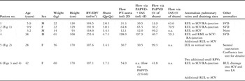
The study was conducted in six patients – female three and male three aged between 3.2 and 42 years (Table 1), who were transferred to our imaging unit with suspected partial anomalous venous drainage after transthoracic echocardiography. The institutional review board approved the study and the patients or guardians of patients gave informed consent.
Patient characteristics and results.

Fig, figure; LA, left atrium; LUL, left upper lobe; n.a., not applicable; PAPVD, partial anomalous pulmonary venous drainage; PT, pulmonary trunk; RA, right atrium; RML, right mid-lobe; RPVs, right pulmonary veins; RUL, right upper lobe; RV-EDV, right ventricular enddiastolic volume; SCV, superior caval vein; SVD, atrial septum defect of the sinus venosus type; 2D, two-dimensional; 4D, four-dimensional
Flow via the maldraining pulmonary veins (PAPVD) and the interatrial septum (IAS) was measured by flow-sensitive four-dimensional velocity-encoded magnetic resonance imaging and calculated as percentage of the overall shunt volume (see text, Method section for details).
Magnetic resonance imaging
In all patients (n = 6), flow-sensitive four-dimensional velocity-encoded magnetic resonance imaging was performed in addition to the previously described standard protocol including cine magnetic resonance imaging in transversal and coronal orientation, magnetic resonance angiography, and standard flow measurements in the ascending aorta and pulmonary trunk.Reference Riesenkampff, Schmitt and Schnackenburg1 The study was conducted in a whole body 1.5 Tesla MR scanner (Achieva R2.6.3, Philips Medical Systems, Best, The Netherlands) using a five-element cardiac phased-array coil (Philips Medical System, Best, The Netherlands). Right ventricular size and function were assessed by volumetric analysis of transversal cine magnetic resonance imaging. Shunt volumes were assessed by subtracting the flow in the aorta from the flow in the pulmonary trunk in millilitres (Table 1) and the ratio of pulmonary to systemic flow (Qp:Qs) was calculated by using flow volumes of standard two-dimensional flow measurements in the aorta and pulmonary trunk.Reference Devos and Kilner9
The study included two patients under 6 years of age who were imaged in conscious sedation free-breathing by intravenous administration of a bolus of midazolam at a dose of 0.1–0.2 milligrams per kilogram body weight followed by continuous infusion of propofol of 2–4 milligrams per kilogram body weight per hour.
Flow-sensitive four-dimensional velocity-encoded magnetic resonance imaging
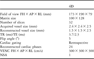
Acquisition: Anisotropic four-dimensional segmented k-space phase contrast gradient echo sequence with retrospective electrocardiographic gating was performed without navigator gating of respiratory motion. In all patients, flow velocities were measured in a three-dimensional volume that covered the thorax from the apex of the heart to the aortic arch in the feet-to-head direction, the sternal border and spine in the anterior-to-posterior direction, and the superior caval vein and the main stem of the right and left pulmonary arteries until further branching in the right-to-left direction. Sequence parameters are shown in Table 2.
Scan parameters of the flow-sensitive four-dimensional velocity-encoded magnetic resonance imaging sequence.

AP, anterior–posterior; FH, feet–head; NSA, number of signal averages; RL, right–left; TE, echo time; TR, repetition time; VENC, velocity encoding; 4D, four-dimensional
Post-processing: Four-dimensional flow parameters were analysed with the software GTFlow (Release 1.3.3, Gyrotools, Zurich, Switzerland). Offline evaluation of the flow-sensitive four-dimensional velocity-encoded magnetic resonance imaging scans was performed using the individual calculation of three-dimensional streamlines inside the maldraining pulmonary veins, the superior and inferior caval vein or vertical vein, and the interatrial communication, if present.
The flow in the maldraining veins and across the interatrial communication was visualised and quantified individually and indicated as a percentage of the overall shunt volume by using the following formulas:

where “PAPVD percentage” is the fraction of the maldraining pulmonary vessel flow of the overall shunt volume, “IAS percentage” is the fraction of the interatrial flow of the overall shunt volume, “flow via PAPVD” stands for the flow of the maldraining pulmonary veins, and “flow via IAS” for the flow across the interatrial septum. “Sp” is the pulmonary shunt volume, calculated by subtracting the flow of the ascending aorta Qs (millilitre) from the flow of the pulmonary trunk Qp (millilitre).
Results
Standard magnetic resonance imaging
In all patients, echocardiographically suspected partial anomalous pulmonary venous drainage was confirmed by standard magnetic resonance imaging and magnetic resonance angiography. In total, ten maldraining pulmonary veins were identified in six patients; multiple maldraining pulmonary veins were found in three patients and interatrial communications were present in three patients. Another patient had an unusual communication between the right upper pulmonary vein to the SCV and the left atrium after surgical closure of an atrial septal defect of unknown morphology.
Draining sites of the maldraining pulmonary veins were the superior caval vein in five patients and the vertical vein in one patient. The ratio of pulmonary to systemic flow (Qp:Qs) varied between 1.4:1 and 4.7:1 (Table 1).
Flow-sensitive four-dimensional magnetic resonance imaging
Visualisation
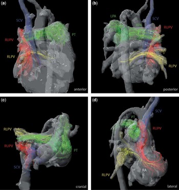
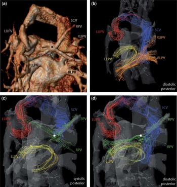
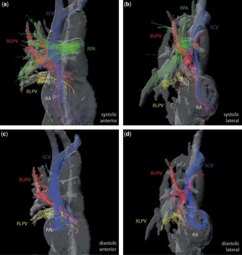
The findings of the standard magnetic resonance imaging were confirmed, as flow visualisation also showed drainage of pulmonary veins into the superior caval vein (Fig 1) in five patients and into the vertical vein (Fig 2) in one patient. Additional, more precise information was gained by selective visualisation of individual pulmonary veins and arteries using flow-sensitive four-dimensional magnetic resonance imaging (Figs 1–3). It revealed, for example, the flow direction of additional right pulmonary veins into the vertical vein and into the left lower pulmonary vein with differing flow during ventricular systole and diastole in one patient (Fig 2c and d), where magnetic resonance angiography had shown a supra-atrial vessel formation without information of flow directions and differences during the cardiac cycle (Fig 2a).
Selective visualisation of streamlines and three-dimensional phase-contrast magnetic resonance angiography (without the use of contrast agent) in a 9.9-year-old child (patient 2, Table 1) diagnosed with anomalous pulmonary venous drainage of the right upper pulmonary vein into the superior caval vein and presence of an interatrial communication in different perspectives at ventricular systole: (a) anterior, (b) posterior, (c) cranial, and (d) lateral view. The streamlines of the different vessels are coloured variably: the red streamlines represent the flow of the maldraining right upper pulmonary vein (RUPV) entering the right atrium together with the blue streamlines of the superior caval vein (SCV, a and b). The green streamlines in the pulmonary trunk (PT), right and left pulmonary arteries (RPA and LPA) allow one to distinguish clearly between peripheral pulmonary arteries (green) and veins (yellow and red, c). The yellow streamlines of the anatomic correctly draining right lower pulmonary vein (RLPV) merge with the red streamlines representing a left-to-right shunt across the interatrial communication and thus functional maldrainage during ventricular systole (d).

Posterior view of anatomy (a, volume rendering mode of contrast-enhanced angiography) and streamlines with three-dimensional phase-contrast magnetic resonance angiography in a 28-year-old patient (patient 5, Table 1) with complex bilateral partial anomalous pulmonary venous drainage of multiple vessels. The left upper pulmonary vein (LUPV, red streamlines) drains via a vertical vein into the superior caval vein (SCV, blue streamlines, b–d). The left lower pulmonary vein (LLPV, yellow streamlines, b–d), the right lower pulmonary vein (RLPV, orange streamlines, b), and the main right upper pulmonary vein (RUPV, orange streamlines, b) drain regularly into the left atrium. In addition, there is a star-shaped vessel confluence cranial to the left atrium (asterisk, a, c, and d) with the drainage of two additional small right pulmonary veins (RPV, green streamlines, c and d). The flow in this confluence changes during the cardiac cycle, as visualised exclusively by the streamlines of flow-sensitive four-dimensional cine magnetic resonance imaging: during ventricular systole, the main flow is directed to the left upper pulmonary vein, thus maldraining to the right atrium (c). During ventricular diastole, these vessels drain to the left atrium together with the left lower pulmonary vein (d).

Streamlines and three-dimensional phase-contrast magnetic resonance angiography of a 42-year-old patient (patient 6, Table 1) with an atypical communication between a maldraining right upper pulmonary vein to the SCV and the left atrium at ventricular systole (a and b) and ventricular diastole (c and d) in anterior (a and c) and lateral view (b and d). Selective visualisation of streamlines represents the flow of the maldraining right upper pulmonary vein (RUPV, red) to the superior caval vein (SCV, blue). The right pulmonary artery (RPA, green) and its branches are clearly distinguished from veins (red) by flow-sensitive four-dimensional cine magnetic resonance imaging (a and b). The right lower pulmonary vein (RLPV, yellow) regularly drains to the left atrium. Note the differences of flow during the cardiac cycle: at ventricular systole, streamlines of the right upper pulmonary vein (red) and the superior caval vein (blue) merge with drainage to the right atrium (RA, a and b). At ventricular diastole, the red streamlines from the right upper pulmonary vein merge with the yellow streamlines of the right lower pulmonary vein entering the left atrium (c and d), representing a left-to-right shunt across the interatrial communication in the presence of a residual or recurring atrial septal defect and a mild stenosis of the superior caval vein after surgery. Note the missing red streamlines to the right atrium during ventricular diastole in the lateral view (d).

In another patient, who had undergone surgery abroad with closure of an atrial septal defect, two maldraining right upper pulmonary veins and a mild stenosis of the superior caval vein were diagnosed. In this patient, one pulmonary vein drained into the superior caval vein approximately five centimetres cranially to the right atrium and another pulmonary vein with partial drainage to the superior caval vein close to the right atrium showed functional drainage to both atria, with predominant drainage to the right atrium during ventricular systole and to the left atrium during ventricular diastole (Fig 3).
Quantification
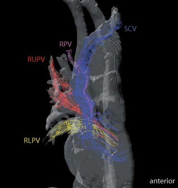
In addition to the flow visualisation, flow-sensitive four-dimensional magnetic resonance imaging was used to quantify the flow volume of each single maldraining pulmonary vein. In six patients, blood flow of the maldraining pulmonary veins accounted for a median of 41.3% with a range from 30.2% to 99.3% (Table 1) of the overall shunt volume. In one patient, an additional small maldraining right upper pulmonary vein into the superior caval vein 56 millimetres cranially to the right atrium was diagnosed during post-processing and could be visualised (Fig 4) and quantified using flow-sensitive four-dimensional magnetic resonance imaging and accounted for 9% of the overall shunt volume.
Anterior view with visualisation of the streamlines of an additional maldraining right pulmonary vein (purple) to the cranial superior caval vein (SCV, blue) during ventricular diastole in the same patient as presented in Figure 3. The red streamlines from the right upper pulmonary vein (RUPV) merge with the yellow streamlines of the right lower pulmonary vein (RLPV) entering the left atrium (see Fig 3 for details).

In patients with an interatrial communication, the flow via the atrial septal defect could be assessed and accounted for 59.3%, 63.6%, and 70.9% of the shunt volume.
Discussion
This study shows that flow-sensitive four-dimensional velocity-encoded magnetic resonance imaging facilitates and improves the accurate diagnosis of partial anomalous pulmonary venous drainage due to its ability to visualise and quantify blood flow selectively in individual vessels in an acquired three-dimensional volume.
Anatomy – visualisation
Diagnosing partial anomalous pulmonary venous drainage using standard magnetic resonance imaging, including magnetic resonance angiography, can be challenging even for experienced investigators, especially when pulmonary arteries and veins are in a close anatomical neighbourhood. Although the connection of the maldraining veins to the corresponding structure, for example the superior caval vein, is visible, the visualisation of the peripheral course of the vessel, the flow direction, and the precise distinction between pulmonary veins and arteries is more demanding. In this study, it was shown that the three-dimensional streamlines acquired by flow-sensitive four-dimensional magnetic resonance imaging elucidate the origin and the connection of the vessels as well as the course of the blood flow from the individual vessels (Figs 1–3). This flow information helps to distinguish adjacent vessels and thus to precisely diagnose maldraining pulmonary veins.
Hence, with this new technique, it is possible for the first time to visualise the blood flow direction of selective maldraining pulmonary veins, for example, to the superior caval vein and into the right atrium (Fig 1), or, in case of a residual atrial septal defect, to the left and right atria (Fig 3). Earlier, this information was only suggested but not visualised. Therefore, this new technique adds important novel information.
Function – quantification
The quantification of blood flow from each maldraining pulmonary vein and across the interatrial communication, if present, renders a more precise diagnostic workup, which could affect the treatment strategy, for example, in patients with multiple maldraining pulmonary veins, which might be distant to the atria and difficult to redirect surgically (Fig 4).
Blood flow quantification of each maldraining vessel and across the interatrial septum could be obtained by standard flow measurements as well, but the examination time would be prolonged due to planning scans for accurate plane acquisition, multiple flow acquisitions, and possible repeat scans.
In the case of the patient with the unusual anatomy of the supra-atrial vessel formation of additional small right pulmonary veins (Fig 2), substantial information on the pathophysiological flow conditions was added which would not have been possible without the new technique.
Acquisition and post-processing
After a single 10–15-minute lasting acquisition scan, flow visualisation and quantification and thus a detailed and accurate diagnosis of maldraining pulmonary veins is possible. This post-processing analysis is made offline without the patient being inside the scanner. Furthermore, if supplemental maldraining vessels exist, which are only diagnosed during post-processing, offline flow analysis and separate shunt quantification can still be carried out without the need for additional examination. The complete volumetric coverage of the areas of interest allows retrospectively positioning analysis planes and quantifying flow at any location within the previously acquired three-dimensional volume.
Furthermore, in the case of significant heart rate variabilities between standard aortic and pulmonary flow measurements during an examination, shunt calculations can be more error-prone than with the new technique, where all flow information is recorded at the same time.
Future prospects
The use of intravenous gadolinium-containing contrast agent for magnetic resonance angiography in children has been shown to be safe as far as renal function is not impaired.Reference Martin10 However, possible long-term side effects cannot be ruled out yet. The use of flow-sensitive four-dimensional magnetic resonance imaging for the diagnosis of partial anomalous pulmonary venous drainage could shorten the examination time, and the need for intravenous application of gadolinium-containing contrast agent could be eliminated with further improvement of the spatial resolution. On the basis of cine images, the anatomical location of maldraining pulmonary veins can be identified, and flow-sensitive four-dimensional magnetic resonance imaging can visualise the streamlines and assist in flow volume calculations. Standard two-dimensional flow measurements and magnetic resonance angiography could become unnecessary and examination time could be shortened.
Conclusions
The selective visualisation and flow quantification of pulmonary veins by using flow-sensitive four-dimensional velocity-encoded magnetic resonance imaging adds clinical value by improving and facilitating the accurate diagnosis of simple and complex cases of partial anomalous pulmonary venous drainage.
In future, with further development of the technique, it might replace contrast-enhanced magnetic resonance angiography and standard flow measurements in these patients.
Limitations
This is a single-centre study in a pre-selected group of six patients with partial anomalous pulmonary venous drainage. Additional research is needed in a larger group of patients.
The acquired three-dimensional volumes do not cover the whole thorax and the three-dimensional streamlines of peripheral vessels are not yet imaged. To achieve this goal, scan time with the present available sequence would be unbearably long for the patient inside the magnetic resonance scanner.
At present, anatomical images derived by flow-sensitive four-dimensional magnetic resonance imaging are less defined than anatomical images derived by standard cine magnetic resonance images. The flow information, however, obtained by the flow streamlines, which refer to the vessel morphology and anatomic course, is nearly comparable to the information obtained by magnetic resonance angiography.
Acknowledgements
The authors thank Alireza Khasheei and Bianca Mandt for technical assistance. This work was supported by the Federal Ministry of Education and Research, Grant 01EV0704. There are no disclosures to be made for any conflicts of interest.