INTRODUCTION
Prader–Willi syndrome (PWS) has a prevalence of 1:10.000–1:30.000 live births. The disorder is caused by the lack of expression of the paternally inherited material located at 15q11-q13. There are three main genetic subtypes: deletion (DEL; 65–75% of cases), maternal uniparental disomy (UPD; 20–30%), and defect of the imprinting center (IC) in 1–3% of cases (Cassidy, Schwartz, Miller, & Driscoll, Reference Cassidy, Schwartz, Miller and Driscoll2012). The main clinical features include neonatal hypotonia, a week suck with feeding difficulties during infancy, hyperphagia with excess of weight gain in childhood, developmental delay and challenging behavior in response to changes in routine (Holm et al., Reference Holm, Cassidy, Butler, Hanchett, Greenswag, Whitman and Greenberg1993). They also present multiple endocrine disorders, which suggest hypothalamic–pituitary axis dysfunction (Moix, Giménez-Palop, & Caixàs, Reference Moix, Giménez-Palop and Caixàs2018) and psychopathology may appear late on in life (Guinovart, Coronas, & Caixàs, Reference Guinovart, Coronas and Caixàs2019) with different degrees of compulsions (Novell-Alsina et al., Reference Novell-Alsina, Esteba-Castillo, Caixàs, Gabau, Giménez-Palop, Pujol and Torrents-Rodas2019) or psychotic features.
The level of intellectual disability (ID) in PWS varies widely across subjects, with IQ usually in the borderline to the mildly moderately impaired range (IQ = 50–80) (Holm et al., Reference Holm, Cassidy, Butler, Hanchett, Greenswag, Whitman and Greenberg1993; Hurren & Flack, Reference Hurren and Flack2016). As a group, relative to healthy individuals, PWS has been associated with disabilities in attention and working memory, language skills, sequential processing, executive functions, and also social cognition (e.g., Chevalère et al., Reference Chevalère, Postal, Jauregui, Copet, Laurier and Thuilleaux2015; Copet et al., Reference Copet, Jauregui, Laurier, Ehlinger, Arnaud, Cobo and Thuilleaux2010; Dykens, Hoddapp, Walsh, & Nash, Reference Dykens, Hodapp, Walsh and Nash1992; Dykens, Roof, Hunt-Hawkins, Daniell, & Jurgensmeyer, Reference Dykens, Roof, Hunt-Hawkins, Daniell and Jurgensmeyer2019; Jauregui et al., Reference Jauregui, Arias, Vegas, Alén, Martínez, Copet and Thuilleaux2007; Koenig, Klin, & Schultz, Reference Koenig, Klin and Schultz2004; Whittington & Holland, Reference Whittington and Holland2011), although findings across studies are not always consistent. When their performance is compared to that of individuals with the ID of different etiologies, the syndrome has been characterized by a distinctive cognitive profile of weaknesses in attention-switching capacities, arithmetic skills, and learning abilities, and certain relative strengths in visuospatial skills, particularly in the deletion subtype (e.g., Bertella et al., Reference Bertella, Girelli, Grugni, Marchi, Molinari and Semenza2005; Copet et al., Reference Copet, Jauregui, Laurier, Ehlinger, Arnaud, Cobo and Thuilleaux2010; Curfs, Wiegers, Sommers, Borghgraef, & Fryns, Reference Curfs, Wiegers, Sommers, Borghgraef and Fryns1991; Dykens, Reference Dykens2002; Foti et al., Reference Foti, Menghini, Orlandi, Rufini, Crinò, Spera and Mandolesi2015; Woodcock, Oliver, & Humphreys, Reference Woodcock, Oliver and Humphreys2009). In the motor domain, some studies have documented deficiencies in PWS in gross and fine motor coordination (Cimolin et al., Reference Cimolin, Galli, Grugni, Vismara, Precilios, Albertini and Capodaglio2011; Lam et al., Reference Lam, Rubin, Duran, Chavoya, White and Rose2016; Reus et al., Reference Reus, Zwarts, van Vlimmeren, Willemsen, Otten and Nijhuis-van der Sanden2010), and articulatory difficulties and deficits in orofacial nonspeech motor functions have also been noted (Defloor, Van Borsel, & Curfs, Reference Defloor, Van Borsel and Curfs2002; Saeves, Asten, Sorthaug, & Bágesund, Reference Saeves, Asten, Sorthaug and Bágesund2011). Beyond these basic motor deficits, parent reports and clinical observations also suggest that more specific, higher order motor functions such as praxis abilities (i.e., the ability to follow commands to perform skilled movements and gesture imitation) may be compromised in PWS. However, to our knowledge, no study has focused on comprehensively assessing praxis performance in this population.
Praxis movements are defined as purposeful and skilled motor actions that may include imitation abilities and tool use pantomimes. Performance of these motor actions requires linking multiple aspects of perception, cognition, and movement (Goldenberg, Reference Goldenberg2014; Leiguarda & Marsden, Reference Leiguarda and Marsden2000). Functional neuroimaging studies in healthy subjects and lesion data have consistently emphasized the involvement of a distributed bilateral parieto–temporal–frontal network subserving gesture processing in praxis-related tasks (Buxbaum, Shapiro, & Coslett, Reference Buxbaum, Shapiro and Coslett2014; Caspers, Zilles, Laird, & Eickhoff, Reference Caspers, Zilles, Laird and Eickhoff2010; Goldenberg, Hermsdörfer, Glindemann, Rorden, & Karnath, Reference Goldenberg, Hermsdörfer, Glindemann, Rorden and Karnath2007; Leiguarda & Marsden, Reference Leiguarda and Marsden2000; Lesourd et al., Reference Lesourd, Osiurak, Baumard, Bartolo, Vanbellingen and Reynaud2018; Niessen, Fink, & Weiss, Reference Niessen, Fink and Weiss2014). This “praxis network” engages the inferior and superior parietal lobes, temporal areas, as well as motor cortices and inferior frontal regions (Foundas & Duncan, Reference Foundas and Duncan2019; Leiguarda & Marsden, Reference Leiguarda and Marsden2000). Subcortical regions, including the basal ganglia and thalamus, have also been implicated (Leiguarda, Reference Leiguarda2001).
Of particular interest to PWS is the frontal-opercular area – encompassing both the inferior frontal and anterior insular cortices – as this region has been implicated in a number of brain functions related to major symptoms of the syndrome, including food intake regulation (Chen, Papies, & Barsalou, Reference Chen, Papies and Barsalou2016; Dagher, Reference Dagher2012), the control of swallowing (Sörös, Inamoto, & Martin, Reference Sörös, Inamoto and Martin2009), language processing and motor aspects of speech production (Brown & Yuan, Reference Brown and Yuan2018; Dick, Garic, Graziano, & Tremblay, Reference Dick, Garic, Graziano and Tremblay2019; Dronkers, Reference Dronkers1996; Maliia et al., Reference Maliia, Donos, Barborica, Popa, Ciurea, Cinatti and Mindruta2018), and response inhibition (Aron, Robbins, & Poldrack, Reference Aron, Robbins and Poldrack2014). Associated with praxis, the caudal part of the pars opercularis has also a role in the so-called mirror neuron system (Cattaneo & Rizzolatti, Reference Cattaneo and Rizzolatti2009; Cross, Torrisi, Reynolds Losin, & Iacoboni, Reference Cross, Torrisi, Reynolds Losin and Iacoboni2013; Kilner, Neal, Wieskopf, Friston, & Frith, Reference Kilner, Neal, Wieskopf, Friston and Frith2009; Liakakis, Nickel, & Seitz, Reference Liakakis, Nickel and Seitz2011; Molnar-Szakacs, Iacoboni, Koski, & Mazziotta, Reference Molnar-Szakacs, Iacoboni, Koski and Mazziotta2005), that is activated by both observation and execution of actions (Iacoboni & Mazziotta, Reference Iacoboni and Mazziota2007), and highly related to imitative and social behavior (Cattaneo & Rizzolati, Reference Cattaneo and Rizzolatti2009). Structure and function of the inferior frontal cortex, adjacent prefrontal, and insular cortex have been highlighted in a number of praxic disorders including buccofacial and limb apraxia (Caspers et al., Reference Caspers, Zilles, Laird and Eickhoff2010; Goldenberg et al., Reference Goldenberg, Hermsdörfer, Glindemann, Rorden and Karnath2007; Lesourd et al., Reference Lesourd, Osiurak, Baumard, Bartolo, Vanbellingen and Reynaud2018; Ozsancak, Auzou, Dujardin, Quinn, & Destée, Reference Ozsancak, Auzou, Dujardin, Quinn and Destée2004).
In this context, the aim of the present study was to assess different aspects of praxis performance in individuals with PWS using tasks standardized for the ID population, and to investigate the relationship of the potential deficits with brain structural abnormalities as measured with voxel-based morphometry (VBM). We hypothesized that PWS will show a more deficient performance in praxis abilities than ID-matched controls, and that these deficits will be associated with anatomical alterations in the inferior frontal region. We also sought to explore differences between genetic subtypes.
METHODS
Study Participants
The sample included 30 patients with genetically diagnosed PWS. Genetic testing showed 20 patients (67%) with DEL and 10 patients (33%) with UPD or IC. One-hundred and thirty-two adult participants with the ID of negative genetically confirmed etiology with equivalent cognitive level, age, sex, and acquired curricular competence were used as a control group for the study of praxis. Participant’s level of education was defined as acquired curricular competence (illiteracy vs. basic education) instead of years of education – as most of the participants went to school for more than 18 years and yet they were illiterate. Thirty healthy subjects matched by age and sex to the PWS group made up the control sample for the anatomical study. To control for the confounding effect of obesity, we included an additional control group of 30 subjects matched by age, sex, and BMI to the PWS group. Table 1 provides characteristics of study participants.
General characteristics of study participants

ID, intellectual disability; M, mean; SD, standard deviation.
All subjects were Caucasian and their weight was stable for at least 3 months before inclusion in the study. PWS patients and obese controls were recruited from the Endocrinology and Nutrition Department of a Reference Center (Hospital Universitari Parc Taulí, Sabadell) and from the Specialized Department in Mental Health and Intellectual Disability, Girona, Spain. The Catalan Association of Prader–Willi Syndrome (Barcelona, Spain) and the Prader–Willi Syndrome Foundation (Madrid, Spain) assisted with the recruitment. Healthy subjects were hospital staff or acquaintances that participated voluntarily. Participants with ID were recruited from local day services. Individuals with the following conditions were excluded: severe ID, sensory impairments precluding proper examination, previous alterations of the central nervous system unrelated to ID (e.g., head injury, stroke, brain tumors, or multiple sclerosis), substance abuse, and untreated diseases with associated cognitive deficits (e.g., hypothyroidism, vitamin B12 deficiency). Four men with PWS were undergoing testosterone replacement for hypogonadism. Four women with PWS were undergoing estrogen and progestin therapy for hypogonadism; all these were studied in the follicular phase. Although 11 subjects with PWS had been treated with growth hormone until puberty, none were receiving growth hormone at the time of the study. Five subjects with PWS had type 2 diabetes mellitus and nine were treated with psychotropic medication (fluoxetine and/or topiramate). One patient among obese subjects had been diagnosed with type 2 diabetes.
Regarding healthy controls, a complete medical interview was carried out to exclude subjects with relevant medical or neurological disorders, psychiatric illness, and history of substance abuse. No subject was undergoing medical treatment or was diagnosed with eating disorders. All participants or their parents/guardians provided written informed consent and all PWS and ID patients who were not able to sign gave drawn assent. The Institutional Ethics Committee of Consorci Corporació Sanitària Parc Taulí approved the protocol, and all investigations complied with the Helsinki Declaration and the Good Clinical Practices.
Neuropsychological Evaluation
ID level was determined according to the Diagnostic and Statistical Manual of Mental Disorders criteria (5th ed.; DSM-5; American Psychiatric Association, 2013). The Kaufman Brief Intelligence Test-2 (KBIT-2; Kaufman & Kaufman, Reference Kaufman and Kaufman2004) was used to determine the participant’s IQ as well as the Adaptive Behavior Scale – Residential and Community, Second Edition (ABS-RC2; Nihira, Leland, & Lambert, Reference Nihira, Leland and Lambert1993). The authors take into account the IQ and the three domains to determine how an individual cope with everyday tasks: conceptual domain (language, reasoning, and memory), the social domain (communication skills, social judgment), and the practical domain (personal care, money management ….). Different components of praxic functioning were assessed by means of seven tasks adapted for the ID population (Esteba-Castillo et al., Reference Esteba-Castillo, Peña-Casanova, Garcia-Alba, Castellanos, Torrents-Rodas, Rodriguez and Novell-Alsina2017) from the Barcelona Test (Peña-Casanova, Reference Peña-Casanova2005), which is a comprehensive neuropsychological battery for the Spanish-speaking population showing good psychometric properties (Serra-Mayoral & Peña-Casanova, Reference Serra-Mayoral and Peña-Casanova2006). The assessment of praxis involved pantomiming the use of objects on verbal command, imitation of meaningful and meaningless gestures with both upper limbs, and imitation of buccofacial nonspeech articulatory movements. Motor sequencing was evaluated using two manual tasks of different motor complexity, a simple bimanual coordination task and the more complex Fist-Edge-Palm test (Luria, Reference Luria1966). As a measurement of constructional praxis, we used the copy of simple geometric figures. The tasks were adapted for individuals with ID to avoid potential floor effects (Esteba-Castillo et al., Reference Esteba-Castillo, Peña-Casanova, Garcia-Alba, Castellanos, Torrents-Rodas, Rodriguez and Novell-Alsina2017).
Assessments were conducted by an expert neuropsychologist in ID person’s evaluation. Gestures and movements were performed slowly by the experimenter in front of the participants for them to reproduce immediately afterward. Table 2 shows the content of each task.
Tasks description

To determine the specificity of the alteration in praxis findings, one neuropsychological test was selected for other major domains: memory, verbal comprehension, visual gnosis, and executive functioning. We used a version of the Rey Auditory-Verbal Learning Test (Geffen, Moar, O’Hanlon, Clark, & Geffen, Reference Geffen, Moar, O’Hanlon, Clark and Geffen1990); comprehension of verbal sentences reflecting different social situations; a Poppelreuter-like test of object recognition (overlapping figures; Ball, Holland, Huppert, Treppner, & Dodd, Reference Ball, Holland, Huppert, Treppner and Dodd2006); and a version of the key search subtest of the Behavioral Assessment of Disexecutive Syndrome (Wilson, Alderman, Burgess, Emslie, & Evans, Reference Wilson, Alderman, Burgess, Emslie and Evans1996), all of them adapted for people with ID (Esteba-Castillo et al., Reference Esteba-Castillo, Peña-Casanova, Garcia-Alba, Castellanos, Torrents-Rodas, Rodriguez and Novell-Alsina2017).
MRI Acquisition
A 1.5 Tesla Signa Excite system (General Electric, Milwaukee, WI, USA) equipped with an eight-channel phased-array head coil was used. The imaging protocol for each subject involved the acquisition of high-resolution anatomical 3D images, based on a T1-weighted fast spoiled gradient inversion recovery prepared sequence. A total of 130 contiguous slices were acquired with the following parameters: inversion time 400 ms; repetition time 11.9 ms; echo time 4.2 ms; flip angle 15°; field of view 30 cm; 256 × 256-pixel matrix; slice thickness 1.2 mm.
Image preprocessing
All the anatomical images were visually inspected before analysis by a trained operator to check for artifacts and motion effects. A total of 7 PWS patients were discarded as a result of poor quality images, and thus the final sample for the 3D anatomical analysis included 23 patients, 30 normal weight controls, and 30 BMI-matched controls.
Gray and white matter tissue volumes were estimated at a voxel level using Statistical Parametric Mapping software (SPM8; http://www.fil.ion.ucl.ac.uk/spm/, The Wellcome Department of Imaging Neuroscience, London, UK) running on MATLAB v14 (The MathWorks Inc., Natick, MA, USA). SPM VBM algorithms with DARTEL registration were used as previously fully described (Pujol et al., Reference Pujol, Fenoll, Ribas-Vidal, Martínez-Vilavella, Blanco-Hinojo, García-Alba and Esteba-Castillo2018). Briefly, native-space anatomical images were segmented and normalized to a common group template and, later, to Montreal Neurological Institute (MNI) space by iteratively registering the individual segmented images with their average. In addition, the Jacobian determinants (estimates of volume changes) derived from the spatial normalization were used to modulate image voxel values to restore volumetric information. Modulated normalized images were finally re-sliced to 1.5 mm resolution in MNI space and smoothed with a 10 mm full-width-at-half-maximum (FWHM) isotropic Gaussian kernel.
STATISTICAL ANALYSIS
Behavioral Data
Visual analyses and Shapiro–Wilk tests showed non-normal distributions in age and the scores of the tasks. Thus, the nonparametric Mann–Whitney test was used to study potential group differences in these variables. Distribution of sex, ID level, and acquired curricular competence, across groups was studied by means of the Pearson’s chi-square test. The significance level for all the statistical tests was set to p ≤ .05, yet the Benjamini–Hochberg procedure was applied to the p-values from the task scores to account for multiple comparisons (Benjamini & Hochberg, Reference Benjamini and Hochberg1995). All the statistical analyses on the behavioral data were conducted with the Statistical Package for the Social Sciences (SPSS v19).
MRI Data
Global gray matter, white matter, and cerebrospinal fluid (CSF) volumes were obtained from the non-normalized image segments for each participant and compared between groups using SPM.
Individual anatomical (Jacobian-modulated white and gray matter) maps were included in second-level (group) analyses in SPM using two-sample t tests to assess group differences between the patient and the control groups for the contrasts Prader–Willi > Controls and Prader–Willi < Controls. In the PWS group, SPM whole-brain regression models were used to map, voxel-wise, the correlation between individual ratings in the selected praxis tests and brain measurements. Global brain volume (gray matter segment plus white matter segment) for each participant was included as a covariate in the volumetric analyses. The Kbit total score was included as a covariate in the correlation analyses.
Results were considered significant with clusters of 1.701 ml (504 voxels with a resolution of 1.5 mm × 1.5 mm × 1.5 mm) at a height threshold of p < .005, which satisfied the family-wise error (FWE) rate correction of pFWE < .05 at the cluster level according to Monte Carlo simulations.
RESULTS
Neuropsychological Assessment
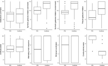
PWS patients and ID participants showed similar distributions in sociodemographic characteristics (Table 1). Figure 1 shows boxplots of task scores for PWS and ID controls. Descriptive statistics and between-group comparisons for all the measures are presented in Table 3. The two groups performed similarly for pantomimes to verbal order, buccofacial praxis, and motor sequencing. In contrast, participants with PWS showed significantly lower scores than their counterparts on imitation of communicative gestures with both the dominant and nondominant hand. They were also worse on the imitation of bimanual pseudo-gestures. On the other hand, participants with PWS were more accurate in the constructive praxis task than control participants (the median score for the former group was actually the maximum possible score on that task). They also used less time to copy the shapes (higher scores on constructive praxis time mean that less time is taken to complete the task). As shown in Table 3, no significant between-group differences were found in the tests for memory, verbal comprehension-abstraction, and executive functioning. However, subjects with PWS scored significantly higher than the control group on the visual discrimination task, showing a ceiling effect.
Boxplots of praxis task scores for the PWS and the ID control group. The median activity is shown by a horizontal bar; the box denotes the upper and lower quartiles. The vertical lines show the full range of the data set.

Scores of the tasks by group

Summary statistics are expressed as means (M) and standard deviations (SD).
In the PWS group, no significant association was found between age or BMI and any of the praxis scores (all p > .335). Likewise, no significant sex differences were found in the performance of the tasks. A significant positive relationship between IQ and scores on imitation of meaningless bimanual hand postures (r = .468 p = .028) and constructional praxis (r = .654 p = .001) were identified.
In order to study the effect of the genetic subtype, PWS patients were split into two groups: DEL (n = 20) and UPD_IC (n = 10). Groups did not differ in age, sex, or BMI. No significant differences in neuropsychological performance were observed between the two groups (data are not shown).
IMAGING RESULTS
Associations of Defective Praxis Performance and Brain Structural Measurements in PWS
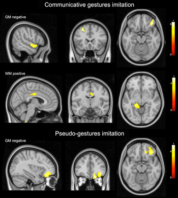
In the whole-brain linear regression analysis with regional volume measurements, the following results were observed: imitation of communicative gestures was negatively associated (i.e., poorer performance, greater tissue volume) with gray matter in the superior frontal, lateral orbital, and superior temporal cortices; similarly, imitation of bimanual pseudo-gestures (meaningless hand postures) was negatively associated with gray matter in the orbitofrontal cortex. In addition, performance in the imitation of communicative gestures, with both hands, was positively (i.e., poorer performance, lower tissue volume) associated with decreased regional white matter volumes involving the left parahippocampal region, and mid-to-posterior cingulate region (Figure 2 and Table 4). No one specific regional white matter volume was significantly related to scores in the imitation of bimanual pseudo-gestures.
Correlation analysis results for deficient praxis tasks. Top and middle row: negative correlation of scores in imitation of symbolic communicative gestures with gray matter (GM) volumes in the temporal, superior frontal, and orbitofrontal cortices and positive correlation with white matter (WM) volume in the parahippocampal region. Bottom row: negative correlation of the scores in the imitation of pseudo-gestures with GM volume in the orbitofrontal cortex. The right hemisphere corresponds to the right side of axial and coronal views. The sagittal views correspond to the right side.

Correlation (linear regression) between regional brain volumes and praxis scores

x y z, coordinates (mm) are given in Montreal Neurological Institute (MNI) space. Statistics at corrected threshold PFWE < 0.05 are estimated using Monte Carlo simulations.
Volume Measurements and Whole-Brain Between-Group Differences
To examine whether these correlated gray and white matter regions exhibited significant abnormalities, global and regional volumetric differences across PWS and a control group of sex- and age-matched healthy subjects were tested using VBM analyses. Additional comparisons were conducted with a BMI-matched control group.
Global brain volumes
In global terms, the PWS group showed significantly reduced mean gray and white matter volumes compared to the healthy (gray matter: PWS mean ± SD: 695 ± 60 ml; controls: 766 ± 60 ml, t = 4.3 p < .0001; white matter: PWS = 375 ± 32 ml; controls = 445 ± 50 ml, t = 5.9 p < .0001) and the BMI-matched control groups (gray matter: mean ± SD: 739 ± 72 ml, t = 2.3 p < .024; white matter: 433 ± 52 ml, t = 5.0 p < .0001). No significant between-group difference was found for CSF spaces.
Regional gray matter volume
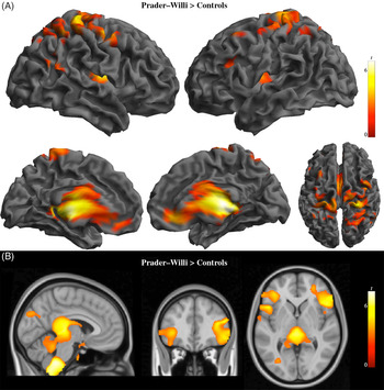
Relative gray matter increases (after controlling for global brain volume) were identified in PWS patients compared to healthy control subjects in dorsal pre- and postcentral cortices bilaterally, premotor areas, and superior parietal regions with right hemisphere predominance. In addition, increases were identified in a wide subcortical region encompassing the upper mesencephalon, bilateral thalamus, and basal ganglia extending to the posterior insula, and rostrally into (subgenual) cingulate and ventromedial prefrontal regions (Figure 3A and Table S1). Conversely, PWS patients showed a relative reduction in gray matter volumes in a small portion of the left caudate nucleus and cerebellum. When compared to BMI-matched controls, the relative increase of gray matter volume in PWS patients was more evident for superior parietal regions than around the central sulcus, while similar results were obtained for subcortical findings (Figure S1 and Table S2).
Regional gray and white matter volume change in patients with PWS as compared to age- and sex-matched control subjects superimposed on three-dimensional (3D) renderings (right and left lateral, medial and top views) in (A) and orthogonal displays (sagittal, coronal, and axial views) in (B). (A) Relative gray matter volume increases. (B) Relative white matter volume reductions. Color bar represents t value. Right side of the figure corresponds to the right hemisphere for coronal and axial views.

Regional white matter volume
Significant relative reductions in white matter volume in patients compared to healthy controls were identified in the brainstem, upper mesencephalon, and posterior aspects of the thalamus, cerebellar vermis, and the splenium of the corpus callosum, as well as in the post-central gyrus and frontal opercular region bilaterally (Figure 3B and Table S1). By contrast, PWS patients showed a relative increase in white matter volume in the right putamen region involving the ventral aspect of the external capsule. Again, similar results were observed in the comparison with the BMI-matched control group (Figure S1 and Table S2).
On the whole, brain regions found in the correlation analysis with praxis scores in PWS did not overlap with regions showing significant structural abnormalities in the current study.
DISCUSSION
The present study investigated, for the first time, praxis performance in PWS, and the relationship between praxis dysfunction and brain structural measurements. The assessment included production and imitation of gestures with testing in upper limbs and oral muscles, motor sequencing, and constructional praxis. Overall, results support specific difficulties in praxis performance in PWS participants. The main finding was a heterogeneous praxic pattern characterized by preserved visuo-constructive praxis and lower performance than the ID-matched control group in tasks involving imitation of gestures (meaningful and meaningless). No differences were found in praxis performance when comparing across PWS genetic subtypes. A significant association of praxis deficits with gray matter volume increases was identified within notably distributed brain areas generally relevant to the cognitive domain of praxis. Yet, these areas did not overlap with volumetric changes found in the comparison with control subjects.
PWS individuals were compared to a control group made up of subjects with equivalent ID level and acquired curricular competence; this comparison reveals that PWS’ praxis deficits are not simply the result of their ID. In our study, both groups had the greatest difficulty with performing gestures to command and performed better with imitation, possibly indicating that subjects had more difficulty with a verbal mode of presentation of the stimulus than a visual mode, as has been shown in other adult populations with ID (Elliot, Weeks, & Gray, Reference Elliot, Weeks and Gray1990; Williams, Whiten, & Singh, Reference Williams, Whiten and Singh2004; Zoia, Pelamatti, & Rumiati, Reference Zoia, Pelamatti and Rumiati2004). Also, both groups showed poorer performance in the imitation of pseudo-gestures than in the imitation of communicative gestures. This result could be related to the fact that, as opposed to imitating nonmeaningful gestures for which no conceptual information was available to participants, imitation of meaningful gestures seems to be mediated by implicit knowledge about the form and meaning of the gesture, which may facilitate its performance (Leiguarda & Marsden, Reference Leiguarda and Marsden2000).
In accordance with our first hypothesis, results revealed that individuals with PWS still showed significantly worse scores than the ID-matched control group on some of the praxis tasks investigated. In particular, a specific difficulty in limb praxis was identified for the first time in PWS with imitation of communicative gestures and of meaningless hand postures in comparison with the control group. However, not all praxis tasks were affected. In the pantomime task, the participants perform the mime of object use as if they are holding the object in their hand (e.g., show me how you would paint a wall with a brush). This gestural description of object use necessarily implicates knowledge about the actions associated with usual objects and tools and the sequence of movements that are appropriate for their use. In our study, this knowledge of tools and objects in terms of their function appeared to be at the expected level according to their ID since the performance in the pantomime was comparable with that of controls. Similarly, the PWS group did not differ from the control group in the more basic motor task involving bimanual coordination nor in the imitation of nonlinguistic oral movements (e.g., blowing, whistling) in contrast to previous findings comparing PWS individuals to healthy non-ID controls (Saeves et al., Reference Saeves, Asten, Sorthaug and Bágesund2011). Moreover, participants with PWS were more accurate and used less time in the constructive praxis in a copying task, and outperformed control subjects in the visual discrimination task when required to identify objects in an overlapping figures configuration, which corresponds to the relative strength in visuospatial abilities previously described in the literature (Curfs et al., Reference Curfs, Wiegers, Sommers, Borghgraef and Fryns1991; Dykens, Reference Dykens2002). Finally, no differences were obtained in Luria’s three-step test, a visuomotor and executive task that requires processing information in a step-by-step manner, which has been previously reported as a specific weakness in individuals with PWS when compared to non-ID controls (Dykens et al.,Reference Dykens, Hodapp, Walsh and Nash1992; Jauregui et al., Reference Jauregui, Arias, Vegas, Alén, Martínez, Copet and Thuilleaux2007).
Within the PWS group, significant and distinct associations between tissue volume and defective praxis performance were identified, such that poor performance in the imitation of limb gestures was associated with a relative increase in gray matter volume in small areas in frontal and temporal cortices. Volumetric differences across PWS and a group of sex- and age-matched healthy subjects were tested. In global terms, significantly reduced brain volumes of gray and white matter were identified in the PWS group, while no difference was found for CSF volumes, which is in accordance with previous reports in young adults and children with PWS (Honea et al., Reference Honea, Holsen, Lepping, Perea, Butler, Brooks and Savage2012; Lukoshe, White, Schmidt, van der Lugt, & Hokken-Koelega, Reference Lukoshe, White, Schmidt, van der Lugt and Hokken-Koelega2013; Ogura et al., Reference Ogura, Fujii, Abe, Hosokai, Shinohara, Takahashi and Mori2011). At a regional level, the PWS group showed a combination of increases and decreases of tissue volume in large regions that are partially coincident with previous results. For instance, our finding of increased gray matter in sensory and motor areas, thalamus, and elements of the basal ganglia is in agreement with results from a recent study in young adults with PWS (Manning, Tait, Suckling, & Holland, Reference Manning, Tait, Suckling and Holland2017), which additionally showed an increased cortical thickness in these same areas. Likewise, smaller white matter volume in the brainstem, thalamus, cerebellum, and inferior frontal cortex largely coincides with results from previous VBM studies (Honea et al., Reference Honea, Holsen, Lepping, Perea, Butler, Brooks and Savage2012; Lukoshe et al., Reference Lukoshe, White, Schmidt, van der Lugt and Hokken-Koelega2013).
Taken together, our anatomic results suggest various possible mechanisms that may underlie our current observation of a deficit in gesture imitation in PWS. First, defective imitation could stem from difficulties in the actual implementation of the action due to alterations in brain (sensori)motor regions. Areas of abnormal tissue volume in our study (e.g., primary motor cortices, premotor areas, thalamus and basal ganglia, brainstem, cerebellum) largely coincide with important elements in the motor system. Functional imaging in PWS has also reported alterations in several of the motor regions showing structural abnormality, including decreased functional connectivity strength in the pre-/postcentral gyri (Zhang et al., Reference Zhang, Zhao, Qiu, Tian, Wen, Miller and Liu2013) and decreased metabolism in the thalamus and cerebellum (Ogura et al., Reference Ogura, Fujii, Abe, Hosokai, Shinohara, Fukuda and Mori2013) during the resting state. Our structural findings of gray matter differences partially overlap with our results from a previous study in which PWS participants exhibited a relevant increase in functional connectivity between the primary sensorimotor cortices and the putamen that correlated with the presence of self-picking behavior (Pujol et al., Reference Pujol, Blanco-Hinojo, Esteba-Castillo, Caixàs, Harrison, Bueno and Novell-Alsina2016). The structural findings are also consistent with reports of motor cortex dysfunction studied using transcranial magnetic stimulation (Civardi, Vicentini, Grugni, & Cantello, Reference Civardi, Vicentini, Grugni and Cantello2004). Nevertheless, no group differences were observed in the motor sequencing or the pantomime tasks, and individuals with PWS demonstrated excellent performance in the constructional praxis task, for which fine motor coordination was required, suggesting that basic sensory and motor disturbances alone may not account for the observed dyspraxic pattern.
Beyond sensorimotor functions, gesture imitation is also associated with a social cognition component. Deficits in imitation have been described in other neurodevelopmental disorders such as autism spectrum disorders (ASD; Vanvuchelen, Van Schuerbeeck, Roeyers, & De Weerdt, Reference Vanvuchelen, Van Schuerbeeck, Roeyers and De Weerdt2013; Williams et al., Reference Williams, Whiten and Singh2004), which are characterized by impairment in social interactions (American Psychiatric Association, 2013). Relevant to imitative and social behavior, the mirror neuron system seems to facilitate social interactions by providing an understanding of the actions of others (Catanneo & Rizzolati, Reference Cattaneo and Rizzolatti2009; Iacoboni & Mazziotta, Reference Iacoboni and Mazziota2007). The pars opercularis of the inferior frontal gyrus, considered to contain mirror neurons (Kilner et al., Reference Kilner, Neal, Wieskopf, Friston and Frith2009), appears to be particularly concerned with coding the intention associated with the observed action (Cattaneo & Rizzolati, Reference Cattaneo and Rizzolatti2009; Iacoboni & Mazziotta, Reference Iacoboni and Mazziota2007). Functional imaging studies in healthy subjects also report activation of this region in the observation and imitation of faces with emotional expressions (Caspers et al., Reference Caspers, Zilles, Laird and Eickhoff2010). Individuals with PWS show deficits in interpreting social information and recognizing emotional facial expressions (Dykens et al., Reference Dykens, Roof, Hunt-Hawkins, Daniell and Jurgensmeyer2019; Koenig et al., Reference Koenig, Klin and Schultz2004; Whittington & Holland, Reference Whittington and Holland2011). Neuroimaging and EEG studies provide evidence for mirror neuron dysfunction in subjects with ASD (Perkins, Stokes, McGillivray, & Bittar, Reference Perkins, Stokes, McGillivray and Bittar2010). Contrary to our hypothesis, we found no evidence for a specific relationship between praxis disability and the structural changes in the inferior frontal gyrus of patients with PWS. This could be due to a lack of statistical power using only 23 patients in these analyses. Nevertheless, abnormal tissue volume was actually identified in this area coinciding with previous findings (Honea et al., Reference Honea, Holsen, Lepping, Perea, Butler, Brooks and Savage2012; Lukoshe et al., Reference Lukoshe, White, Schmidt, van der Lugt and Hokken-Koelega2013). Abnormalities noted in this area may be also involved in the performance of praxis in PWS via deficits in mirror neuron function as they have been proposed to be in ASD (Iacoboni & Dapretto, Reference Iacoboni and Dapretto2006; Yang & Hofmann, Reference Yang and Hofmann2015), although direct measures of differences in action recognition may be needed to confirm this possibility.
Lastly, in contrast to acquired apraxia, in which there’s a loss of previously acquired skilled actions, usually with an identifiable brain lesion associated with the praxis deficit, in the presence of developmental disorders, the term developmental dyspraxia is used to represent a failure to acquire motor skills in a normal fashion (Dewey, Reference Dewey1995; Steinman, Mostofsky, & Denckla, Reference Steinman, Mostofsky and Denckla2010). Specific impairments in gesture imitation have been described in a number of neurodevelopmental disorders (Vanvuchelen et al., Reference Vanvuchelen, Van Schuerbeeck, Roeyers and De Weerdt2013; Williams et al., Reference Williams, Whiten and Singh2004; Zoia et al., Reference Zoia, Pelamatti and Rumiati2004) and in progressive diseases like Alzheimer’s disease in the absence of a focal brain lesion (Rousseaux, Rénier, Anicet, Pasquier, & Mackowiak-Cordoliani, Reference Rousseaux, Rénier, Anicet, Pasquier and Mackowiak-Cordoliani2012; Sanin & Benke, Reference Sanin and Benke2017), but the relationship between impaired gestural performance and brain abnormalities in these conditions has not yet been well established (Dewey, Reference Dewey1993; Steinman et al., Reference Steinman, Mostofsky and Denckla2010). In our study, structural correlates of deficient praxis performance did not appear to be abnormal in quantitative terms, as both groups showed similar gray and white matter volumes in the regions showing a significant association with praxis scores (i.e., left superior frontal cortex, right temporal and orbitofrontal cortex, and left parahippocampal region). It is also possible that the dyspraxic pattern observed in our PWS group could be attributed to diffuse and distributed brain structural abnormalities as has been previously suggested in other populations showing developmental intellectual deficits (Dewey, Reference Dewey1993). In this case, dyspraxic features in PWS might be considered as one sign among several other neuropsychological deficits due to the basis of widespread neuroanatomic anomalies.
The authors consider these findings relevant from the interventional point of view. Many adaptive skills need to be performed in the everyday’s life of a patient with PWS. It should also be taken into account that people with PWS will be trained to carry out practical and manipulative tasks in employment or occupational centers. In all of them, work tasks imply the constant use of those altered praxic components. As clinicians, we need to ensure that adaptive changes in the environment will be taken into consideration to enable them to perform better on their day-to-day tasks. On the other hand, we should start working with neuropsychological training techniques in the early stages of life when the patient is still a child.
A limitation of our study could be the small number of patients included, especially when we split for genetic subtypes, which might prevent us from drawing general conclusions. Other potential confounding factors associated with PWS, either directly or through the clinical management of its features, such as BMI or IQ were taken into account and were well matched. However, the use of GH (during childhood) and sexual hormones treatment may have influenced brain development. Lastly, although authors think it’s quite improbable, the use of different psychotropic medication may have affected the performance, however, less than one-third of patients were under this kind of drugs, all of them with antidepressants (selective serotonin reuptake inhibitors) and/or topiramate.
In summary, our results revealed how praxis is a domain that is specifically compromised in people with PWS. We found that PWS participants show poor gesture imitation abilities evident in the comparison with an ID-matched group, whereas praxis related to visuospatial capacities appear relatively preserved. The fact that not all praxic tasks were affected suggests that common features in the PWS phenotype, such as hypotonia, or other basic sensory or motor disturbances may not account for the dyspraxic pattern. Large regional gray and white matter abnormalities were found in the PWS group compared with a group of healthy subjects. However, structural correlates of deficient praxis performance did not overlap with volumetric changes, but praxis scores correlated with regional measures in distributed apparently normal brain areas. Altered imitation gestures could, therefore, be a consequence of widespread brain dysfunction. However, the specific contribution of key brain structures (e.g., areas containing mirror neurons) should be more finely tested in future research.
SUPPLEMENTARY MATERIAL
To view supplementary material for this article, please visit https://doi.org/10.1017/S1355617721000060.
ACKNOWLEDGEMENTS
We thank the Associació Catalana de Síndrome de Prader-Willi and all the patients and their families for kindly agreeing to participate.
CONFLICT OF INTEREST
The authors have nothing to disclose.
FINANCIAL SUPPORT
This project was supported by three grants from Fondo de Investigación Sanitaria del Instituto Carlos III and Fondo Europeo de Desarrollo Regional (PI-10/00940, PI-14/02057, and PI-18/0734) and by three grants from Beques Taulí de Recerca i Innovació 2010/006, 2011/004, and 2014/002 Institut d’Investigació I Innovació Parc Taulí (I3PT) and the grant Intensifica’t al Taulí from I3PT_Banc Sabadell 2016–17, all to Assumpta Caixàs.
ETHICS STATEMENT
All participants or their parents/guardians provided written informed consent and all PWS and ID patients who were not able to sign gave drawn assent. The Institutional Ethics Committee of Consorci Corporació Sanitària Parc Taulí approved the protocol, and all investigations complied with the Helsinki Declaration and the Good Clinical Practices.