Introduction
Obesity and unhealthy nutrition are increasing even in women of childbearing age. Reference Hedley, Ogden and Johnson1 Maternal obesity and malnutrition during gestation and lactation are well recognized to increase the risk of obesity and metabolic disorders in the offspring across the lifespan. Reference Drake and Reynolds2–Reference Hochner, Friedlander and Calderon-Margalit4 Feeding a dam with a high-fat diet (HFD) is the most common method to induce maternal obesity and overnutrition in rodents. Reference Kretschmer, Schulze-Edinghausen and Turnwald5,Reference Song, Chen and Gao6 However, the gestational period during which maternal HFD consumption has a decisive effect on offspring metabolism is not known yet.
Adipose tissues are classified as white adipose tissue (WAT) and brown adipose tissue (BAT). WAT stores energy, whereas BAT consumes energy as heat. Owing to its high efficiency in oxidizing glucose and fatty acids and the recent discovery of its abundance in adult humans, BAT plays a critical role in energy metabolism. The fetal and neonatal stages are critical for BAT development Reference Zhang, Xiao and Zheng7 and are considered to exert long-term effects on BAT function in the offspring. Reference Yu, Lv and Yan8 The objective of this study was to determine the impact of maternal HFD consumption only during pregnancy on BAT and energy metabolism in offspring mice.
Materials and methods
Study 1: Effect of HFD on maternal and fetal parameters
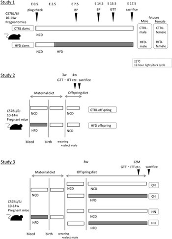
As outlined in Fig. 1, pregnant C57BL/6J mice (10–14 weeks old) were purchased from CLEA Japan (Tokyo, Japan) at embryonic day 1.5 (E1.5) and kept in a humidity-controlled room with a 12/12-h light–dark cycle. Food and water were provided ad libitum. Dams were matched by weight and divided into two diet groups at E2.5. The dams were fed either normal chow diet (NCD) (3.57 kcal/g with 14% of total calories as fat and 25% as protein; Charles River CRF-1, Oriental Yeast Japan, Tokyo, Japan) during the entire pregnancy (CTRL dams) or HFD (5.08 kcal/g with 57% of total calories as fat and 20% as protein, containing high oleic acid; Clea High Fat Diet 32, CLEA Japan, Tokyo, Japan) from E2.5 till euthanasia (HFD dams). NCD contained 55.3% carbohydrates, 21.9% protein, and 5.4% fat. The carbohydrates in NCD were mainly derived from corn and wheat; the proteins were derived from soybeans, fish, and skim milk; and the fats were derived from soybean oil. HFD contained 29.4% carbohydrates, 25.5% protein, and 32% fat. The sources of carbohydrates in HFD were maltodextrin, lactose, and sucrose; proteins were derived from milk casein and egg whites; and fats were derived from beef tallow and high oleic acid type safflower oil.
Experimental paradigm for maternal and offspring diets in mice. HFD dams were fed with HFD from E2.5 to E18.5. CN/HN groups were fed with NCD until sacrifice. CH/HH groups were fed with HFD from 8 weeks of age until sacrifice. NCD is indicated by white bars, and HFD is indicated by gray bars.

The dams were individually housed during gestation, and their body weight and food intake were recorded. The food weight of each cage was measured every two or three days, and the daily food intake was calculated from the amount of food remaining in the cage. The daily intake of each nutrient was calculated from the carbohydrate, protein, and fat contents of the NCD or HFD. The intake from E2.5 to E14.5, during which HFD feeding was initiated, was integrated for each mouse and statistically analyzed between groups. Furthermore, the mice underwent a glucose tolerance test (GTT) at E15.5 and blood pressure measurement at E7.5 and E14.5, as described below, and were euthanized at E17.5. The weights of the fetus and placenta were recorded separately. The sex of fetuses was confirmed using Sry genotyping.
Study 2: Effect of intrauterine HFD exposure on offspring at 4 weeks of age
Another set of dams, as described in Study 1, was prepared. The HFD dams were switched to NCD at E18.5 and were allowed to deliver on E19.5. The body weights of all offspring were measured on the day of birth. On postnatal day 1 (PND1), litter sizes were balanced to 6–8 pups per dam. Offspring were weaned on a NCD on PND21. To avoid confounding effects of sex, only male offspring were used in Study 2. Offspring body weight and food intake were recorded weekly. Glucose metabolism, thermogenesis, and energy expenditure were assessed at 4 weeks of age, as described later. Thereafter, the offspring were euthanized. Subcutaneous and visceral WAT and interscapular BAT were collected and stored at −80 °C until further use.
Study 3: Effect of intrauterine HFD exposure on offspring at 12 months of age
To investigate the effect of intrauterine HFD exposure and feeding with HFD in adults, another set of dams and offspring was prepared as described in Studies 1 and 2. At 8 weeks of age, male offspring born from the CTRL dams were randomly assigned to NCD (CN) or HFD (CH), and male offspring from HFD dams were randomly assigned to NCD (HN) or HFD (HH) for 10 months. Body weight was recorded weekly until 12 weeks of life and then monthly until the end of the study. The offspring were allowed to grow until 12 months of age and then assessed for glucose metabolism, thermogenesis, and energy expenditure. Four groups of offspring were euthanized at 12 months of age.
All animal experiments were conducted in accordance with the guidelines of the Animal Care and Use Committee of the Kyoto Prefectural University of Medicine.
Blood pressure measurements
Systolic and diastolic blood pressure of dams were measured on at E7.5 and E14.5, using a computerized tail-cuff system (BP-98A system; Softron Co., Tokyo, Japan). Briefly, the mice were placed in a holder with a built-in temperature controller. The blood pressure was measured multiple times with a minimum of three valid measurements recorded for each mouse. The average of three recordings was calculated for each measurement time.
Glucose and insulin tolerance tests
For the GTT, the pregnant mice and offspring were fasted for 6 and 16 h overnight, respectively. Blood glucose levels at different time points (0, 30, 60, and 120 min) were measured using a glucometer (Glutest Neo alpha, SANWA KAGAKU KENKYUSYO, Nagoya, Japan) before and after intraperitoneal (IP) administration of glucose (0.5 g/kg body weight). The insulin tolerance test (ITT) was conducted after fasting for 4 h. Blood glucose was measured before (0 min) and after (15, 30, 60, 90, and 120 min) IP injections of insulin (0.75 U/kg). Blood glucose levels during GTT and ITT were determined using tail blood. Blood samples were collected after fasting to measure fasting blood glucose and insulin levels using a commercial enzyme-linked immunosorbent assay kit (Ultra Sensitive Mouse Insulin ELISA Kit, Morinaga Institute of Biological Science, Japan). The area under the curve (AUC) was calculated during GTT and ITT.
Oxygen consumption measurement
Oxygen consumption (VO2) was measured using a O2/CO2 metabolism measuring system (MK-5000; Muromachikikai, Tokyo, Japan). Briefly, mice were placed in plastic chambers at 22 °C on a 12/12-h light-dark cycle with ad libitum access to food and water. This experiment was performed on 7 mice each in the CTRL and HFD groups at 4 weeks of age and on 10 mice each in the CN, HN, CH, and HH groups at 12 months of age.
Determination of body temperature and cold exposure
Body temperature was measured using a rectal temperature probe. For experiments of cold-induced thermogenesis, mice were kept individually at an ambient temperature of 4 °C for 4 h.
Histology
Hematoxylin-eosin staining was performed for BAT and placenta as previously described. Reference Kawabe, Mori and Morimoto9 All images were captured using an all-in-one fluorescence microscope BZ-X710 (Keyence, Osaka, Japan). The area of lipid droplets in BAT cells was measured using the BZ Analyzer software (Keyence).
Quantitative real-time PCR
Quantitative real-time PCR was performed, as previously described. Reference Tsuma, Mori and Ota10 The expression of the target genes (Table S1) was examined using TB Green Premix Ex Taq II (Tli RNaseH Plus; Takara, Shiga, Japan) on an AB 7500 Real-Time PCR System (Applied Biosystems, Tokyo, Japan). Beta-actin was used as an internal standard.
Western blotting analysis
Western blotting analysis was performed, as previously described. Reference Morimoto, Mori and Nakajima11 The following antibodies were used: anti-uncoupling protein1 (UCP1, ab10983; Abcam, Tokyo, Japan), anti-total hormone-sensitive lipase (HSL; no. 4107, Cell Signaling Technology, Tokyo, Japan), anti-phospho-HSL (Ser660; no. 4126, Cell Signaling Technology), anti-adipose triglyceride lipase (ATGL; no.2138, Cell Signaling Technology), anti-perilipin (no. 9349, Cell Signaling Technology), proliferator-activated receptor-γcoactivator-1α (PGC-1α, ab54481; Abcam), or anti-β-actin (no. 3700, Cell Signaling Technology).
GC/MS analysis of placental fatty acid composition
The fatty acid composition of the placenta was analyzed using gas chromatography–mass spectrometry (GC/MS) on an Agilent 7890B/7000D System (Agilent Technologies, Santa Clara, CA, USA). The tissue sample (15 mg) was methylated using a fatty acid methylation kit (Nacalai Tesque, Kyoto, Japan). Each fatty acid methyl ester was detected in the selected ion monitoring mode. All results were normalized to the peak height of the C17:0 internal standard.
Statistical analysis
All data are expressed as the mean ± standard error of the mean (SEM) and were analyzed using the Student’s t-test or analysis of variance (two-way ANOVA), where appropriate, using GraphPad Prism version 9.0 (GraphPad Prism Software, San Diego, CA, USA). For comparisons between two groups, Student’s t-test was performed with the CTRL group as the control. In comparisons between four groups, the statistical analysis was performed with two-way ANOVA, using maternal diet and postnatal diet as variables. In GTT, ITT, and cold induced test, statistical analysis was conducted using two-way ANOVA for the CTRL and HFD groups, the CN and HN groups, and the CH and HH groups. Differences were considered statistically significant at P ≤ 0.05.
Results
Study 1: Effect of HFD on body weight and nutrition intake in pregnant mice
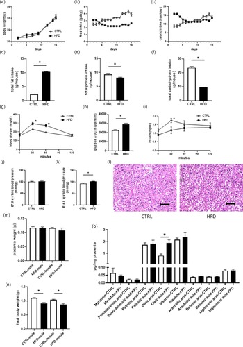
The CTRL and HFD dams showed similar body weight gain during pregnancy (Fig. 2a). The daily food intake of the HFD dams was lower than that of the CTRL dams (Fig. 2b); however, their energy intake did not differ (Fig. 2c). In contrast, the fat intake of the HFD dams was significantly higher than that of the CTRL dams (p < 0.0001; Fig. 2 d). The total fat intake of the HFD dams was 4.5 times higher than that of the CTRL dams. Moreover, the protein (p = 0.0016; Fig. 2e) and carbohydrate (p < 0.0001; Fig. 2f) intake of the HFD dams were significantly lower than those of the CTRL dams.
Effect of HFD on body weight and metabolism in pregnant mice and fetuses. a: Change in the body weight of CTRL or HFD dams during pregnancy (n = 12–14). b: Time course of food intake per day (n = 12–14). c: Time course of calorie intake per day (n = 12–14). d: Total fat intake per mouse (n = 12–14). e: Total protein intake per mouse (n = 12–14). f: Total carbohydrate intake per mouse (n = 12–14). i and h: Levels and area under the curve (AUC) of blood glucose during IP glucose tolerance test (IPGTT, n = 12–13). i: Levels of serum insulin during IPGTT (n = 4–5). j and k: Systolic blood pressure in dams at E7.5 and E14.5 (n = 8–9). l: Representative histological images of placenta. Scale bar, 100 µm. m: Placental weight of male and female fetuses exposed to NCD or HFD group (n = 9–17). n: Weight of male and female fetuses from CTRL or HFD dams (n = 9–17). o: Analysis of fatty acids with GC/MS in placenta (n = 5–6). CTRL, control; HFD, high-fat-diet; NCD, normal chow diet. Values are shown as the mean ± SE. *p < 0.05

HFD during pregnancy induces glucose intolerance and hypertension in dams
To determine whether HFD induces glucose intolerance in pregnant mice, we performed GTT in CTRL and HFD dams at E15.5. Basal glucose levels were not significantly different in both groups. However, the blood glucose levels at 30 and 60 min after glucose administration (p = 0.002 and p = 0.0008; Fig. 2g) and glucose AUC during the GTT (p = 0.0003; Fig. 2h) were significantly higher in the HFD dams than in the CTRL dams. The serum insulin levels in the HFD dams at 30 min after glucose administration were lower than those in the NCD dams at the same timepoint (p = 0.0369; Fig. 2i). The systolic blood pressure was not significantly different between CTRL and HFD dams at E7.5 (Fig. 2j). However, the systolic blood pressure of the HFD dams was significantly higher than that of the CTRL dams at E14.5 (p = 0.0162; Fig. 2k).
HFD during pregnancy lowers fetal weight without affecting placental weight
There was no histological difference between the placenta of CTRL and HFD dams (Fig. 2 l). Placental weight was not different between male and female fetuses in CTRL dams, and HFD did not affect placental weight in either male or female fetuses (Fig. 2m). Fetal weights of mice born to HFD dams were significantly lower than those born to CTRL dams, irrespective of sex (male, p < 0.0001; female, p = 0.0001; Fig. 2n). Maternal HFD did not affect placental weight but affected fetal weight. The amount of oleic acid was significantly higher in the placenta of HFD dams than in the placenta of CTRL dams (p < 0.0001; Fig. 2o).
Study 2: Effect of intrauterine HFD exposure on offspring at 4 weeks of age
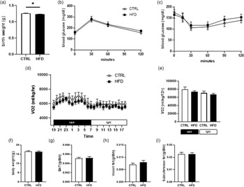
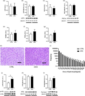
On the day of birth, the body weight of offspring born to HFD dams was significantly lower than that of offspring born to CTRL dams (p = 0.0087; Fig. 3a). At 2 weeks of age, offspring born to HFD dams reached a body weight similar to that of offspring born to CTRL dams. At 4 weeks of age, glucose intolerance and insulin sensitivity were similar between the groups (Fig. 3b, c). The oxygen consumption of the two groups in both the dark and light phases was not significantly different (Fig. 3d, e). There was no significant difference in the body weight (Fig. 3f), BAT (Fig. 3g), visceral fat (Fig. 3h), and subcutaneous fat weight (Fig. 3i) between the male offspring of the CTRL and HFD dams. We analyzed the mRNA and protein levels of the differentiation markers of BAT. The mRNA and protein expression levels of both UCP1 (Fig. 4a, b) and PGC1α (Fig. 4c, d) were not different between the offspring of CTRL and HFD dams; the levels of other differentiation markers, namely PRDM16, PPARγ, and Cidea, were also not different (Fig. 4e-g). The lipid droplet size in BAT from offspring born to HFD dams was lower than that in BAT from offspring born to CTRL dams (Fig. 4h, i), concomitant with the increased phosphorylation of hormone-sensitive lipase (HSL) (p = 0.0255; Fig. 4j) without altering ATGL (Fig. 4k) and perilipin levels (Fig. 4l).
Effect of intrauterine HFD exposure on offspring at birth and 4 weeks of age. a: Body weight of offspring at birth (n = 81–99). b: Levels of blood glucose during IP glucose tolerance test (IPGTT, n = 10). c: Levels of blood glucose during IPITT (n = 9–11). d: 24 h oxygen consumption (n = 7). e: Total amount of oxygen consumption for 12 h dark or light phase (n = 7). f: Body weight of male offspring at 4 weeks of age (n = 8). g: Weight of brown adipose tissue in male offspring at 4 weeks of age (n = 8). h and i: Weight of visceral fat and subcutaneous fat in male offspring at 4 weeks of age (n = 8). Values are shown as the mean ± SE. * p < 0.05. HFD, high-fat diet

Effect of intrauterine HFD exposure on differentiation and lipolysis in interscapular brown adipose tissue (BAT) on offspring at 4 weeks of age. a: mRNA expression of UCP1 (n = 5–6). b: Protein expression of UCP1 (n = 6). c: mRNA expression of PGC1α (n = 5–6). d: protein expression of PGC1α (n = 6). e: mRNA expression of PRDM16 (n = 6). f: mRNA expression of PPARγ (n = 6). g: mRNA expression of Cidea (n = 6). h: representative histological images of BAT. Scale bar, 100 µm. i: Frequency distribution of lipid droplet in BAT (n = 6). j: Phosphorylation of HSL (n = 6). k: Protein expression of ATGL (n = 6). l: Protein expression of perilipin (n = 6). Values are shown as the mean ± SE. *p < 0.05

Study 3: Effect of intrauterine HFD exposure on offspring at 12 months of age
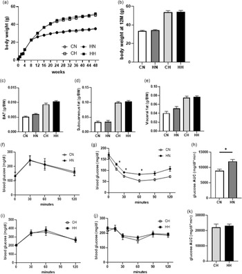
The body weight at 12 months of age was similar between the CN and HN groups (Fig. 5a, b). The body weight of mice fed HFD was significantly higher than that of mice fed NCD; however, there was no difference in the body weights of the CH and HH groups (Fig. 5a, b). BAT, visceral, and subcutaneous adipose tissue from CN and HN mice had similar weights (Fig. 5c-e). There was no difference in those from CH and HH mice (Fig. 5c-e). The GTT results demonstrated that blood glucose levels in 12-month-old HN mice were comparable to those in CN mice (Fig. 5f). Twelve-month-old HN mice had a reduced response to insulin compared with CN mice, indicating that 12-month-old HN mice had reduced insulin sensitivity (15 min p = 0.0384, 30 min p = 0.0126, 60 min p = 0.0068, AUC p = 0.0011; Fig. 5g, h). However, there were no differences in the GTT and ITT results between the CH and HH groups (Fig. 5i-k).
Effect of intrauterine HFD exposure on offspring at 12 months of age. a: Body weight change in offspring fed NCD or HFD born to CTRL or HFD dams (n = 13–14). b: Body weight of offspring at 12 months of age (n = 11–12). c: Weight of brown adipose tissue in male offspring at 12 months of age (n = 11–12). d and e: Weight of visceral fat and subcutaneous fat in male offspring at 12 months of age (n = 11–12). f: Levels of blood glucose during IP glucose tolerance test (IPGTT) on NCD-fed mice born to CTRL or HFD dams (n = 10). g and h: Levels and area under the curve (AUC) of blood glucose during IPITT on NCD-fed offspring born to CTRL or HFD dams (n = 9–11). i: Levels of blood glucose during IPGTT on HFD-fed offspring born to CTRL or HFD dams (n = 10). j and k: Levels and AUC of blood glucose during IPITT on HFD-fed mice born to CTRL or HFD dams (n = 10–11). Values are shown as the mean ± SE. *p < 0.05

HFD after birth exacerbates oxygen consumption and cold-induced thermogenesis at 12 months of age
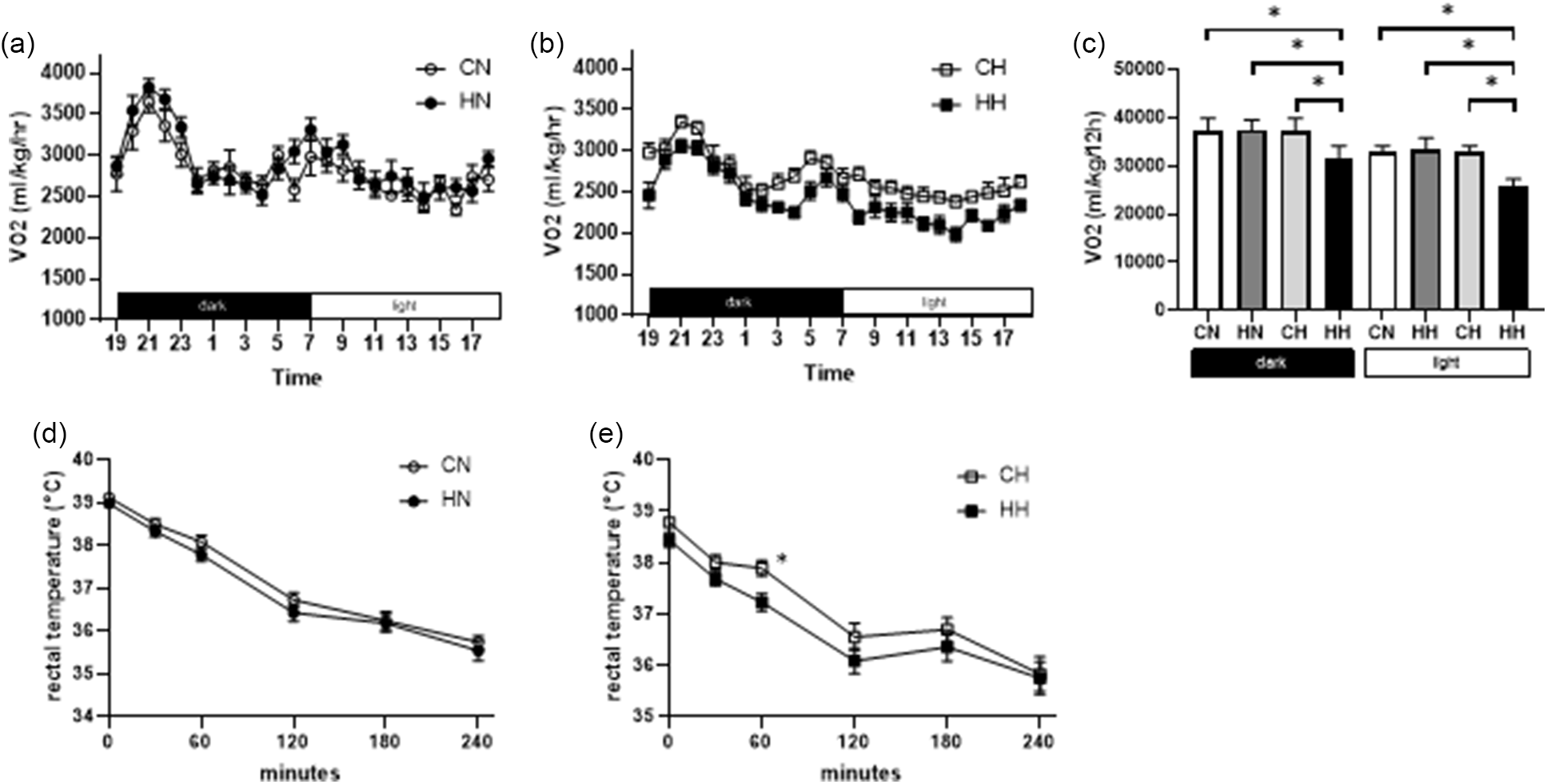
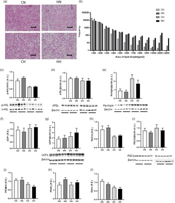
Oxygen consumption of HH mice in both dark and light phases was significantly lower than that of CN, HN, and CH mice (p < 0.0001, respectively; Fig. 6a-c). We measured cold-induced changes in body temperature in mice. The rectal temperature of the HH group was lower than that of the CH group (60 min p = 0.0491, Fig. 6d, e). The lipid droplet size in BAT from HN mice was smaller than that from CN mice (Fig. 7a, b). HFD-fed mice had increased lipid droplet size compared with that in NCD-fed mice. Furthermore, HH mice had increased lipid droplet size compared with that in CH mice (Fig. 7a, b), with no alteration in the extent of phosphorylation of HSL (Fig. 7c), ATGL (Fig. 7d), and perilipin (Fig. 7e). mRNA expression and protein levels of UCP1, PGC1α, PRDM16, PPARγ, and Cidea did not differ significantly between CN and HN and between CH and HH (Fig. 7j-l).
Effect of intrauterine HFD exposure on offspring at birth and 4 weeks of age. a: Effect of 24 h oxygen consumption on NCD-fed offspring born to CTRL or HFD dams (n = 5). b: Effect of 24 h oxygen consumption on HFD-fed offspring born to CTRL or HFD dams (n = 5). c: Total amount of oxygen consumption for 12 h dark or light phase (n = 5). d: Effect of cold-induced rectal temperature on NCD-fed offspring born to CTRL or HFD dams (n = 12). e: Effect of cold-induced rectal temperature on HFD-fed offspring born to CTRL or HFD dams (n = 10–12). Values are shown as the mean ± SE. *p < 0.05

Effect of intrauterine HFD exposure on differentiation and lipolysis in interscapular brown adipose tissue (BAT) on offspring at 12 months of age. a: Representative histological images of BAT. Scale bar, 100 µm. b: Frequency distribution of lipid droplet in BAT (n = 6). c: Phosphorylation of HSL (n = 6). d: Protein expression of ATGL (n = 6). e: Protein expression of perilipin (n = 6). f: mRNA expression of UCP1 (n = 6). g: Protein expression of UCP1 (n = 6). h: mRNA expression of PGC1α (n = 6). i: Protein expression of PGC1α (n = 6). j: mRNA expression of PRDM16 (n = 6). k: mRNA expression of PPARγ (n = 6). l: mRNA expression of Cidea (n = 6). Values are shown as the mean ± SE. *p < 0.05

Discussion
Maternal nutrition before and during pregnancy and/or lactation has been reported to affect the health of the offspring. Reference Langley-Evans12,Reference Zhang, Zhang and Didelot13 This study showed that maternal HFD consumption during pregnancy reduces offspring body weight; this finding was consistent with that reported by Zheng et al., Reference Zheng, Xiao and Zhang14 who used obese dams (mated with HFD-fed male mice) fed an HFD during pregnancy and lactation. Reference Zheng, Xiao and Zhang14 However, recent evidence indicates that maternal HFD consumption before and during pregnancy and lactation does not reduce offspring body weight. Reference Lin, Gao and Tian15 This discrepancy in the reported effects of HFD on offspring weight might be due to the difference in the timing of the HFD loading. That is, HFD consumption during pregnancy decreases offspring body weight, and HFD consumption before pregnancy increases offspring body weight. Dams fed an HFD are subjected to high-fat loading, as well as protein restriction. Maternal protein restriction is also known to result in the offspring being small-for-gestational-age (SGA). Reference Fernandez-Twinn, Ozanne and Ekizoglou16,Reference Jansson, Pettersson and Haafiz17 It is difficult to determine whether fat loading or protein restriction causes the SGA phenotype. However, maternal fat loading was associated with elevations in the induction of placental inflammation and placental insufficiency. Placental inflammation induced by the HFD exposure of dams impaired uteroplacental blood flow and reduced nutrient supply to the fetus, thus inhibiting optimal growth. Reference Reina, Katelyn and Jordan18 However, the expression of inflammation markers, such as TNFα, IL-6, CCR2, MCP-1, and TGFß, was not increased in the present study (data not shown). The concentration of oleic acid in the placenta of HFD-fed dams increased as the HFD used herein was abundant in oleic acid. The fatty acid profile is known to play a role in the SGA phenotype. Reference Gómez-Vilarrubla, Mas-Parés and Díaz19 The accumulation of fatty acids in the placenta by HFD may have influenced the offspring body weight. In this study, maternal HFD consumption during pregnancy also induced maternal hypertension and glucose intolerance. Maternal exposure to HFD impairs spiral vascular remodeling and relaxation. Consequently, HFD-exposed dams develop maternal hypertension and placental dysfunction associated with fetal growth inhibition. Reference Sato, Fudono and Imai20,Reference Emilly, Daniel and Michael21 In contrast, glucose intolerance is unlikely to be the cause of low birth weight in offspring, as intrauterine hyperglycemia is known to increase offspring fat mass. Reference Chung, Moon and Lim22
Intrauterine exposure to malnutrition or hypertension is associated with the development of obesity and metabolic disorders in adulthood. Reference Wang, Bhatta and Moen23–Reference Sellayah, Dib and Anthony25 BAT is a central organ of energy metabolism because it contributes to energy expenditure by activating nonshivering thermogenesis. BAT activity is inversely related to body mass index (BMI) and body fat percentage. Reference van Marken Lichtenbelt, Vanhommerig and Smulders26 A previous study showed that maternal HFD induced early onset obesity concomitant with lipid accumulation in BAT. Reference Almeida, Dias-Rocha and Souza27 Thus, we evaluated BAT in mice aged 4 weeks before HFD loading. Interestingly, lipid droplet size in BAT from the offspring of HFD-fed dams was smaller than that in BAT from the offspring of CTRL dams, even though oxygen consumption was not different between the offspring of HFD-fed and CTRL dams. This decreased lipid droplet size in BAT from offspring of HFD dams was accompanied by an increase in the phosphorylation of HSL, which is essential for lipolysis in BAT. Reference Morimoto, Mori and Nakajima11 The increased phosphorylation of HSL might have a protective effect against HFD loading. BAT dysfunction due to intrauterine malnutrition becomes apparent with age. Reference Dumortier, Roger and Pisani28 After 10 months of NCD feeding, the offspring of HFD dams presented with increased BAT lipid droplet size (accompanied by glucose intolerance) compared with the offspring of CTRL dams. These data show that the effects of HFD loading during pregnancy on BAT persist until adulthood, although BAT dysfunction is not apparent. Furthermore, after 10 months of HFD loading, the offspring of HFD dams showed increased BAT lipid droplet size, accompanied by decreased oxygen consumption and lower cold-induced thermogenesis, compared with the offspring of CTRL dams. These data indicate that the deterioration of BAT function becomes apparent upon long-term HFD loading. A previous study using HFD loading before pregnancy reported increased BAT weight in NCD-fed offspring of HFD dams compared with that in NCD-fed offspring of CTRL dams. Since the HFD loading in this study was milder, BAT function did not decrease compared with that in the normal diet.
The limitation of this study is that we did not determine the mechanism underlying the effects of HFD on offspring. Epigenetic mechanisms, such as DNA methylation, are thought to be involved in metabolic programming. Reference Lim, Chia and Jin29 Hypomethylation of the promoter region plays an important role in the transcription of genes associated with BAT functions, such as fatty acid oxidation. Reference Chung, Moon and Lim22 A recent study showed that maternal HFD consumption before and during pregnancy disturbs fatty acid oxidation in BAT via hypermethylation of genes involved in fatty acid oxidation. It is speculated that DNA methylation is involved in the effect of HFD consumption only during pregnancy on the offspring, but this effect was not observed in the present study. Therefore, it is necessary to consider this possibility in the future.
In conclusion, maternal HFD consumption during pregnancy causes the fetal SGA phenotype and results in histological changes in offspring BAT. The effect on BAT is long-lasting and causes glucose intolerance in adulthood. Furthermore, the HFD-fed offspring of HFD-fed dams showed BAT dysfunction. These findings suggest the importance of nutrition during pregnancy in terms of the energy metabolism of offspring; thus, pregnant women may need to ensure proper nutrition to ensure normal energy metabolism in their offspring.
Supplementary material
To view supplementary material for this article, please visit https://doi.org/10.1017/S2040174422000551.
Acknowledgement
The authors thank Editage (http://www.editage.com) for proofreading, editing, and reviewing this manuscript.
Author contributions
M.Y. and J.M. conceived and designed the research; M.Y., N.N., S.M., Y.K., T.O., H.M., T.O., T.O., N.N., M.H., and H.N. acquired the data; M.Y., J.M., H.N., M.F., and T.I. analyzed and interpreted the data; M.Y. and J.M. drafted the manuscript; J.M., M.F., and T.I. critically revised the manuscript; M.Y., J.M., N.N., S.M., Y.K., T.O., H.M., T.O., T.O., N.N., M.H., H.N., M.F., and T.I. approved the final version of the manuscript.
Financial support
This research received no specific grant from any funding agency, commercial, or not-for-profit sectors.
Conflicts of interest
None.
Ethical standards
The authors assert that all procedures contributing to this work comply with the ethical standards of the relevant national guides on the care and use of laboratory animals (Guidelines for Proper Conduct of Animal Experiments) and has been approved by the institutional committee (Comittee of Kyoto Prefectural University of Medicine).