Introduction
Titanium alloys, stainless steel (SS), and CoCr alloys are the commonly used metallic biomaterials in biomedical implantology [Reference Likibi, Jiang and Li1]. Among these biomaterials, CoCr alloys are widely used as orthopedic implants, particularly in dentistry and total joint replacements (TJR) because of their robust mechanical properties and wear resistance [Reference Sahasrabudhe, Bose and Bandyopadhyay2, Reference Cetiner, Paksoy, Tazegul, Baydogan, Guleryuz, Cimenoglu and Atar3]. However, the ions (Co2+, Cr3+, and Cr6+) that release because of the excessive corrosion of CoCr alloys induce allergic reactions and can even cause DNA damage, necrosis, cell apoptosis, and chromosome breakage [Reference Pradhan, Wren, Misture and Mellott4, Reference Luo, Petit, Antoniou, Zukor, Huk, Liu, Winnik and Mwale5]. Despite the advancements in medical and surgical therapies, the osteomyelitis (bone infection) rates after TJR procedures are still noticeable with 0.5–6% for total hip replacement (THR) and about 1.5% in total knee replacement (TKR) [Reference Darwiche, Barsoum, Klika, Krebs and Molloy6, Reference Garvin and Konigsberg7]. It has been predicted that by the year 2030, there will be a rise of 174% and 673% in THR and TKR procedures, respectively [Reference Logan, Sherif, Cross, Collins, Traynor, Bozec, Parkin and Brett8]. Therefore, the need for improvements in the performance of the current generation of biomaterials is of prime importance and interest.
Calcium phosphate–based coating, especially HA (Ca10(PO4)6(OH)2), plays a crucial role in enhancing the corrosion resistance and biologic response of metallic biomaterials [Reference Singh, Singh and Sidhu9, Reference Ratha, Anand, Chatterjee, Kundu and Kumar10]. There are various methods to produce HA coating on a metallic substrate, among which plasma spray technique is commercially well proven and approved by Food and Drug Administration (FDA, USA) [Reference Campbell11]. In the earlier studies, HA coating had been deposited on CoCr alloys, mainly through sol–gel method and electrodeposition, to enhance their performance for bioimplant applications [Reference Moskalewicz, Łukaszczyk, Kruk, Kot, Jugowiec, Dubiel and Radziszewska12, Reference ibrahim Coşkun, Karahan and Yücel13, Reference Shirdar, Izman, Taheri, Assadian and Abdul Kadir14, Reference Kheimehsari, Izman and Shirdar15, Reference Barry, Cowley, McNally and Dowling16]. Several ceramic, polymer, and metallic secondary phases, such as ceria (CeO2), zirconia (ZrO2), calcium phosphate (CaP), silica (SiO2), titania (TiO2), polycaprolactide (PCL), magnesium oxide (MgO), zinc, strontium, and silver, etc., were also incorporated in HA to improve its properties for biomedical applications [Reference Singh, Singh and Sidhu17, Reference Singh, Singh and Singh18, Reference Xiong, Hu and Song19, Reference Singh, Singh and Sidhu20, Reference Huang, Zhang, Qiao, Hao, Zhang, Xu, Zhang, Pang and Lin21, Reference Anusha Thampi and Subramanian22, Reference Singh, Singh and Sidhu23, Reference Ke, Robertson, Dernell, Bandyopadhyay and Bose24, Reference Fielding, Roy, Bandyopadhyay and Bose25]. However, Nb has been scarcely studied in combination with HA as compared to other combinations mentioned above. Nb is considered as one of the safest metal that can be tolerated by the human body and it possesses excellent corrosion resistance in aggressive environments [Reference Pauline and Rajendran26, Reference Sowa, Kazek-Kęsik, Krząkała, Socha, Dercz, Michalska and Simka27, Reference Robin and Rosa28]. Nonetheless, Nb is mechanically weak in its pure form [Reference Fathi and Azam29]. Utilization of Nb as a coating constituent along with HA is one possible way to improve the corrosion resistance and biologic response of metallic biomaterials. The surface properties such as surface hardness, roughness, and wettability influence corrosion behavior and biological response of bioimplants; thus, the investigation of these properties is also very crucial [Reference Sun, Berndt, Gross and Kucuk30].
In this study, the CoCr alloy was coated with HA, Nb, and HA-Nb coatings by varying the reinforcement of Nb at three levels, i.e., 10, 20, and 30 wt% (HA-10Nb, HA-20Nb, and HA-30Nb). Corrosion behavior and cytotoxicity of uncoated and coated samples were investigated along with the analysis of surface properties.
Results and discussion
Characterization
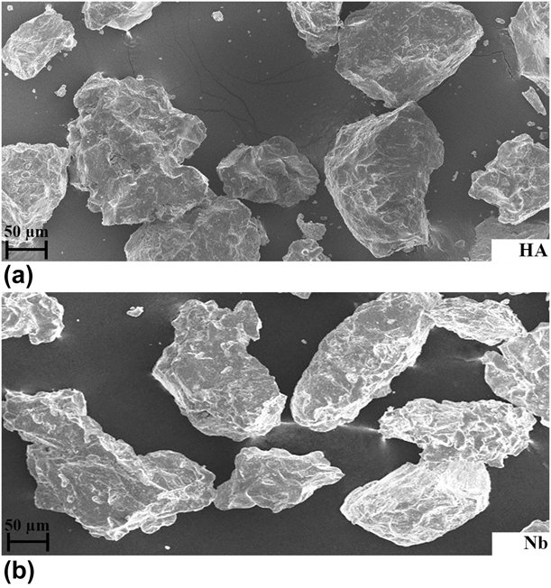
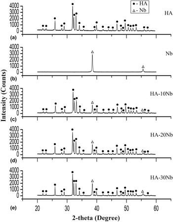
The scanning electron micrographs of HA and Nb powders are shown in Fig. 1. Particles of both the powders [Figs. 1(a) and 1(b)] had an irregular morphology. Figure 2 presents the X-ray diffraction (XRD) patterns of the feedstock powders. The analysis of the XRD patterns demonstrated the presence of HA and Nb powders in crystalline phases. The phases were identified by comparing the XRD patterns of HA and Nb with JCPDS card 9-432 and 35-0789, respectively. With an increase in the Nb content, the peak intensities corresponding to Nb were pronounced, whereas the HA phase maintained its identity.
Scanning electron micrographs of (a) HA and (b) Nb powders.

XRD patterns of (a) HA, (b) Nb, (c) HA-10Nb, (d) HA-20Nb, and (e) HA-30Nb spray powders.

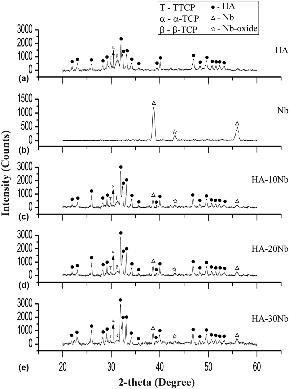
Figure 3 shows the XRD patterns of different as-sprayed coatings. The amorphous hump corresponding to β-tricalcium phosphate (β-TCP; JCPDS card 9-169), peaks corresponding to tetracalcium phosphate (TTCP; JCPDS card 25-1137), and peaks corresponding to α-tricalcium phosphate (α-TCP; JCPDS card 9-348), as observed in the diffraction pattern HA-based coatings [Figs. 3(a) and 3(c)–3(e)], were also reported in earlier studies [Reference Sun, Berndt and Grey31, Reference Singh, Singh and Prakash32]. During the plasma spraying process, the HA powder is melted at a very high temperature due to which HA is dehydroxylated as well as decomposed and amorphous phases commonly exist in the HA coating [Reference Ntsoane, Topic and Bucher33]. The presence of an amorphous phase is reported to be advantageous for the early integration of bioimplant to bone/cell tissue [Reference Sun, Berndt, Gross and Kucuk30].
XRD patterns of as-sprayed (a) HA, (b) Nb, (c) HA-10Nb, (d) HA-20Nb, and (e) HA-30Nb coatings.

The peaks of Nb oxide phase (JCPDS card 30-0873) appear in the diffraction pattern of the Nb coating and in HA-Nb coatings [Figs. 3(b)–3(e)]. The earlier studies on Nb-based coatings revealed that the Nb oxide content was beneficial for the enhanced protection ability and biological response [Reference Ardelean, Frateur and Marcus34, Reference Nagarajan, Raman and Rajendran35, Reference Pauline and Rajendran36]. With the increase of Nb reinforcement in HA, the peaks of TTCP were reduced and the amorphous hump related to β-TCP also lowered. When the Nb reinforcement was increased, the overall intensity of HA peaks increased marginally. HA coating had 74.6% crystallinity of the HA phase, whereas HA-10Nb, HA-20Nb, and HA-30Nb coatings represented crystallinity of 77.4%, 80.5%, and 82.9%, respectively. The crystallinity value of the HA and HA-Nb coatings obtained in this study are consistent with the crystallinity values reported for plasma-sprayed HA coatings by other researchers [Reference Sun, Berndt, Gross and Kucuk30, Reference Ntsoane, Topic and Bucher33, Reference Prevéy37]. Moreover, the proportion of crystalline HA phase in pure HA coating and HA-Nb coatings is in accordance with the ISO 13779-2 standard [Reference Zhang, Xu, Geng, Wang, Xiao and Zhu38]. The variation in crystallinity of the HA and HA-Nb coatings could be due to the difference between the thermal conductivity of HA and Nb. The value of thermal conductivity for HA and Nb is 0.7 W/m K and 52 W/m K, respectively [Reference Williams, Butler, Graves and Moore39, Reference Rapacz-Kmita, Ślósarczyk, Paszkiewicz and Paluch40]. It has been reported that when the reinforcement possesses significantly higher thermal conductivity, it allows slower cooling rate to neighboring HA region [Reference Chen, Zhang, Gong, Zhou and Li41]. After plasma spraying, the decline in the crystallinity of HA phase is mainly caused by the rapid cooling rate [Reference Sun, Berndt, Gross and Kucuk30]. The slower cooling rate because of the presence of Nb is a possible explanation for the higher proportion of crystalline HA phase in HA-Nb coatings.
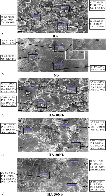
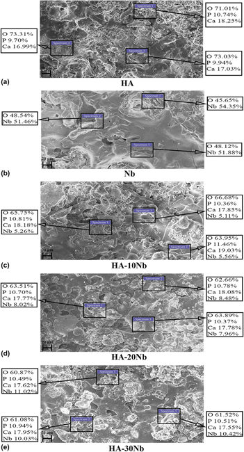
Figure 4 presents the scanning electron micrographs of the surface of as-sprayed coatings. Figures 4(a) and 4(c) show the morphology of pure HA and HA-10Nb coating that mainly is composed of melt-resolidified, spheroidized and fine particles and a few accumulated splats. When the Nb content in HA was increased, well-flattened splats were observed at the surface of HA-20Nb and HA-30Nb coatings [Figs. 4(d) and 4(e)] and spheroidized particles were also reduced. The surface of the Nb coating [Fig. 4(b)] exhibited a relatively flat morphology but some microcracks were noticed at the surface. However, no prominent microcracks appeared at the surface of HA and HA-Nb coatings. The existence of the microcracks in the coating could result in rapid corrosion of the substrate material and also coating failure due to delamination because it facilitates direct contact between the substrate and body fluids [Reference Sun, Berndt, Gross and Kucuk30].
Scanning electron micrographs from the surface of as-sprayed (a) HA, (b) Nb, (c) HA-10Nb, (d) HA-20Nb, and (e) HA-30Nb coatings.

The calcium–phosphorus ratio (Ca:P) of as-sprayed HA coating was 1.74 as determined by EDX analysis. The Ca:P did not fluctuate much with the reinforcement of Nb in HA, and Ca:P values of 1.71, 1.69, and 1.68 were observed for HA-10Nb, HA-20Nb, and HA-30Nb coatings, respectively. Ellies et al. [Reference Ellies, Nelson and Featherstone42] reported that the Ca:P ratio ranging from 1.67 to 1.76 is ideal for HA-coated bioimplants. Furthermore, it is evident from the EDX analysis that the Nb reinforcement in HA-Nb coatings was uniformly distributed and lump formation of Nb reinforcement did not occur.
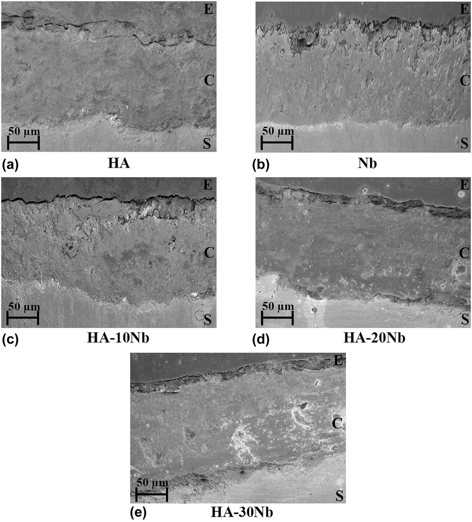
Figure 5 shows the scanning electron micrographs from the cross-section of as-sprayed coatings. In this figure, the area marked as ‘S,’ ‘C,’ and ‘E’ represents the substrate, coating, and epoxy region, respectively. The coatings exhibited almost uniform thickness of ∼125 µm. The HA and Nb coating [Figs. 5(a) and 5(b)] were intact with the substrate as the substrate–coating interface was free from microcracking. Similarly, Nb-reinforced HA coatings [Figs. 5(c)–5(e)] revealed strong bonding with the substrate because no prominent microcrack appeared at the substrate–coating interface.
Cross-sectional scanning electron micrographs of as-sprayed (a) HA, (b) Nb, (c) HA-10Nb, (d) HA-20Nb, and (e) HA-30Nb coatings.

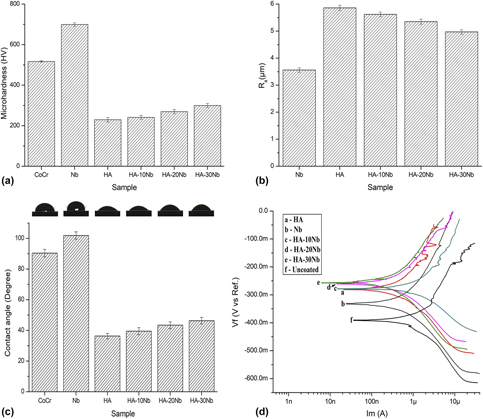
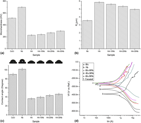
Among the mechanical properties, the hardness of the surface plays an important role in determining the quality of a coating [Reference Hasan, Wang and Berndt43]. Figure 6(a) demonstrates the value of microhardness for different surfaces. The surface of uncoated CoCr alloy and the Nb coating revealed significantly higher microhardness value (P < 0.05) as compared to HA and HA-Nb coatings. Among all the samples, the microhardness value of HA coating was the lowest. With the Nb reinforcement, the value of microhardness of the surface enhanced significantly (P < 0.05) as compared to HA coating. The increment in the microhardness value could be attributed to the formation of a layered framework as a result of the uniform distribution of the Nb reinforcement. A similar increase in surface hardness of HA coating has also reportedly observed in the earlier investigations of reinforced HA coatings [Reference Singh, Singh and Prakash32, Reference Gopi, Karthika, Rajeswari, Kavitha, Pramod and Dwivedi44]. The microhardness values of HA and HA-Nb coatings are comparable to the microhardness of HA coatings that have been reported in the earlier studies [Reference Singh, Singh and Prakash32, Reference Chen, Zhang, Gong, Zhou and Li41, Reference Hasan, Wang and Berndt45].
(a) Microhardness values of samples. (b) R a values for coated samples. (c) Contact angle and droplet profile on the surface of samples. (d) Potentiodynamic polarization curves for different samples.

Figure 6(b) presents the average surface roughness (R a) value for the coated samples. The Nb-coated sample had the lowest surface roughness. HA coating had higher R a value in comparison to Nb coating. The R a values of HA and HA-10Nb coatings did not differ much. With the gradual increment of Nb content in HA, the R a value was further decreased (P < 0.05) for HA-20Nb and HA-30Nb. It can also be observed in Figs. 4(a)–4(c) that with the increase of Nb reinforcement, the melt-resolidified particles lessened and the surface was relatively flattened. The earlier studies which investigated the relationship of surface roughness and the biologic response endorse that an adequate roughness at the surface of implants enhances its protein absorption, which results in better osteoconduction [Reference Yamashita, Machigashira, Miyamoto, Takeuchi, Noguchi, Izumi and Ban46, Reference Shah, Johansson, Omar, Simonsson, Palmquist and Thomsen47]. The R a values of HA and HA-Nb coatings are within the range for R a values of plasma-sprayed HA coatings (2–6 µm) [Reference Gross and Babovic48].
Wettability of the samples was evaluated from the contact angle values of the Ringer’s solution droplet [Fig. 6(c)]. The CoCr alloy and Nb coating surface demonstrated hydrophobic nature, whereas the nature of the surface of HA coating was hydrophilic. The contact angle values increased (P < 0.05) for HA-Nb coatings; however, the surface retained hydrophilic nature. The presence of a large amount of hydroxyl groups in the coating facilitates rapid absorption of the fluid that comes in contact with the surface [Reference Xie, Wang, Berndt, Xie, Wang and Berndt49], which could be the reason of hydrophilic nature of HA and HA-Nb coatings. During the early stage of the contact between the cellular tissue and surface of an implant, the cells can be considered as a simple viscous fluid [Reference Geng, Wang, Zhuo, Li, Huang, Ma, Cui, Zhu, Liang, Liu, Bao, Li, Huo, Liu and Yang50]. After the implantation, the adherence of the cells starts after the absorbance of blood proteins at the surface of the implant [Reference Marashi-Najafi, Khalil-Allafi and Etminanfar51]. As per the literature, the contact angle value less than or equal to 60° is classified as the threshold value of hydrophilicity, and cell adherence was found to be favored for hydrophilic surfaces [Reference Durdu, Korkmaz, Aktuğ and Çakır52].
Corrosion behavior analysis
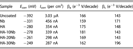
Potentiodynamic polarization curves for different samples are displayed in Fig. 6(d). The electrochemical parameters derived from these curves through Tafel extrapolation are listed in Table I. Corrosion potential (E corr) and corrosion current density (I corr) are crucial parameters through which corrosion behavior of different samples is analyzed. A nobler E corr value and lower I corr value indicate superior corrosion resistance [Reference Poorraeisi and Afshar53, Reference Wang, Zheng, Jiang, Li and Fu54].
Corrosion parameters obtained through Tafel extrapolation.

The uncoated CoCr alloy and Nb-coated sample revealed lower E corr as compared to the other samples. The E corr value increased with HA coating, and with Nb reinforcement in HA, E corr further shifted toward more noble values. The uncoated CoCr alloy exhibited the highest I corr value among all the samples. The I corr value of Nb-coated sample was lower in comparison to the uncoated sample but higher than the HA-coated sample. Earlier studies that investigated the corrosion behavior of HA coatings on CoCr alloy also endorse that HA coating enhances passivation ability of the surface [Reference Campbell11, Reference Moskalewicz, Łukaszczyk, Kruk, Kot, Jugowiec, Dubiel and Radziszewska12, Reference ibrahim Coşkun, Karahan and Yücel13, Reference Shirdar, Izman, Taheri, Assadian and Abdul Kadir14]. The lower E corr and higher I corr values of Nb coating could be due to the presence of microcracks [Fig. 4(b)]. A structural defect in the coating, such as microcracking, promotes corrosion underneath the coating and at the surface, which results in the release of ions into the corrosive media and protection ability of the surface against corrosion [Reference Kiahosseini, Afshar, Mojtahedzadeh Larijani and Yousefpour55]. Because of the incorporation of Nb in HA, the I corr values declined further and the sample with HA-30Nb coating demonstrated lowest I corr, which is about one order of magnitude lesser than the uncoated CoCr alloy. Enayati et al. [Reference Enayati, Fathi and Zomorodian56] have also reported that a noteworthy increase in corrosion resistance of SS 316L was observed after HA-Nb coating. In this study, the fraction of crystalline HA phase increased and the value of I corr decreased with the increase of Nb content in HA. Reportedly, the corrosion resistance of the implants improves with the increase in the phase purity of HA coating [Reference Mohajernia, Pour-Ali, Hejazi, Saremi and Kiani-Rashid57]. Thus, the lower I corr values, i.e., superior corrosion resistance of HA-Nb coatings could be attributed to their higher crystallinity of HA phase. The results obtained through electrochemical investigation are evident that the Nb reinforcement in HA increased the protection ability of the surface against corrosion, which could enhance long-term survivability of CoCr implants.
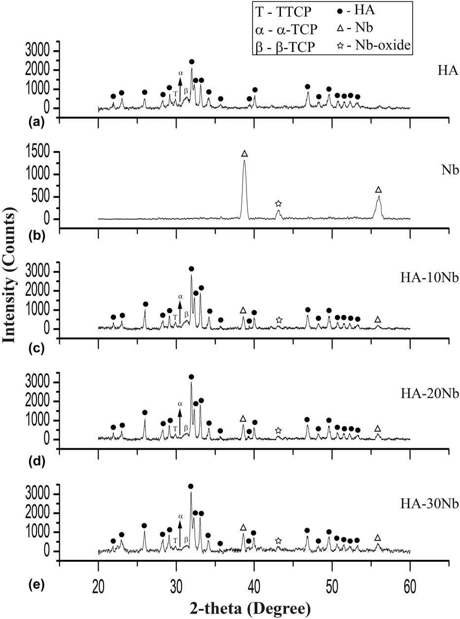
After the corrosion testing, XRD analysis of the coatings was done to analyze phase constituents [Fig. 7]. The results of XRD analysis demonstrated that formation/vanishing of any prominent phase did not occur after exposure to the Ringer’s solution indicating that phase purity was retained by all the coatings. A marginal increment in the overall peak intensities of HA and HA-Nb coatings was observed. After corrosion testing, the crystallinity of HA phase in HA, HA-10Nb, HA-20Nb, and HA-30Nb coatings was 78.8%, 79.7%, 82.1%, and 84.2%, respectively. After the immersion in Ringer’s solution, an increment in the phase purity of HA-based coatings is generally observed because of the dissipation of amorphous phases [Reference Gu, Khor and Cheang58, Reference Yang, Wang, Chang and Wu59]. The initial dissolution of HA enhances the growth of biological equivalents that promote mineralization of bone structure and bone formation [Reference Ntsoane, Topic and Bucher33].
XRD patterns of (a) HA, (b) Nb, (c) HA-10Nb, (d) HA-20Nb, and (e) HA-30Nb coatings after corrosion testing in Ringer’s solution.

Figure 8 presents the scanning electron micrographs of the coatings after corrosion testing. The microstructure of the pure HA coating comprised primarily of the accumulated splats with a few flattened particles of asymmetrical morphology [Fig. 8(a)]. The microcracks became more notable at the surface of Nb coating [Fig. 8(b)] after corrosion testing. Morphology of the surface of HA-10Nb, HA-20Nb, and HA-30Nb coatings [Figs. 8(c)–8(e), respectively] after corrosion testing was dominated by well-flattened splats without any noticeable microcracking. It has been reported that the existence of a microcrack in the coating reduces corrosion protection ability of the surface and even coating failure/delamination can occur because of the microcracking [Reference Sun, Berndt, Gross and Kucuk30, Reference Zhang, Dunn, Xiao, Tomsia and Saiz60]. EDX analysis of all the samples was conducted to analyze the elemental composition of the coating surface after corrosion testing. After the exposure to the Ringer’s solution, a decrease in Ca:P was observed for HA and HA-Nb coatings. HA coating revealed Ca:P value of 1.72; whereas HA-10Nb, HA-20Nb, and HA-30Nb coatings had Ca:P values 1.69, 1.68, and 1.66, respectively. The proportion of oxygen was increased for HA, HA-10Nb, HA-20Nb, and HA-30Nb coatings from 69.9%, 63.2%, 62%, and 59.7% to 72.4%, 65.5%, 63.4%, and 61.2%, respectively. Previous studies of HA coating have reported a similar decrease in Ca:P and increment in oxygen content as observed in the present investigation [Reference Singh, Singh and Singh18, Reference Singh, Singh and Sidhu20, Reference Zhang, Dunn, Xiao, Tomsia and Saiz60]. The osseointegration has also reportedly enhanced by the increase in oxygen content of the coating [Reference Wang, Zheng, Jiang, Li and Fu54].
Scanning electron micrographs from the surface of (a) HA, (b) Nb, (c) HA-10Nb, (d) HA-20Nb, and (e) HA-30Nb coatings after corrosion testing in Ringer’s solution.

Biocompatibility analysis
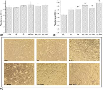
The biocompatibility of the samples was tested on MG-63 cells. Cells were directly exposed to the samples as indicated. Cell proliferation (MTS) data are shown in Figs. 9(a) and 9(b) in which asterisk (*) denotes significance, i.e., P-value < 0.05. After 72-h incubation [Fig. 9(a)], no cytotoxic effect was observed for coated samples as compared to CoCr alloy, and cell proliferation for the uncoated and coated CoCr samples was not significantly different. Further, after 96 h [Fig. 9(b)], an increase in cell proliferation was observed for HA-coated sample. The pure Nb-coated sample revealed almost same cell proliferation as the uncoated CoCr alloy. These results demonstrate that the surface of HA coating was more conducive to cell proliferation than the CoCr alloy and Nb coating. Reportedly, HA coating outperforms the metallic surfaces in biocompatibility because it possesses similarities to the mineral phase of the human bone [Reference Kheimehsari, Izman and Shirdar15]. With the Nb reinforcement in HA coating, no cytotoxic effect was observed on MG-63 cells, and a significant increase in cell proliferation was observed for HA-10Nb, HA-20Nb, and HA-30Nb coatings as compared to CoCr alloy. The crystallinity of HA phase is a critical parameter, which influences the biologic response of the coating surface [Reference Balani, Anderson, Laha, Andara, Tercero, Crumpler and Agarwal61]. The crystallinity of HA phase in HA-Nb coatings was higher than the pure HA coating, which could be the reason that HA-Nb coatings revealed relatively higher cell proliferation as compared to pure HA coating. Microscopic studies of cells exposed to the samples for 96 h revealed no changes in the morphology of the cells [Fig. 9(c)], indicating that the coatings do not have cytotoxic effect.
Cell proliferation (MTS) data after (a) 72-h incubation and (b) 96-h incubation; (c) cell health after 96-h incubation with different samples.

All in all, the results of the present investigation showed that the Nb reinforcement in HA increased the protection ability of the surface against corrosion and promoted cell proliferation, which could enhance long-term survivability and biologic response of the CoCr implants.
Conclusions
In conclusion, plasma spray technique was used to deposit HA, Nb, HA-10Nb, HA-20Nb, and HA-30Nb coatings on the CoCr alloy ASTM F-1537. XRD results demonstrated that the fraction of crystalline HA phase increased as the Nb content was increased in the HA. Scanning electron microscopy (SEM) analysis revealed that microcracking was not observed at the surface and cross-section of as-sprayed HA and HA-Nb coatings, whereas microcracks appeared at the surface of Nb coating. The surface hardness of the HA coating was increased with the incremental increase of Nb reinforcement and the surface roughness decreased as the Nb reinforcement increased. The surface of CoCr alloy and Nb coating demonstrated hydrophobic properties; however, HA and HA-Nb coatings displayed hydrophilic nature of the surface. Both HA coating and Nb coating enhanced the corrosion resistance of CoCr alloy, whereas Nb-coated sample revealed the lowest corrosion resistance among all the coated samples. The values of I corr decreased and E corr became nobler with the incremental increase of Nb reinforcement in HA. HA-30Nb coating exhibited preeminent corrosion resistance with the noblest E corr and lowest I corr values, which were about one order of magnitude lower as compared to the uncoated CoCr alloy. HA-Nb coatings revealed a significant increase in cell proliferation as compared to the uncoated CoCr alloy and demonstrated no cytotoxic effect on MG-63 cells. The results of this study suggest that Nb-reinforced HA coatings could be potentially used to enhance the corrosion resistance and biocompatibility of CoCr implants and thus deserve further biological investigation.
Experimental setup
Deposition of Coatings
Plasma-sprayed coatings of HA, Nb, HA-10Nb, HA-20Nb, and HA-30Nb were prepared using the MF4 plasma spray gun (MEC Pvt., Ltd., Jodhpur, India). An arc current of 600 amperes with 63 volts of arc voltage was used for plasma spraying. Argon and hydrogen were used as primary and secondary gas with the flow rate of 40 SLPM and 5 SLPM, respectively. Spraying distance for the process was kept 75 mm. Spray powders of HA and Nb were supplied by Medicoat, Etupes, France and BGY Science and Technology Co., Ltd., Beijing, China, respectively. A jar mill was used to prepare the mixtures of HA and Nb powders, which were used as spray feedstock. The medical grade CoCr alloy ASTM F-1537 (Zapp Precision Metals, Schwerte, Germany) of dimensions Ø 15 mm × 5 mm was used as substrate material. To generate a rough surface, the substrates were grit blasted with alumina particles before the plasma spraying, and then air blasting of the substrates was performed to remove residual grit. The surface of CoCr substrates had an average surface roughness (R a) value of 1.16 ± 0.2 after air blasting.
Characterization techniques
Phase identification of the coatings was conducted using X-ray diffraction (XRD) analysis (X'Pert Pro Panalytical PN-3040/80, Almelo, Netherlands) over 20°–60° two-theta (2θ) range. The percentage amount of crystalline HA phase in different coatings was estimated as in an earlier study [Reference Sun, Berndt and Grey31]. Scanning electron microscopy (SEM; JEOL JSM-6610LV, Akishima, Japan) equipped with EDX was used for the microstructural and compositional analyses of the coated surfaces before and after corrosion testing. The samples were coated with a thin layer of gold before the SEM/EDX analysis to obtain the desired conductivity of the surface. The substrate–coating interface was also analyzed through the SEM analysis. The sample preparation for cross-sectional SEM analysis was done by cutting the coated samples across the cross-section using a low-speed precision saw. Afterward, the samples were mounted in epoxy resin and polished with emery papers up to 2000 grade. Subsequently, buffing of the samples was conducted using alumina slurry on the napped cloth to obtain a mirror finish.
A microhardness tester (Wolpert Wilson 402MVD, Aachen, Germany) was used to determine the value of microhardness from the polished cross-section of the coatings. The microhardness testing was conducted using a load of 50 gf for a dwell time of 15 s. The contact type profilometer (Mitutoyo SJ-210, Kamasaki, Japan) was used to analyze the roughness of the surfaces. The wettability of the samples was evaluated with a goniometer (First Ten Angstroms FTA2000, Portsmouth, New Hampshire) by measuring the contact angle of Ringer’s solution droplet. For each surface property, an average of fifteen readings has been reported, and the data are expressed in the form of mean ± standard deviation. The statistical analysis of the data has been conducted using Student’s two-tailed t-test with a P-value < 0.05 as statistical significance.
Corrosion behavior analysis
The electrochemical corrosion testing was performed using a Potentiostat/Galvanostat/ZRA (Gamry G-750, Warminster, Pennsylvania). The potentiodynamic polarization studies were conducted using a standard three-electrode cell with Ag/AgCl (saturated KCl), graphite rod, and uncoated/coated sample serving as the reference, counter, and working electrode, respectively. As an electrolyte, Ringer’s solution (Nice Chemicals Pvt., Ltd., Kochi, India) was used for simulating fluid conditions of the human body. To attain a relatively steady state, each sample was immersed in Ringer’s solution for 24 h before the corrosion testing. Potentiodynamic scan was executed from an initial potential of −0.25 V to final potential 0.25 V with scan rate of 0.5 mV/s. The values of electrochemical parameters were acquired from the potentiodynamic curve using Echem Analyst software (Gamry Instruments, Warminster, Pennsylvania).
Biocompatibility investigation
The absence of cytotoxicity for all the samples was analyzed via in vitro cytotoxicity studies. Cytotoxicity of the samples was analyzed on MG-63 cells (osteoblast-like adherent cells), which were procured from the National Center for Cell Science (NCCS), Pune, India. Cells were cultured in Gibco RPMI 1640 media supplemented with 10% (V/V) fetal bovine serum (Gibco -Thermo Fisher, Waltham, Massachusetts) and 1% Penicillin-Streptomycin (Gibco -Thermo Fisher, Waltham, Massachusetts) at 37 °C in a humidified incubator at 5% CO2. The samples were autoclaved before the analysis. Cells were grown at a seeding density of 8000 cells/well for 72-h incubation and of 6000 cells/well for 96-h incubation along with the samples in a 12-well plate. The uncoated CoCr alloy sample was used as the control. Media and samples were removed from the 12-well plate after 72- or 96-h incubation, and cells were washed with 2 mL DPBS solution (Gibco -Thermo Fisher, Waltham, Massachusetts). After washing, the cells were trypsinized with 80 µL of 0.25% Trypsin–EDTA (Gibco -Thermo Fisher, Waltham, Massachusetts) and neutralized with 300 µL of fresh media/well. The suspended cells were then seeded into 96-well plate as triplicates (100 µL/well). MTS assay (Promega Celltiter 96, Madison, Wisconsin) was done after 12-h incubation in 96-well plate. Spectrophotometrical absorbance was determined by a microplate reader (BMG LABTECH CLARIOstar, Ortenberg, Germany) at 490 nm. The qualitative analysis of cell health was conducted through the digital inverted microscope (Thermo Fisher EVOS XL, Waltham, Massachusetts).
Acknowledgment
Zapp Precision Metals, Germany and Medicoat, France are gratefully acknowledged for sponsoring CoCr alloy substrate material and HA powder, respectively. The authors are also grateful to the Mechanical Engineering Department and Center for Biomedical Engineering, IIT Ropar, India for providing research facilities.