Introduction
The circadian and pulsatile secretion of growth hormone (GH) from the anterior pituitary gland plays a crucial role in postnatal longitudinal growth and development, tissue growth, lactation, reproduction, as well as protein, lipid and carbohydrate metabolism (Ohlsson et al., Reference Ohlsson, Bengtsson, Isaksson, Andreassen and Slootweg1998; Ayuk and Sheppard, Reference Ayuk and Sheppard2006). The secretion of GH is down-regulated by somatostatin (SS), which is secreted by the hypothalamus (Vance et al., Reference Vance, Hartman and Thorner1992). The SS has broad inhibitory effect on animals, especially on animal growth and immune response; thus, exhausting or immuno-neutralizing the SS level could lead to increase in GH and promote animal growth (Ohlsson et al., Reference Ohlsson, Bengtsson, Isaksson, Andreassen and Slootweg1998). Therefore, as an alternative approach to direct GH injection, immunization against SS that inhibits GH secretion has been suggested as a method to promote the growth of animals by inactivating SS in the plasma.
SS is a 14-peptide-hormone with a small molecular weight and a very short half-life. Synthetic SS conjugated to a carrier protein promotes the growth rates of immunized animals, including lambs, steers and chickens (Buonomo et al., Reference Buonomo, Sabacky, Della-Fera and Baile1987; Van Kessel et al., Reference Van Kessel, Korchinski, Hampton and Laarveld1990). However, native SS preparation is limited in source.
Chemical synthesis is inefficient for polypeptides longer than about 300 amino acids, and the synthesized proteins may not readily assume their native tertiary structure. Besides, these programs are time-consuming and relatively expensive, which limit the application of SS immunization to promote animal growth.
DNA immunization represents a novel vaccine strategy (Tang et al., Reference Tang, DeVit and Johnston1992). So far, there have been several reports on DNA vaccines encoding the SS gene that can promote the growth of immunized animals in our lab (Liang et al., 2008). However, the potency of naked DNA vaccines is limited by their inability to amplify and spread in vivo; in primates and other large animals the responses are less impressive and the quantities of DNA required may be large (Calarota et al., Reference Calarota, Bratt, Nordlund, Hinkula, Leandersson, Sandstrom and Wahren1998; Macgregor et al., Reference MacGregor, Boyer, Ugen, Lacy, Gluckman, Bagarazzi, Chattergoon, Baine, Higgins, Ciccarelli, Coney, Ginsberg and Weiner1998; Wang et al., Reference Wang, Doolan, Le, Hedstrom, Coonan, Charoenvit, Jones, Hobart, Margalith, Ng, Weiss, Sedegah, de Taisne, Norman and Hoffman1998). It was reported that the tegument protein VP22 of bovine herpesvirus-1 (BHV-1) could increase the immunogenicity of antigens to which it was fused (Oliveira et al., Reference Oliveira, Harms, Afonso and Splitter2001; Wills et al., Reference Wills, Atencio, Avanzini, Neuteboom, Phelan, Philopena, Sutjipto, Vaillancourt, Wen, Ralston and Johnson2001; Hung et al., Reference Hung, He, Juang, Lin, Ling and Wu2002). VP22 of BHV-1 is a 258 amino acid tegument protein (Liang et al., Reference Liang, Chow, Li, Raggo, Yoo, Attah-Poku and Babiuk1995) that can transport proteins to neighboring cells from the cells where they were originally produced (Harms et al., Reference Harms, Ren, Oliveira and Splitter2000). In addition, the intercellular trafficking capacity of VP22 can disseminate the expressed antigen to promote the efficiency of transfection (Zheng et al., Reference Zheng, Babiuk and van Drunen Littel-van den Hurk2005).
Therefore, we hypothesize that VP22 can be a promising gene adjuvant to enhance the immunogenicity and speed up the spread of antigen of SS DNA vaccine. In the present work, we investigated the effects of DNA immunization on growth and anti-SS-specific antibody response in mice immunized with a plasmid DNA encoding the SS and VP22 fusion gene.
Material and methods
Animals and cell lines
Four-week-old female Kunming mice were purchased from Hubei Disease Control Center (Wuhan, China). All animals were performed according to the National Institutes of Health Guides for the care and use of laboratory animals. Hela cells were purchased from Shanghai Shenergy Biocolor BioScience & Technology Co., Ltd.
The primer of VP22 and somatostatin gene
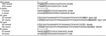
According to the UL49 sequence of BHV-1 (GenBank Z54206), the primers were designed for PCR amplification of the VP22 fragment using the genomic DNA of BHV-1 as a template (Table 1). The amplified VP22 encoded 258 amino acids and its product had the full transport activity of the native protein. The PCR reaction volume was 50 μl, which included 2 × GC buffer 25 μl, 10 mM dNTP mix 2 μl, 5 U/μl Taq 1.5 μl, 1 μg/μl BHV-1 template 1 μl, 10 μM forward primer 1 μl, 10 μM reverse primer 1 μl and autoclaved distilled water 9.5 μl. Cycling conditions began with an initial hold at 95°C for 5 min, followed by 35 cycles consisting of 94°C for 1 min, 65°C for 45 s and 72°C for 1 min, and then by a final extension at 72 °C for 10 min.
Primers and somatostatin (SS) gene sequence

Construction of the pEGS2SS and pEGS2SS-Vplasmid
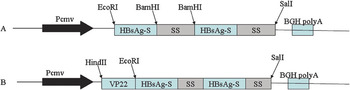
The Hepatitis B surface antigen gene was amplified by PCR with the S primers (Table 1) from the vector pCMV-S (Davis et al., Reference Davis, Weeratna, Waldschmidt, Tygrett, Schorr and Krieg1998), which was kindly supported by Aldevron LLC with restriction tails EcoRI and KpnI and then cloned into the pEGFP-N1. Two SS gene fragments, which were synthesized by Shanghai Sangon Biological Engineering Technology & Service Co., Ltd (Song Jiang, Shanghai, China) were inserted into the pEGFP-N1 SS1 was inserted into the C termination of the HbsAg-S gene with KpnI and SalI; SS2 was inserted into the middle of the S gene at the 112 amino acid with BamHI (Table 1). Then the constructed recombinant plasmid was named as pEGS2SS.
To evaluate the VP22 for enhancement of DNA vaccine against SS, the VP22 gene was inserted into the N termination of the HbsAg-S gene with HindIII and EcoRI, and the resulting vector was named as pEGS2SS-V. All recombinant plasmids were verified by restriction endonuclease analysis and the integrity of coding sequences was checked by sequence analysis.
Detection of the recombinant expressed protein by fluorescence microscope and RT-PCR
The recombinant expressed protein was measured by a fluorescence microscope according to the manufacturer’s protocol. Plasmids of pEGS2SS-V, pEGS2SS and pEGFP-N1 were transfected into Hela cells with the transfection reagent liposome 2000™ according to the manufacturer’s protocol. After 3 days, the positive colony of the cells after selecting for 2 weeks was harvested. A fluorescence microscope was used to detect the expression of the plasmids.
The expression of the SS was detected at the transcription level using the reverse transcription (RT)-PCR. Total RNAs were extracted from the transfected HeLa cells with SV total RNA Isolation System (Promega Corporation, Madison, WI, USA) and then cDNAs were synthesized using the RevetAid First Strand cDNA Synthesis Kit (Fermentas). Each cDNA sample was used as a template for PCR amplified of SS and the PCR products were verified by automated sequencing (Sangon).
Animals immunization
To study the effect of SS immunization on the BW and the P/N value of the antibody against SS, five groups of 45 mice were injected with 100 μg of SS fusion plasmid (pEGS2SS-V) DNA, 100 μg pEGS2SS and 100 μl 0.85% saline. After 2 weeks a booster injection was given. All mice were anesthetized with bupivacaine hydrochloride 24 h before DNA administration.
Blood collecting and weight gain of mice
Sera were recovered from the tail vein on the 2nd, 4th, 6th and 8th week after the first immunization under anesthesia with ether and anticoagulation with heparin sodium salt. Animals were weighed prior to the collection of blood samples.
Detection of anti-somatostatin antibody by ELISA
ELISA plates were used for detecting mouse anti-SS antibody. The wells were coated with 0.5 μg/ml of SS antigens (Sigma) at 4°C overnight at 100 μl per well. These wells were subsequently blocked with 1.5% of BSA-PBST for 2 h at 37°C and 1 : 100 dilutions of sera samples were added in the wells. After three washes, the plates were incubated for 1 h at 37°C with a 1 : 1000 dilution of horseradish peroxidase-labeled goat anti-mouse IgG antibody. To develop the ELISA result, 10 mg of 3,3′,5,5′-tetramethylbenzidine (TMB tablets; Sigma) was dissolved in 0.025 M phosphate–citrate buffer and 50 μl of the resulting solution was added to each well. The reaction was stopped by the addition of 0.2 M H2SO4 and the resulting optical density (OD) at 450 was analyzed with a plate reader (Thermo Electron Corporation). The results were judged by the P/N value, where P is the optical density of the detected sample and N is the optical density of the negative control sample (Han et al., 2007). If the P/N value was more than 2, it was regarded as positive, otherwise as negative.
Results
PCR amplification of the VP22 fragment and the construction of the plasmids
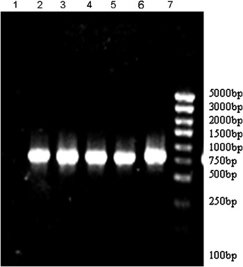
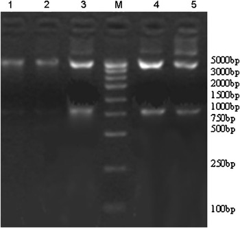
The VP22 gene was amplified from BHV-1 by PCR, and the amplified product was of length 795 base pair (bp) (Figure 1). The DNA sequence was verified by auto sequencing, and the recombinant plasmids were detected by double digestion by the endogenous enzyme. The exogenous VP22 gene was of length about 790 bp when cut by double digestion with HindIII and EcoRI sites. The exogenous HBsAg and SS recombinant gene was of length about 790 bp when cut with EcoRI and SalI sites (Figures 2 and 3).
VP22 gene product by PCR: Lane 1: the negative product from water as a template; Lanes 2 to 6: PCR product of VP22 gene from BHV-1 as a template; Lane 7: DNA marker: DL-5000.

Results of agarose gel electrophoresis of restriction enzyme digestion analysis. M: DNA molecular weight markers (DL-5000); Lane 1: the plasmid pEGS2SS; Lane 2: the plasmid pEGS2SS-V; Lane 3: the plasmid pEGS2SS digested with EcoRI and SalI Restriction enzymes; Lanes 4 and 5: the plasmid pEGS2SS-V digested with EcoRI and HindIII restriction enzymes.

Map of construction of somatostatin (SS) DNA vaccine encoding HBsAg-S and SS. A: the map of construction of SS DNA vaccine named pEGS2SS. B: the map of construction of SS DNA vaccine named pEGS2SS-V.

Expression of SS in transfected HeLa cells
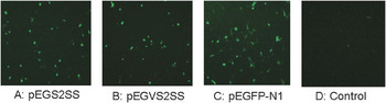
Fluorescence microscope and RT-PCR analysis were applied to verify the expression of the recombinant protein in transfected HeLa cells. All transfected HeLa cells showed distinct green fluorescence while no fluorescence could be observed from control cells (Figure 4). This indicated the expression of the fused gene.
Expression of the somatostatin (SS) recombinant protein in PK15 cells at 48 h post transfection. A: expression of the SS recombinant protein. B: expression of the SS-VP22 recombinant protein. C: The cells with pEGFP-N1 plasmids transfection. D: The cells without plasmids transfection.

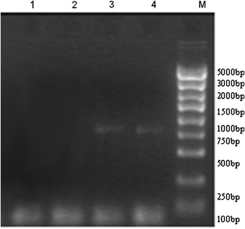
PCR products of about 790 bp were gained using template RNA extracted from pEGS2SS or pEGS2SS-V transfected HeLa cells while no specific band was detected from pEGFP-N1 transfected cells (Figure 5). This indicated that the SS could be expressed in transfected HeLa cells at least in the transcriptional level.
Reverse transcription-PCR analysis of transcription of transfected HeLa cells with the plasmids. Lane 1: HeLa cells transfected without plasmid; Lane 2: HeLa cells transfected by pEGFP-N1 amplified with the RT primers; Lane 3: HeLa cells transfected by pEGS2SS amplified with the RT primers; Lanes 4 and 5: HeLa cells transfected by pEGS2SS-V amplified with the RT primers.

Enhancement of DNA vaccine in mice
All immunized mice grew quicker than in the control group, and the mice in the pEGS2SS-V-immunized group grew fastest (Table 2). At week 2 after the first immunization, the average weight gain of mice immunized with pEGS2SS-V and pEGS2SS increased 48.4% and 34.3% compared to the control group, respectively. At week 2 after the booster immunization, average weight gain of pEGS2SS-V and pEGS2SS increased 12.3% and 16.4%, respectively, while there was no difference between the pEGS2SS-V group and the pEGS2SS group and there was no difference between the experimental group and the control group. At week 6 and week 8 after the first immunization, the tendency of the weight gain of every group is same (Table 2). These indicated that the SS DNA vaccine could benefit the growth rate of mice, and the pEGS2SS-V could induce a faster growth rate than pEGS2SS.
Weight gain (g) of the mice at different weeks after immunization (mean ± s.d.)

In the horizontal row, significance level was P < 0.05 between values with different lowercase letters, and not significant with same lowercase letters. In the vertical row, significance level was P < 0.05 between values with different uppercase letters and not significant with same uppercase letters. Weight gain (g): mean ± s.e.
Effects on the antibody against somatostatin in mice after immunization
Sera from more than 40% (6/15) mice in both vaccine groups were tested positive by ELISA, while no mice in the control group were tested positive (Table 3). The mice immunized with the pEGS2SS-V showed the highest positive sera ratio of 40% (6/15) at week 4 and the mice immunized with the pEGS2SS showed the highest positive sera ratio of 40% (6/15) at week 6. These indicated that the DNA vaccine could develop the antibody against SS in mice, and the pEGS2SS-V could supply faster antibody than pEGS2SS
The P/N value of antibody against somatostatin in mice at different weeks after immunization (mean ± s.d.)

In the horizontal row, significance level was P < 0.05 between values with different lowercase letters, and not significant with same lowercase letters. In the vertical row, significance level was P < 0.05 between values with different uppercase letters and not significant with same uppercase letters. P/N: mean ± s.e.
Discussion
In this study, the VP22 gene fragment was difficult to amplify because of the high GC percent (76%). Many reports showed the 2 × GC buffer I (Takara) was more suitable for amplification of this kind of high GC content sequences, because the additional reagents in this buffer could destroy the secondary structure of the template and thus increase the specificity.
The immunogenic activity of SS is very weak, and direct immunization cannot achieve the ideal effect. Our previous study showed that HBsAg could enhance the immunogenicity of inhibin, which is a weak antigen as well (Han et al., 2007). In the present study, the HBsAg was used to enhance the DNA vaccine of SS. Mice in both vaccinated groups grew faster than in the control group, and mice from only both immunized groups were tested positive by SS-ELISA while there was no positive result from the control group. These indicated that the vaccine of SS can speed up animal’s growth in mice, and the HBsAg could improve the immunogenicity of the SS. Though faster growth was achieved in mice, enhancement of the growth rate in animals still needs further study.
More recently, the VP22 gene of BHV-1 has been used as a gene adjuvant to enhance the immunogenic activity and thus make the antigen easily recognized and processed by the organism immunity system, and VP22 can enhance immune responses in vivo in a variety of applications (Hung et al., Reference Hung, Cheng, Chai, Hsu, He, Ling and Wu2001; Wills et al., Reference Wills, Atencio, Avanzini, Neuteboom, Phelan, Philopena, Sutjipto, Vaillancourt, Wen, Ralston and Johnson2001; Saha et al., Reference Saha, Yoshida, Ohba, Matsui, Matsuda, Takeshita, Umeda, Tamura, Okuda, Klinman, Xin and Okuda2006). In this work, VP22 was used as a gene adjuvant to study the enhancement of the SS DNA vaccine in mice. Mice immunized with pEGS2SS-V grew faster than in the groupthat was immunized with pEGS2SS. The antibody against SS from the pEGS2SS-V-immunized group can be detected much earlier. Forty percent positive rate was detected in the pEGS2SS-V-immunized group within 4 weeks while it was 6 weeks for the pEGS2SS-immunized group. These indicated that pEGS2SS-V was better than pEGS2SS in mice. These results clearly demonstrate that BHV-1 VP22 can be used as a gene adjuvant in the SS DNA vaccine to increase weight gain and enhance immune responses. Our results proved that VP22 could make the SS more easily recognized and processed by the mouse immunity system, and then enhanced the immunogenicity of antigen. Moreover, it is consistent with the results of Michel and Kim (Michel et al., Reference Michel, Osen, Gissmann, Schumacher, Zentgraf and Muller2002; Kim et al., Reference Kim, Hung, Kim, Juang, Chen, He, Boyd and Wu2004). They proved that VP22 may improve immune presentation in different ways, either through enhanced direct priming by transfected antigen-presenting cells (APCs) or by amplified cross-presentation after release from transfected nonprofessional APCs.
In conclusion, this study found VP22 gene fragment can be a gene adjuvant to enhance immune response against SS. The VP22 gene adjuvant-immunized group had higher and quicker peaks in immunization than in the group without this adjuvant. Further studies on the mechanism by which VP22 enhances the effects of SS gene immunization are needed in order to better understand the biotherapeutic potential of VP22 in gene immunization. The VP22 gene can be a useful adjuvant to enhance the efficacy of gene vaccine.
Acknowledgments
The authors thank Professor Guo Ai-zhen (National Key Laboratory of Agricultural Microbiology, Huazhong Agricultural University) for providing us with the genomic DNA of BHV-1. This work was partially supported by National natural Science Foundation of China (30771549).