Introduction
Alzheimer's disease (AD) is the leading cause of late-onset dementia worldwide. Although the first neuropsychological deficit in AD is episodic memory loss (Perry and Hodges, Reference Perry and Hodges1999; Perry et al., Reference Perry, Watson and Hodges2000), the presence of frontal or executive deficits in patients with even mild AD is now widely recognized (Perry et al., Reference Perry, Watson and Hodges2000). Current evidence suggests that after an initial amnestic stage in AD, attention and executive functions are the first non-memory domains to be affected, before deficits in language and visuospatial functions occur (Perry and Hodges, Reference Perry and Hodges1999; Perry et al., Reference Perry, Watson and Hodges2000). Moreover, perseveration errors are frequently found even in mild AD (Pekkala et al., Reference Pekkala, Albert, Spiro and Erkinjuntti2008). However, brain perfusion imaging evidence suggests the relative preservation of the frontal lobes in AD (Johnson et al., Reference Johnson, Mueller, Walshe, English and Holman1987). It is, therefore, somewhat surprising that AD produces marked impairment in the attentional and executive functions, which have been linked with frontal lobe function, before deficits in language and visuospatial function occur (Perry and Hodges, Reference Perry and Hodges1999).
The Wisconsin Card Sorting Test (WCST) has long been used to investigate deficits in executive function in humans (Milner, Reference Milner1963; Nelson, Reference Nelson1976). The subject is asked to match test cards to reference cards according to the color, shape, or number of stimuli on the cards. After a fixed number of correct matches, the sorting rule is changed without notice, and the subject must shift to a new mode of classification. Thus, the WCST measures cognitive flexibility, that is, the ability to alter a behavioral response mode in the face of changing contingencies (set-shifting) (Monchi et al., Reference Monchi, Petrides, Petre, Worsley and Dagher2001). Perry et al. found that 15% of minimally impaired patients with AD (Mini-Mental State Examination (MMSE) score 24–30) and 50% of mildly demented patients with AD (MMSE score 18–23) were impaired on the modified WCST (mWCST) (Perry et al., Reference Perry, Watson and Hodges2000). The WCST is a complex problem-solving task that probably requires multiple cognitive processes rather than a single unitary function (Nagahama et al., Reference Nagahama2003). The majority of studies that investigated deficient mWCST performance in AD focused on the number of categories achieved (CA) and the number of perseverative errors of the Nelson (PEN) type (Perry et al., Reference Perry, Watson and Hodges2000; Nagahama et al., Reference Nagahama2003). However, there is insufficient evidence that these two measures reflect the same neural deficit and that both measures equally indicate executive dysfunction (Takeda et al., Reference Takeda2010). On the contrary, it has been reported that the CA and PEN scores of dementia patients are each related to regional cerebral blood flow (rCBF) in different brain regions (Takeda et al., Reference Takeda2010).
As far as we could find, there have been only two studies of the relationship between WCST scores and rCBF in patients with dementia (Nagahama et al., Reference Nagahama, Okina, Suzuki, Nabatame and Matsuda2005; Takeda et al., Reference Takeda2010). The first report by Nagahama et al. showed that the “stuck in set” type perseveration error (similar to the perseverative errors defined by Nelson) was associated with reduced rCBF in the rostrodorsal prefrontal cortex (PFC) in 72 elderly subjects including 51 patients with AD (Nagahama et al., Reference Nagahama, Okina, Suzuki, Nabatame and Matsuda2005). They used stereotaxic volume of interest analysis, and examined only four small areas in the frontal lobes (rostrodorsal PFC, posterior PFC, ventrolateral PFC and anterior cigulate area). Moreover, they did not include the effects of MMSE scores and CA scores of patients.
The second study (Takeda et al., Reference Takeda2010) reported that PEN scores correlate with rCBF in the right thalamus of 77 subjects including 31 patients with AD. They analyzed the data using the rCBF quantification software program 3DSRT (Takeuchi et al., Reference Takeuchi, Yonekura, Matsuda and Konishi2002). The usefulness, validity, and objectivity of 3DSRT have been reported previously (Takeuchi et al., Reference Takeuchi, Yonekura, Matsuda and Konishi2002). However, these two studies include normal subjects and patients with dementias other than AD. There have been no studies on the relationship between PEN scores and rCBF in AD patients.
This study aimed to compare the rCBF of AD patients with high and low PEN scores on the mWCST, and to investigate the neural substrate important for the PEN scores on the mWCST of AD patients. Because functional neuroimaging studies of the WCST have confirmed the involvement of the PFC (Berman et al., Reference Berman1995; Monchi et al., Reference Monchi, Petrides, Petre, Worsley and Dagher2001; Konishi et al., Reference Konishi, Hayashi, Uchida, Kikyo, Takahashi and Miyashita2002), we predicted a relationship between PEN scores and prefrontal hypoperfusion among AD patients.
Methods
Modified Wisconsin Card Sorting Test
Owing to the long time required for administration of the original WCST and the refusal of some patients to complete the test due to frustration or fatigue, a number of modifications to the WCST have been developed (Nagahama et al., Reference Nagahama2003). We used this modification of Nelson's version using 48 (2 × 24) response cards that share one and only one attribute with the stimulus cards (Nelson, Reference Nelson1976). Patients were not informed of the correct sorting principle, nor were they told when the principle would shift during the test, but they were informed of the three possible categories before testing. The patients were required to determine which one was correct based solely on the feedback indicating whether each response was right or wrong. When the patient maintained a correct progression through six trials, the rule was changed without warning. The testing in this study continued until 48 cards were sorted (Nagahama et al., Reference Nagahama, Okina, Suzuki, Nabatame and Matsuda2005).
For the mWCST, the number of CA and PEN were evaluated. In mWCST, the mean of the PEN score was reported to be 4.8 ± 5.6 among the normal elderly (mean age, 70.8) (Nagahama et al., Reference Nagahama2003). In this study, patients with a PEN score ≥16 (mean + 2SD among normal elderly) were placed in the high PEN score group, while patients with a PEN score ≤10 (mean + 1SD among normal elderly) were placed in the low PEN group.
Subjects
Twenty AD patients with dementia severity of 0.5 (suspicious) or 1 (mild) based on the Clinical Dementia Rating (CDR) (Hughes et al., Reference Hughes, Berg, Danziger, Coben and Martin1982) and whose PEN score on the mWCST was over 15, were selected from 227 consecutive Japanese patients who were fully examined at the Memory Clinic of Okayama University Hospital between April 2004 and March 2008. In addition, 20 AD patients at the Memory Clinic with low PEN scores on the mWCST, matched for age, sex ratio, education, Addenbrooke's Cognitive Examination (ACE) scores (Mathuranath et al., Reference Mathuranath, Nestor, Berrios, Rakowicz and Hodges2000; Yoshida et al., Reference Yoshida2011), and CA scores on mWCST, were also selected.
They all (i) underwent general physical and neurological examinations and extensive laboratory testing, including thyroid function tests, serum vitamin B12, and syphilis serology; (ii) took the MMSE (Folstein et al., Reference Folstein, Folstein and McHugh1975), ACE (Mathuranath et al., Reference Mathuranath, Nestor, Berrios, Rakowicz and Hodges2000), and frontal assessment battery (FAB) (Dubois et al., Reference Dubois, Slachevsky, Litvan and Pillon2000; Kugo et al., Reference Kugo2007) within one week of scanning; (iii) underwent brain SPECT as well as head MRI; and (iv) were diagnosed with probable AD according to the criteria formulated by the NINCDS-ADRDA (McKhann et al., Reference McKhann, Drachman, Folstein, Katzman, Price and Stadlan1984); (v) had a dementia severity of 0.5 or 1 on the CDR (Hughes et al., Reference Hughes, Berg, Danziger, Coben and Martin1982); and (vi) they or their nearest relatives gave written informed consent.
The exclusion criteria were: (i) complications from other neurological diseases or illnesses, (ii) history of mental illness or substance abuse prior to the onset of dementia, (iii) any evidence of focal brain lesions on head MRI, (iv) treatment with cholinesterase inhibitors, antipsychotics, antidepressants or anxiolytic drugs, and (v) left handedness or ambidexterity.
Information was also gathered from caregivers familiar with the patient's daily behavior. To serve as an informant, the caregiver was required to be living with the patient. The profile of each subject (age, sex, years of education, and years of disease duration) was recorded. The Physical Self-Maintenance Scale (PSMS) and the Lawton Instrumental Activities of Daily Living (IADL) scale (Hokoishi et al., Reference Hokoishi2001) were scored based on the information from caregivers.
Other instruments
ACE was developed to provide a brief test sensitive to early stage dementia, and is capable of differentiating between dementia subtypes including AD, frontotemporal dementia, progressive supranuclear palsy, and other parkinsonian syndromes (Mathuranath et al., Reference Mathuranath, Nestor, Berrios, Rakowicz and Hodges2000). ACE includes MMSE but extends it to encompass important areas not covered by MMSE, such as frontal-executive function and visuospatial skills. ACE has a sensitivity comparable to the Dementia Rating Scale (Mathuranath et al., Reference Mathuranath, Nestor, Berrios, Rakowicz and Hodges2000), a well-established dementia screening tool that is widely used in research, but not in clinical practice because of its length and difficulty of administration. For this study, we used the Japanese version of the ACE described by Yoshida et al. (Reference Yoshida2011). The reliability of the Japanese version of ACE is excellent, and its validity is, to some extent, established (Yoshida et al., Reference Yoshida2011).
The FAB consists of six items, and the score on each item ranges from 0 to 3. A lower score indicates a greater degree of executive dysfunction. The six subtests of FAB explore (1) similarities (conceptualization), (2) lexical fluency (mental flexibility), (3) Luria motor sequences (programming), (4) conflicting instructions (sensitivity to interference), (5) a go/no-go test (inhibitory control), and (6) prehension behavior (environmental autonomy). For this study, we used the Japanese version of FAB described by Kugo et al. (Dubois et al., Reference Dubois, Slachevsky, Litvan and Pillon2000; Kugo et al., Reference Kugo2007). The reliability of the Japanese version of FAB is good to excellent, and its validity is, to some extent, established (Kugo et al., Reference Kugo2007).
The Physical Self-Maintenance Scale (PSMS) and Instrumental Activities of Daily Living Scale (IADL) are validated scales for the assessment of activities of daily living (ADL) (Hokoishi et al., Reference Hokoishi2001). The PSMS is a six-item scale that rates self-care ability in toileting, feeding, dressing, personal hygiene and grooming, locomotion (physical ambulation), and bathing. The IADL scale assesses patients’ ability to perform eight complex daily tasks: ability to use the telephone, shopping, food preparation, household tasks, laundry, mode of transportation, responsibility for medications, and ability to manage finances.
Ethics
The study protocol was approved by the Committee of Okayama University Hospital on Human Research. After providing participants with a complete description of the study, written informed consent was obtained.
Brain perfusion SPECT imaging
All subjects were examined by brain perfusion single-photon emission computed tomography (SPECT). Patients were examined in a comfortable supine position with their eyes closed in quiet surroundings. First, the passage from the heart to the brain was monitored after intravenous administration of 99mTc-ethylcysteinate dimer (ECD, 600 MBq, Daiichi Radioisotope Laboratories Ltd., Tokyo, Japan). Ten minutes after the angiography, SPECT images were obtained using a triple-head, rotating gamma camera interfaced to a minicomputer (GCA9300A/ DI; Toshiba, Tokyo, Japan) equipped with a fanbeam, low-energy, high-resolution collimator. Sixty projection images over a 360° angle in a 128 × 128 matrix were acquired. All images were reconstructed using ramp-filtered back-projection and then three-dimensionally smoothed with a Butterworth filter (order 8, cutoff 0.12 cycles/cm). The reconstructed images were corrected for gamma ray attenuation using the Chang method (μ = 0.09).
Data analysis
Spatial reprocessing and statistical analysis of images was performed on a voxel by voxel basis using Statistical Parametric Mapping 2 (SPM2, Wellcome Department of Imaging Neuroscience, UK) running on MATLAB (The Mathworks, Inc., Natick, MA, USA). All SPECT images of each subject were normalized to the standard brain of the Montreal Neurological Institute (MNI), and spatial normalization was performed with 12-parameter affine and non-linear transformations (Friston et al., Reference Friston, Ashburner, Poline, Frith, Heather and Frackowiak1995). The voxel sizes of the reslice option were 2 mm × 2 mm × 2 mm. The non-linear parameter was set at 25 mm cut-off basis functions and 16 iterations. All the normalized SPECT images were then smoothed with an isotropic gaussian kernel filter (12-mm full-width at half-maximum). To examine the images for specific regions showing differences in perfusion, two sample t-tests were performed. Global normalization was performed by proportional scaling with the mean voxel value. Masking was applied using the threshold method (0.8 times the global value). The analysis used a threshold of p < 0.01 at the voxel level and results were considered significant at p < 0.05 (corrected) at the cluster level.
Statistical analysis
Statistical analysis was performed using the SPSS 14.0J software program (SPSS Inc., Chicago, IL). The two groups were compared using independent sample t-tests. A value of p < 0.05 was accepted as significant.
Results
Clinical characteristics and neuropsychological tests
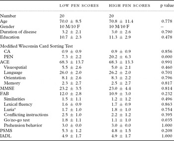
No significant differences were found between high and low PEN score groups with respect to age (p = 0.778), disease duration (p = 0.790), or years of education (p = 0.478) (Table 1).
Demographic characteristics and neuropsychological tests

All data are means ± SD. p value, unpaired and two-tailed t tests.
PEN = perseverative errors of the Nelson type; CA = categories achieved.
ACE = Addenbrooke's cognitive examination.
MMSE = Mini-Mental State Examination.
FAB = frontal assessment battery; Luria* = Luria motor sequences.
PSMS = Physical Self-Maintenance Scale.
IADL = Lawton Instrumental Activities of Daily Living.
With the exception of PEN scores on mWCST (p < 0.001), no significant differences were found between high and low PEN score groups in CA scores on the mWCST (p = 0.856), ACE (p = 0.991), MMSE (p = 0.814), FAB (p = 0.230), PSMS (p = 0.208), or IADL (p = 1.000) (Table 1). There were no significant differences between high and low PEN groups in the four subscale scores on ACE. Comparison of FAB subscales revealed that AD patients with high PEN scores got lower scores than those with low PEN scores in the subscale score of the go/no-go test (Table 1).
Regional cerebral blood flow (rCBF)
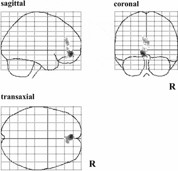
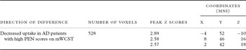
The areas of hypoperfusion in the high PEN score group that were not present in the low PEN score group according to the SPM analysis are shown in Figure 1 and Table 2. A similar test for areas of hypoperfusion in the low PEN score group that were not present in the high PEN score group found no cluster of voxels. Figure 1 shows the z score for each voxel in this cluster superimposed onto a 3-way glass brain view. Figure 1 shows a significant cluster of voxels in the left rectal gyrus extending to the right rectal gyrus and bilateral orbital gyrus (in the ventromedial PFC). Table 2 shows the probability results of the SPM analysis and the location of peak z scores in terms of MNI coordinates.
SPM (z) map of rCBF decrease in AD patients with high PEN scores on the modified WCST compared with AD patients with low PEN scores. Three-way-glass view of the area of significant hypoperfusion.

Significant regional uptake differences between AD patients with low and high PEN scores on mWCST

AD = Alzheimer's disease; MNI = Montreal Neurological Institute; mWCST = modified Wisconsin Card Sorting Test.
Discussion
Ventromedial PFC and WCST
We predicted hypoperfusion in the PFC among AD patients with high PEN scores, and we revealed in this study that rCBF in the bilateral rectal and orbital gyrus among AD patients with high PEN scores was lower than that among AD patients with low PEN scores. Therefore, the results are in accordance with our hypotheses. It was reported that all patients with lesions involving regions of the ventromedial PFC (vmPFC) were impaired on WCST, and that most patients without ventral prefrontal damage performed normally on WCST (Schnyer et al., Reference Schnyer, Maddox, Ell, Davis, Pacheco and Verfaellie2009). Our results also suggest the importance of the vmPFC in WCST.
The vmPFC is reported to play a key role in implementing probabilistic rules (Bhanji et al., Reference Bhanji, Beer and Bunge2010), and patients with vmPFC damage show deficits in learning reward-punishment rules (Hartstra et al., Reference Hartstra, Oldenburg, Van Leijenhorst, Rombouts and Crone2010). Moreover, vmPFC is involved in guiding decision-making when strategies change (de Wit et al., Reference de Wit, Corlett, Aitken, Dickinson and Fletcher2009). Therefore, difficulty in changing a strategy when the rules are altered might cause the increase in PEN scores on WCST as a matter of course.
WCST and rCBF in AD and other neurodegenerative diseases
In this study, the PEN score of WCST was significantly associated with rCBF in the bilateral rectal and orbital gyrus. However, the results of other studies are not always consistent with our findings (Nagahama et al., Reference Nagahama, Okina, Suzuki, Nabatame and Matsuda2005; Takeda et al., Reference Takeda2010). What causes the difference?
Nagahama et al. showed that the “stuck in set” type perseveration error was associated with reduced rCBF in the rostrodorsal PFC (Nagahama et al., Reference Nagahama, Okina, Suzuki, Nabatame and Matsuda2005). They did not examine rCBF in the ventromedial PFC and calculated the correlation of perseveration errors to rCBF without adjusting the CA scores. Therefore, if MMSE scores or CA scores correlate with rCBF in the rostrodorsal PFC, the correlation of perseveration error to rCBF in the rostrodorsal PFC is not specific to perseveration error. The CA score was reported to correlate with rostrodorsal PFC (Takeda et al., Reference Takeda2010). Therefore, the correlation of perseveration error to rCBF in the rostrodorsal PFC might be affected by the correlation of CA score to rCBF in the rostrodorsal PFC.
Our previous report showed that PEN scores correlate with rCBF in the right thalamus (Takeda et al., Reference Takeda2010). Twenty-nine of 40 AD patients in this study were also included in the previous study. In that study, the data were analyzed, using the rCBF quantification software program 3DSRT (Takeuchi et al., Reference Takeuchi, Yonekura, Matsuda and Konishi2002). The 3DSRT divides each hemisphere into 12 arbitrary segments and each segment includes a relatively broad area. For example, the callosomarginal segment includes the superior frontal, medial frontal, and paracentral lobules, anterior cingulate, and subcallosal, orbital, and rectal gyri (Takeuchi et al., Reference Takeuchi, Yonekura, Matsuda and Konishi2002). Therefore, if PEN scores correlate strongly with only a small region in the frontal lobe, the correlation of PEN scores to a broad segment such as the callosomarginal segment in the 3DSRT could obscure statistically significant results in smaller regions. In this study, rCBF in whole voxels of brains of high and low PEN score groups were compared. This approach facilitates identification and analysis of smaller, discrete regions of interest. Moreover, both Nagahama et al. (Reference Nagahama, Okina, Suzuki, Nabatame and Matsuda2005) and Takeda et al. (2002) studied a mixture of subjects including different etiologies, MCI, and elderly subjects, whereas this study included only patients with AD. The difference in subjects might influence the results.
Ventromedial PFC and AD
We found a decrease in rCBF in the ventromedial PFC among AD patients with high PEN scores. Significant metabolic decreases in ventromedial prefrontal areas are reported to begin early in the course of AD (Fouquet et al., Reference Fouquet2009). In support of this contention, the same two medial prefrontal areas showed specific perfusion decreases from the entorhinal to the limbic neuropathologic Braak stages (Braak and Braak, Reference Braak and Braak1991), corresponding to amnestic mild cognitive impairment and early AD, respectively (Bradley et al., Reference Bradley2002). Our results suggest that some AD patients suffer from frontal neuropsychological deficits and frontal hypoperfusion in the early stage, and that those patients might be included in a so-called frontal variant of AD (Johnson et al., Reference Johnson, Head, Kim, Starr and Cotman1999; Woodward et al., Reference Woodward, Brodaty, Boundy, Ames, Blanch and Balshaw2010).
WCST and the go/no-go task
In this study, AD patients with high PEN scores got lower scores on the go/no-go task on FAB than those with low PEN scores. The go/no-go task, in which the participant must inhibit a response that was previously given to the same stimulus, can assess the difficulty in controlling impulsiveness (Drewe, Reference Drewe1975). Therefore, the go/no-go task is commonly used to investigate processes of selective attention or response inhibition (Ambach et al., Reference Ambach, Stark, Peper and Vaitl2008). Response inhibition is difficult for patients with damage to the ventral part of the frontal lobes (Rolls et al., Reference Rolls, Hornak, Wade and McGrath1994). Thus, the PEN score on mWCST may be a promising index to detect dysfunction of the orbitofrontal area among mildly affected AD patients, and equally these results suggest that the go/no-go task from the FAB may possess particular utility as a bedside test of orbitofrontal dysfunction in AD.
Limitations of this study
The results in this study should be interpreted with some caution: (1) the relatively small sample size, comprising only 20 AD patients with high CA scores and 20 AD patients with low CA scores, may limit the sensitivity to actual group differences and associations between neuropsychological functions and neural activation; (2) the low spatial resolution of SPECT scans may limit detection of alterations in small regions, thus increasing the risk of false negatives and interference from surrounding regions as well (Takeuchi et al., Reference Takeuchi, Yonekura, Matsuda and Konishi2002); (3) PEN and CA scores on mWCST were evaluated, but other subscales of WCST were not investigated and therefore we cannot discuss the relationship of other subscales with the PEN scores; and (4) stereotypical behaviors among AD patients were not scored. Future studies including such data will be important in relating or translating SPECT findings into clinically relevant understanding of behavior.
Conclusions
We compared rCBF between patient groups with high and low PEN scores on mWCST, and found that the high PEN score group showed lower rCBF in the bilateral rectal and orbital gyrus. Hypoperfusion of the orbitofrontal area might therefore be related to perseveration in AD patients. Further studies on the other scores on mWCST will shed light on the neural substrate of mWCST.
Conflict of interest
None.
Description of authors' roles
S. Terada designed the study, analyzed the data and wrote the paper. S. Sato assisted in preparing the statistical design and supervised the statistical analysis. H. Honda collected the data and was involved in data analysis. O. Yokota, E. Oshima, Y. Kishimoto and N. Takeda collected data. All authors discussed the results and conclusions. Y. Uchitomi supervised the study design, participated in data analysis and assisted with writing the paper.
Acknowledgments
We thank Ms. Ido, Ms. Horiuchi, Ms. Imai and Ms. Yabe for their skillful assistance in this study. The work was supported by a grant from the Japanese Ministry of Education, Culture, Sports, Science and Technology (21591517), and the Zikei Institute of Psychiatry.