Introduction
The smalleye smooth-hound (Mustelus higmani) is a small, demersal tropical shark, inhabiting coastal waters from the continental shelf to middle slopes, especially on muddy bottoms (Compagno, Reference Compagno1984). The distribution of M. higmani is restricted to the central western Atlantic, from the Gulf of Mexico to southern Brazil, including Venezuela, Trinidad and Tobago, Guyana, Suriname and French Guiana (Compagno, Reference Compagno and Carpenter2002). Throughout its distribution range, M. higmani is exploited by several coastal fisheries, being captured by shrimp seines, bottom longline and gillnets, and marketed fresh or dried salted for human consumption (Compagno, Reference Compagno and Carpenter2002; Tavares et al., Reference Tavares, Sanchez and Medina2010). Off Venezuela, the smooth-hound sharks (Mustelus spp.) were commonly captured as bycatch by industrial trawl fleets targeting shrimp (FAO, 2001) until the prohibition of this fishery in 2005. Mustelus higmani is an abundant shark in the north-eastern region and around Margarita Island where it is the species most frequently caught by the artisanal shark fishery, accounting for 36.2–40.8% of the shark landings numerically (Tavares et al., Reference Tavares, Sanchez and Medina2010; Márquez et al., Reference Márquez, Tavares and Ariza2019). Nevertheless, due to the small size of Mustelus species, this group represents ~6% of the total elasmobranch production in the eastern Venezuelan Caribbean (Tavares, Reference Tavares2019).
Despite the commercial importance of M. higmani as a food resource in the Venezuelan Caribbean, its life history traits are not well known and need further investigation. Moreover, the lack of biological and fishing information has prevented stock assessment and population modelling of this species in the region. However, some information regarding biological descriptions, size structure and reproduction is available for M. higmani off the northern coast of South America (Heemstra, Reference Heemstra1997; Tavares et al., Reference Tavares, Sanchez and Medina2010; Tagliafico et al., Reference Tagliafico, Hernández-Ávila, Rangel and Rago2015). Mustelus higmani is a viviparous placentotrophic species having 3–5 young (Compagno, Reference Compagno1984). Currently, this species is classified as ‘Least Concern’ (LC) according to the global status of the International Union for Conservation of Nature (IUCN, Faria & Furtado, Reference Faria and Furtado2006). In Venezuela, however, the national assessment classified this species as ‘Vulnerable’ (VU) due to the decreasing abundance trend as a consequence of excessive fishing and the absence of management measures (Tavares, Reference Tavares, Rodriguez, García-Rawlins and Rojas-Suarez2015). Although Venezuela has a National Plan of Action for sharks, management and conservation of shark stocks have not been a priority for the governmental institutions with responsibility in fishery administration.
Overfishing and habitat degradation are recognized among the main causes of large population declines in coastal and pelagic shark species (Stevens et al., Reference Stevens, Bonfil, Dulvy and Walker2000; Camhi et al., Reference Camhi, Valenti, Fordham, Fowler and Gibson2009). At present, 25% of shark species are considered threatened with an elevated risk of extinction based on conservation assessments by the IUCN (Dulvy et al., Reference Dulvy, Simpfendorfer, Davidson, Fordham, Bräutigam, Sant and Welch2017). These negative trends, combined with the commercial and ecological importance of sharks, continue to attract attention from researchers, fishery managers and conservation organizations. The implementation of management and conservation strategies needs to regard all aspects of elasmobranch reproductive systems and processes, particularly because population modelling requires specific information on reproductive parameters (Conrath & Musick, Reference Conrath, Musick, Carrier, Musick and Heithaus2012). The objective of the present study was to examine the size structure and several aspects of the reproductive biology of M. higmani; in particular, length at maturity, fecundity and reproductive cycle. In addition, observations on the gross morphology of the transition from lecithotrophy to placentotrophy are provided, including both embryos and extra-embryonic membranes.
Materials and methods
Study area
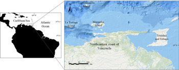
The artisanal fishing fleets based in Margarita Island (Venezuela) operate in a wide extent of the south-eastern Caribbean (10°35′–10°38′N 65°50–61°51′W; Figure 1). The fishing area extends from the marine zone near the continental coastline to ~50 km north of Margarita Island, and from the areas surrounding La Tortuga Island to the boundary with Trinidad and Tobago. The shark fishery is part of the multispecies fisheries exploiting seasonally abundant elasmobranchs and teleosts. The artisanal vessels involved in the fishery are primarily wooden, 8–14 m in length, and are equipped with either outboard or central motors (48–150 hp). The fishing gears used to capture sharks include mainly longlines, bottom-set gillnets and driftnets (Tavares et al., Reference Tavares, Sanchez and Medina2010).
Map of the study area, south-eastern Caribbean, where Mustelus higmani individuals were captured by artisanal shark fishery between 2015 and 2016. (Map sources: Esri, GEBCO, NOAA, National Geographic, Garmin, Here and Geinames.org).

Data collection
Between January 2015 and December 2016, 84 surveys (3–4 per month) were carried out at the fishing ports of Juan Griego and Los Cocos, two important localities for the fish trade in Margarita Island. Mustelus higmani individuals were sampled immediately after being landed and each specimen was examined and measured for total length (TL cm; with the caudal fin in a natural position) and sexed. Neonates were identified by the presence of an open umbilical scar. When possible, a random subsample of sharks was used for reproductive assessment. In males, the clasper length (CL cm) was measured along the inner margin from the anterior end of the cloaca to the tip of the clasper, and its calcification degree was also registered. Individuals were dissected in situ and the reproductive tracts were stored at −5°C and taken to the laboratory for analyses.
Size and sex composition
Size composition by sex was described by grouping the individuals in size classes of 2.0 cm TL. Seasonal variation in sex ratio by month (years combined) was evaluated using a χ2-test. Mean size at birth of M. higmani was estimated based on the TL of all sharks identified as neonates. Statistical analyses were performed using R software version 3.5.2 (R Core Team, 2018).
Reproductive parameters
In order to assess male and female sexual development and the onset of maturity a macroscopic maturity scale of proposed criteria by ICES (2013) for viviparous chondrichthyan species was applied. Slight modifications in the number of categories of this scale were done; the sixth stage for females (regenerating), and the fourth stage for males (regressing) were excluded. The maturity stages considered were: immature (I), developing (II), mature (III), pregnant (IV) and post-partum (V). Stages I–III are common for both sexes, and stages III–V considered as ‘mature’ for deriving estimates of the length at maturity.
For estimating the length at which 50% of individuals were sexually mature (TL50%), a logistic model was fitted to binomial maturity data (0 = unable to reproduce; 1 = able to reproduce), using TL classes of 5 cm in both sexes. The logistic function is as follows:  $y = \lpar {1 + e^{-\lpar {{\rm \beta }_0 + {\rm \beta }_1x} \rpar }} \rpar ^{{-}1}$; where, y is the proportion of mature individuals, x is the TL classes, β0 is an intercept term, β1 is the effect of size in terms of TL, and the value of TL50% is given by −β0/β1. Model parameters were estimated through maximum likelihood method using the binomial generalized linear model function in R software (R Core Team, 2018). Confidence intervals (95% CI) of the logistic regression curves were estimated by applying bootstrap resampling techniques (N = 1000) from fits of the logistic model to the maturity data. Variation in uterus width (UW mm), oviductal gland weight (OGW g), ovary weight (OW g), clasper length (CL cm) and testis weight (TW g), relative to total length (TL cm), were used to assess sexual development in both sexes. Other reproductive organ measurements (e.g. diameter of vitellogenic follicles, oviductal width, testis length and liver weight) did not provide conclusive information, and were then excluded from this relationship analysis.
Embryos found in pregnant females were examined, measured for TL (cm) and sexed. Uterine and ovarian fecundity were determined by quantifying the number of embryos occupying the uterus (UF) and the number of vitellogenic follicles in the ovary (OF), respectively. Linear regression techniques were applied to the relationships between maternal TL, and UF and OF in order to determine if fecundity increases with maternal size. To examine the reproductive cycle of females, the embryo size (TL cm) and diameter of the largest vitellogenic follicle (FD mm) were plotted against months (years combined). This information, together with the occurrence of neonates was also used to infer parturition time and ovulation. Additionally, embryo size and follicle diameter data were log-transformed and mean values were statistically compared between months by applying one-way analysis of variance (ANOVA-I; R Core Team, 2018).
Embryonic development
Pregnant (IV) (early, mid and late-term pregnant) and post-partum (V) females were randomly chosen for observation and photographic record of the gross morphology of the embryonic development. Descriptive embryology also included observation of the extraembryonic membranes, i.e. the yolk sac (YS) and the YS stalk and later the yolk sac placenta (YSP) and umbilical cord. The descriptions agree with terminology and concepts of Hamlett et al. (Reference Hamlett, Kormarik, Storrie, Serevy, Walker and Hamlett2005). Early pregnant female uteri were examined for the presence of uterine eggs, the length of which were measured (cm) along the cranial-caudal axis, considering their in utero position. Pre- and post-implantation stages were described according with the aspect of embryos and extraembryonic membranes in early, mid, late and term pregnant females. Particular attention was devoted to those embryonic development instances that provided morphological information on the processes of placentotrophy, especially when evidencing the transitional stages from lecithotrophy to placentotrophy/histotrophy.
Results
Size and sex composition
For the study period (January 2015 to December 2016) captures of M. higmani were comprised of 813 females between 23.2 and 72.5 cm TL, and 960 males between 22.6 and 62.5 cm TL (Figure 2). Size composition by sex also indicated that females reach a larger TL than males. Regarding sex proportion analysis (Figure 3), a significant difference was not observed from November to March (χ2 test, P > 0.05). However, females significantly predominated during April and May (χ2 test, P < 0.05), and males from June to October (χ2 test, P < 0.05). In all, 53 neonates ranging from 22.6–33.8 cm TL (mean: 28.7 ± 2.6 SD) were recorded during the study period, representing 3.0% of the total sample. These neonates were observed throughout the year, in April (N = 2), June (N = 5), July (N = 1), September (N = 2) and October (N = 2), but with a higher occurrence from November to February (N = 38).
Size composition by sex of Mustelus higmani individuals captured by artisanal shark fishery in the south-eastern Caribbean.

Monthly sex ratio (years combined) of Mustelus higmani individuals captured by artisanal shark fishery in the south-eastern Caribbean.

Reproductive parameters
A subsample of 143 females (34.5–72.5 cm TL) and 189 males (27.6–59.8 cm TL) was examined for reproductive state. The distributions of maturity stages for females were 2.1% immature (I), 4.9% developing (II), 26.6% mature (III), 55.2% pregnant (IV) and 11.2% post-partum (V), and for males were 15.3% immature (%), 20.1% developing (II) and 64.6% mature (III).
In females, the smallest mature individual was 44.0 cm TL and the largest immature specimen was 50.7 cm TL (Figure 4A–C). Uteri width (Figure 4A) starts active development from TL of 35.0 cm onward. Immature females (N = 3) had UW of up to 4.0 mm, undeveloped OGW and OW of up to 1.0 g. Developing females (N = 6, TL range of 39.5–50.7 cm) had UW of 4.0–12.2 mm, OGW of 0.08–0.18 g and OW of 0.1–0.8 g (Figure 4A–C). Over 47.0 cm of TL, a high overlap in the mature, pregnant and post-partum females was observed, for the three variables under study. Within this range, UW widely varied from 8.1–48.5 mm, with an abrupt growth from TL of 49.0 cm (Figure 4A). Differently, the growth of OGW and the OW with TL was steady but quite gradual (Figure 4B & C). Mature females (N = 38) had UW ranging from 8.1–39.6 mm, OGW from 0.1 to 1.1 g and OW from 1.1 to 9.2 g (Figure 4A–C). In pregnant females (N = 79) UW ranged from 10.0–48.5 mm, the latter corresponding to term pregnant females (Figure 4A). In this category, OGW varied from 0.1–1.2 g and OW from 0.4–12.6 g (Figure 4B & C). In post-partum females (N = 16) UW ranged from 14.3–37.3 mm; on the other hand, OGW varied from 0.4–1.2 g and OW from 1.2–9.6 g (Figure 4A–C). The length at maturity (TL50%) for females was estimated in 47.8 cm TL (95% CI: 45.7–50.0 cm), equating to 66.1% of maximum TL recorded (Figure 5A).
Relationship between the total length (TL) of female Mustelus higmani and (A) uterus width, (B) oviductal gland weight and (C) ovary weight according to maturity stages (immature, I; developing, II; mature, III; pregnant, IV; and post-partum, V).

Length at maturity for (A) females and (B) males of Mustelus higmani individuals captured by artisanal shark fishery in the south-eastern Caribbean. Shadow areas correspond to the 95% confidence intervals.

In males, the smallest mature individual was 45.0 cm TL and the largest immature specimen was 49.1 cm TL; with most individuals beginning to mature at ~47.0 TL (Figure 6A & B). Clasper length varied from 1.1–5.3 cm in immature (N = 29), 4.0–6.7 in developing (N = 38) and 5.0–6.5 in mature males (N = 122, Figure 6A). In immature males, TW ranged from 0.2–2.5 g, developing males from 1.0–3.6 g and mature from 1.7–5.3 g (Figure 6B). The length at maturity (TL50%) for males was estimated in 47.5 cm TL (95% CI: 46.1–48.6 cm), which equates to 80.5% of maximum TL recorded (Figure 5B).
Relationship between the total length (TL) of male Mustelus higmani and (A) clasper length and (B) testis weight according to maturity stages (immature, I; developing, II; and mature, III).

The number of embryos (UF) per female varied between 1 and 8 (mean: 3.8 ± 1.9 SD), and the TL of the embryos ranged from 3.0–26.5 cm. A weak relationship (r 2 = 0.24) between maternal TL and UF was found, but with a significant positive slope (t-test; t = 2.6, P < 0.05). The analysis to compare sex proportion of embryos did not show significant difference between females and males (χ2 test, P > 0.05). Mean embryo size by month varied between 6.6 (± 1.0 SD) and 22.6 (± 1.1 SD) cm TL, with maximum values observed from September to February (Figure 7A). Comparison between monthly mean values was highly significant (ANOVA-I, F = 41.7, P < 0.000). The number of vitellogenic follicles (OF) ranged between 1 and 9 (mean: 4.7 ± 1.8 SD). The relationship between TL and OF was also weak (r 2 = 0.30) with a highly significant positive slope (t-test; t = 6.3, P < 0.000). Regarding the seasonal variation in follicle growth, the analysis showed that mean diameter by month varied between 9.5 (± 3.2 SD) and 11.7 (± 1.1 SD) mm FD, with maximum values recorded at the end of the year, between September and December (Figure 7B). However, the analysis to test for statistical difference between mean follicle diameters by month was not significant (ANOVA-I, F = 1.4, P > 0.05). The information on embryo and follicle development, together with the occurrence of neonates showed that time of parturition and mating season of M. higmani may occur throughout the year, though both processes may be more frequent between November and February. Six pregnant females (51.5–65.1 cm TL) simultaneously bearing vitellogenic follicles and embryos in an advanced stage of development were recorded in February (N = 2), June (N = 1), August (N = 1), September (N = 1) and December (N = 1), indicating an annual reproductive cycle. However, the length of gestation could not be determined as both folliculogenesis and gestation are not synchronized to the population level.
Seasonal variation in (A) embryo size (TL) and (B) diameter of the largest vitellogenic follicle (FD mm) by months (years combined) of Mustelus higmani individuals captured by artisanal shark fishery in the south-eastern Caribbean.

Descriptive gross morphology of placentotrophic embryonic development
Pre-implantation stages
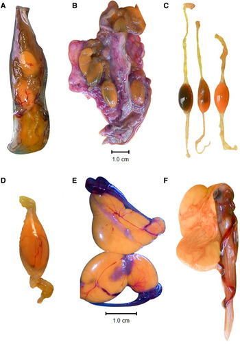
Uterine eggs were observed in seven early pregnant females and had lengths ranging from 2.1–2.5 cm (N = 3). The encapsulated eggs were ellipsoid in shape. The number of uterine eggs per female varied from 1–6 (mean: 2.9 ± 1.7 SD). External eggs colouration varied from yellow to amber (Figure 8A–D). The diagonal position of the uterine eggs inside the uterus indicates that compartments formation may be in early progress (Figure 8A). In uterine eggs bearing females, the uterine epithelium was highly vascularized (Figure 8B). The tertiary egg envelope was light yellow and folded on the egg edges, forming the egg envelope reservoir; and once stretched; the egg occupied the centre of the envelope (Figure 8C & D).
Instances of early and mid-gestation during the pre-implantation stages of the placentotrophic embryonic development of Mustelus higmani, captured by artisanal shark fishery in the south-eastern Caribbean. (A) An early pregnant uterus of a 62.1 cm TL female captured on 20 June 2016, soon after fertilization, with two uterine eggs. (B) A complete set of posterior oviducts extracted from a 62.0 cm TL pregnant female captured on 30 August 2016. The dissection of both early pregnant uteri revealed three uterine eggs per uterus. Compartments are not evident but the uterine epithelium shows rich vascularization. (C) Three uterine eggs with posterior and anterior tertiary egg envelope edges stretched (egg envelope reservoir), extracted from the uterus of a 51.5 cm TL female captured on 20 June 2016. The cause of the variation of colouration on the uterine eggs (yellow to amber) is unknown. (D) A single intrauterine egg extracted in utero from a 62.1 cm TL early pregnant female captured on 20 June 2016. The uterine egg had 2.1 cm of length. The egg envelope reservoir was photographed in its natural intrauterine position (folded), visible on both envelope edges. (E) Two embryos extracted from a 59.0 cm TL early pregnant female captured on 24 June 2016. At the embryos' (~3.0 cm TL) cranial end, external gill filaments are visible. Yolk sac at this stage becomes withered. (F) A 6.0 cm TL embryo extracted in utero from a 57.5 cm TL early-mid-pregnant female captured on 6 September 2016 presenting an irregularly shaped YS. The external YS stalk has turned into a conspicuous umbilical cord visible around the embryo.

The smallest visible recorded embryos were 3.0 cm TL and bore external highly vascularized gill filaments (Figure 8E). In these embryos, the YS withered and started to lose the typical round shape to assume an irregular one, as visible in embryos of 6.0 cm TL onward (Figure 8F). Embryos of 3.0–6.0 cm TL presented conspicuous, lengthened YS stalks, vitelline artery and vitelline vein visible by transparency through the omphalopleure (Figure 8E & F).
Post-implantation stages
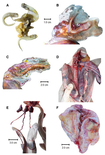
On a 6.0 cm TL embryo the external yolk sac was no longer visible as such and the umbilical cord was connected to the proximal aspect of the YSP, indicating that implantation had occurred (Figure 9A). The umbilical cord was formed by an umbilical artery and umbilical vein. On the other hand, a 5.0 cm TL embryo (the other uterus contained a 5.7 cm embryo) showed a YS anchored to the uterus epithelium, suggesting the recent formation of the placental connection.
Instances of mid and late gestation during the pre- and post-implantation stages of the placentotrophic embryonic development of Mustelus higmani, captured by artisanal shark fishery in the south-eastern Caribbean. (A) Three 6.0 cm TL embryos extracted from a 55.4 cm TL female captured on 23 February 2016. Yolk sacs are no longer visible and the umbilical cords are connected to the proximal aspect of the YSP. (B) A 5.0 cm TL embryo lying on the dissected uterus, extracted from a 48.5 cm TL female, captured on 16 June 2016. The YS has turn into the embryonic portion of the YSP, anchored to the uterus epithelium. (C) A 10.6 cm TL embryo (the other uterus contained an embryo of 10.9 cm TL) extracted from a 52.5 cm TL mid to term pregnant female captured on 20 June 2016. The embryo is lying on the dissected uterus. The jelly umbilical cord shows a conspicuous ductus vitellointestinalis filled with yolk. Note the abundant periembryonic fluids in both (B) and (C). (D) A dissected 56.8 cm TL term pregnant female, bearing two embryos per uterus (TL of 23.3, 22.7, 23.2 and 23.3 cm) captured on 12 November 2015. The embryos can be seen by transparency across the thin uterus wall. Uteri width was 3.2 cm. Left ovary is visible, presenting ripe vitellogenic follicles. (E) Three term embryos extracted from a term pregnant female (62.0 cm TL) captured on 19 February 2016. The embryos, with full term placenta (embryonic aspect) and smooth umbilical cord, had TL of 25.6, 26.1 and 25.9 cm. (F) Empty uteri of a post-partum 63.5 cm TL female captured on 12 September 2016. The stretched, distended uterine wall is internally embedded by a yellowish histotrophic secretion which avoids visualizing the umbilical scars.

The jelly, diaphanous umbilical cord of a 10.6 cm TL embryo, revealed a conspicuous ductus vitellointestinalis filled with yolk and a term YSP. The female bearing this embryo bore another one, on the other uterus, with TL of 10.9 cm. Abundant periembryonic fluids were noted in the two latter cases (Figure 9B & C). Four term embryos (two per uterus) with TL of 23.3, 22.7, 23.2 and 23.3 cm, were attached to full term placentas (Figure 9D). Term placentas were also recorded in three term embryos measuring 25.6, 26.1 and 25.9 cm TL. The umbilical cord of M. higmani was smooth along its whole surface, with no ornamentations (Figure 9E). Vitellogenic follicles in the ovaries were present in term pregnant and post-partum females (Figure 9E). The term placenta had a diameter of around 2.0 cm. Post-partum uteri had distended uterine walls and were internally embedded by an abundant yellowish histotrophic secretion (Figure 9F).
Discussion
The commercial landings of M. higmani in the study area comprised a wide range of lengths, 23.2–72.5 cm TL in females and 22.6–62.5 cm TL in males, but with a predominance of captures in the ranges of 52.0–66.0 and 46.0–56.0 cm TL for females and males, respectively. The pattern of size composition observed in this study is similar to those previously reported for the same species in other studies conducted in the north-eastern coast of Venezuela (Tavares et al., Reference Tavares, Sanchez and Medina2010; Marquez et al., Reference Márquez, Tavares and Ariza2019). The sexually dimorphic maximum sizes (favouring M. higmani females) appear to be a common characteristic of mustelid sharks, having also been reported for M. canis from the east coast of the USA (Conrath & Musick, Reference Conrath and Musick2002) and northern Brazil (Zagaglia et al., Reference Zagaglia, Damiano, Hazin and Broadhurst2011), M. schmitti in common waters of Uruguay and Argentina (Oddone et al., Reference Oddone, Paesch and Norbis2005; Belleggia et al., Reference Belleggia, Figueroa, Sánchez and Bremec2012), M. henlei from the Gulf of California, Mexico (Perez-Jimenez & Sosa-Nishizaki, Reference Perez-Jimenez and Sosa-Nishizaki2008) and M. asterias in the north-eastern Atlantic (Farrell et al., Reference Farrell, Mariani and Clarke2010; McCully Phillips & Ellis, Reference McCully Phillips and Ellis2015). The low number of M. higmani caught at a small size (22.0–36.0 cm TL) was likely related to lower gear selectivity (samples were mainly from gillnets; mesh size >7.6 cm) and/or to fishing areas. The capture of small specimens (with lengths around the mean size at birth, 28.7 cm TL) by the recreational reel and rod fishery conducted from land in Margarita Island (Tavares, pers. obs.) suggests that the primary nursery areas of M. higmani are distributed near the coast; and then, these shallow areas are out of the depth range for artisanal fishing operations.
Spatial and sex segregation is a widespread behaviour exhibited by several animal groups, including elasmobranchs, and is commonly associated with reproduction, movement patterns and nutritional requirements (Klimley, Reference Klimley1987; Wearmouth & Sims, Reference Wearmouth and Sims2008; Heist, Reference Heist, Carrier, Musick and Heithaus2012). In several shark populations, seasonal variation in sex proportion appears to be related to differences in the degrees of site fidelity (philopatry) or dispersal by sex (Heist, Reference Heist, Carrier, Musick and Heithaus2012; Portnoy et al., Reference Portnoy, Puritz, Hollenbeck, Gelsleichter, Chapman and Gold2015). However, during reproductive periods, the receptivity of females towards males increases and this generally leads to observing similar sex proportions (Magurran & Garcia, Reference Magurran and Garcia2000). In the present study, the observed sex proportion did not differ from the expected ratio (1:1) during the months of November–March, suggesting that this is the main mating period of M. higmani.
Females and males of M. higmani matured at similar sizes in the study area with individuals beginning to develop at ~44.0 cm TL. The TL50% estimates obtained in this study were 47.8 cm TL for females and 47.5 cm TL for males. The first TL50% estimates reported by Tavares et al. (Reference Tavares, Sanchez and Medina2010) were higher for both females (59.9 cm TL) and males (52.5 cm TL), and this discrepancy might be a consequence of differences in sample sizes. In another recent study, Tagliafico et al. (Reference Tagliafico, Hernández-Ávila, Rangel and Rago2015) reported TL50% of 46.7 cm TL for females and 47.6 cm TL for males, similar values to those obtained in our study but in contrast these authors found a TL50% of males over the females. Although female M. higmani reached higher maximum sizes than males, the lengths at maturity observed in the present study were close between sexes. The phenomenon of bimaturism in sharks (Cortes, Reference Cortes, Carrier, Musick and Heithaus2004), where males mature at a smaller size/age than females has been commonly found for congeneric species such as M. canis (Conrath & Musick, Reference Conrath and Musick2002; Zagaglia et al., Reference Zagaglia, Damiano, Hazin and Broadhurst2011), M. schmitti (Oddone et al., Reference Oddone, Paesch and Norbis2005), M. henlei (Perez-Jimenez & Sosa-Nishizaki, Reference Perez-Jimenez and Sosa-Nishizaki2008) and M. asterias (Farrell et al., Reference Farrell, Mariani and Clarke2010; McCully Phillips & Ellis, Reference McCully Phillips and Ellis2015).
The fecundity of M. higmani in terms of both uterine fecundity (1–8 embryos) and ovarian fecundity (1–9 vitellogenic follicles) seemed to be relatively low. The similarity between uterine and ovarian fecundity suggests that most fully-yolked oocytes ovulate during the process and settle towards the uterus. Litter size estimates previously documented for M. higmani are within the range of 1–10 (Heemstra, Reference Heemstra1997; Tavares et al., Reference Tavares, Sanchez and Medina2010; Tagliafico et al., Reference Tagliafico, Hernández-Ávila, Rangel and Rago2015). Most Mustelus species exhibit higher fecundity compared with M. higmani; with maximum litter sizes >18 embryos (Capapé, Reference Capapé1983; Conrath & Musick, Reference Conrath and Musick2002; Perez-Jimenez & Sosa-Nishizaki, Reference Perez-Jimenez and Sosa-Nishizaki2008; Saidi et al., Reference Saidi, Bradai and Bouain2008; McCully Phillips & Ellis, Reference McCully Phillips and Ellis2015; Gonzalez-Pestana et al., Reference Gonzalez-Pestana, Alfaro-Shigueto and Mangel2019). Our findings showed an increase in fecundity (UF and OF) with maternal size for M. higmani, a reproductive characteristic that is commonly observed among elasmobranch species (Conrath & Musick, Reference Conrath, Musick, Carrier, Musick and Heithaus2012).
The information on the capture of neonates, the seasonal development of embryos and follicles, and the sex proportion suggest that time of parturition, ovulation and mating of M. higmani occurred during a main period of several months, between November and February. An annual and continuous reproductive cycle was supported by the capture of pregnant females carrying concomitantly pre-ovulatory vitellogenic follicles and embryos in an advanced stage of development. This pattern is typical of placentotrophic species that reproduce annually, like Rhizoprionodon terraenovae and M. canis (Hamlett et al., Reference Hamlett, Kormarik, Storrie, Serevy, Walker and Hamlett2005). Only the left ovary is present in M. higmani, in contrast to other species of the genus Mustelus (Daniel, Reference Daniel1928; TeWinkel, Reference TeWinkel1950). Although the gestation period could not be determined due to the wide variability in the ovarian cycle and embryo development, it probably encompasses between 11 and 12 months taking into account the findings related to the main season of parturition and the characteristics of the reproductive cycle. For most studied species of the genus Mustelus, an annual reproductive cycle with gestation periods ranging from 10–12 months has been reported (Conrath & Musick, Reference Conrath and Musick2002; Oddone et al., Reference Oddone, Paesch and Norbis2005; Perez-Jimenez & Sosa-Nishizaki, Reference Perez-Jimenez and Sosa-Nishizaki2008; Saidi et al., Reference Saidi, Bradai and Bouain2008). However, other species with non-placentotrophic reproduction displayed a biennial reproductive cycle, for example M. manazo in waters of Japan and Taiwan (Yamaguchi et al., Reference Yamaguchi, Taniuchi and Shimizu2000), M. antarticus off southern Australia (Walker, Reference Walker2007) and M. asterias from the north-east Atlantic (Farrell et al., Reference Farrell, Mariani and Clarke2010).
The lack of a defined seasonal pattern in relation to the development of embryos and follicles in M. higmani could be explained in part by the stability of the environmental conditions in the study area, compared with subtropical and temperate regions. The north-eastern shelf of Venezuela is located in a central tropical region of the Caribbean, and is characterized by a wide diversity of fishing resources (FAO, 2020). However, the apparent main season for parturition and mating registered during November–February appears to be related with the seasonal upwelling system that influences the north-eastern coast of Venezuela. This oceanographic event has a major influence during the first months of the year (January–April) when strong winds drive the mixing of water masses and nutrients (Castellanos et al., Reference Castellanos, Varela and Muller-Karger2002; Rueda-Roa & Muller-Karger, Reference Rueda-Roa and Muller-Karger2013); and then contributes to the high marine productivity by increasing primary production and abundance of marine fauna in the region (Freon & Mendoza, Reference Freon and Mendoza2003). In the study area, water temperature varies between 20.5 and 29.7°C during the principal and relaxation upwelling periods, respectively (Castellanos et al., Reference Castellanos, Varela and Muller-Karger2002; McConnell et al., Reference McConnell, Thunell, Lorenzoni, Astor, Wright and Fairbanks2009). The synchronization of parturition with the upwelling period is advantageous because it provides abundant food for neonates and juvenile sharks. In the Venezuelan Caribbean, the distribution and abundance of important groups of clupeid fish and oceanic sharks also seem to be influenced by the seasonal upwelling system (Tavares & Arocha, Reference Tavares and Arocha2008; Rueda-Roa et al., Reference Rueda-Roa, Mendoza, Muller-Karger, Cárdenas, Achury and Astor2017).
Within the genus Mustelus, placentotrophic and non-placentotrophic (previously referred to as ‘aplacental’) embryonic development strategies can be found (Teshima, Reference Teshima1981). Non-placentotrophic strategies in Mustelus include strict lecithotrophy (Yamaguchi et al., Reference Yamaguchi, Taniuchi and Shimizu2000) and minimal histotrophy (Storrie et al., Reference Storrie, Walker, Laurenson and Hamlett2009; Orlando et al., Reference Orlando, Pereyra, Silveira, Paesch, Oddone and Norbis2015). The morphology of the yolk sac placenta of M. canis has been largely studied among Mustelus species (TeWinkel, Reference TeWinkel1963a, Reference TeWinkel1963b). However, for the remaining Mustelus species declared as placentrotrophic, including M. higmani, the placenta have received little or no attention regarding the general gross morphology, changes during the embryonic developmental stages or other basic aspects. TeWinkel (Reference TeWinkel1950) observed that the spherical shape of the vitellogenic follicles of M. canis is lost during its passage through the anterior oviduct and oviducal gland, where it is compressed into an ellipsoid shape, just as observed in M. higmani. This author also reported brownish colouration in M. canis uterine eggs, after fertilization. Our observations demonstrated that the YSP implantation in M. higmani occurs when embryos attain a TL of about 5.0–6.0 cm. From this size onward, the YSP develops until term, when attaining a diameter of around 2.0 cm and embryos have grown up to about 20.0 cm TL.
Despite the same embryological origin, there is a wide morphological diversity in the shark YSP. Basically, there are two major types (Hamlett et al., Reference Hamlett, Kormarik, Storrie, Serevy, Walker and Hamlett2005); the first group including those placentae with two morphological distinct portions (proximal and distal), as seen in Carcharhinus plumbeus and R. terranovae. The second group includes placentae with no distinct proximal portion, as described for M. canis (Hamlett et al., Reference Hamlett, Musick, Hysell and Sever2002). The YSP of M. higmani belongs to the second group. Moreover, as in most placentotrophic sharks, the umbilical cord of M. higmani is smooth-surfaced, unadorned (Lombardi, Reference Lombardi1998), with no appendiculae (Compagno, Reference Compagno1988).
Periembryonic fluids were observed in all embryonic stages of M. higmani on record. However, uterine secretion seemed to intensify and change in composition by the end of gestation, according with the observation of late pregnant and post-partum females' uterine internal lining, with abundant yellowish, thicker secretion. In spite of the fact that further studies are needed (coupled with histology), our observations lead us to hypothesize that M. higmani histotrophy intensifies late in gestation. According to Hamlett (Reference Hamlett1989), during the initial lecithotrophic phase the uteri of placentotrophic shark species are able to produce a histotroph (mucoid secretion), which nourishes the embryos during the period between yolk depletion and placenta establishment.
In M. higmani 3.0 cm TL embryos had external gills, which represent nutrient uptake sites, as in other matrotrophic development modes (Hamlett et al., Reference Hamlett, Kormarik, Storrie, Serevy, Walker and Hamlett2005). Also, once the YSP has formed, paraplacental sites are able to produce histotroph with nutritive or uterine lubricant functions (Hamlett, Reference Hamlett1989). In M. canis, the lecithotrophic phase takes 12 weeks. Immediately after (experimentally demonstrated), paraplacental histotrophy takes place, and the yolk sac placenta is formed two weeks later, 14 weeks after fertilization (Hamlett et al., Reference Hamlett, Kormarik, Storrie, Serevy, Walker and Hamlett2005). Even though these time estimates cannot be assessed for M. higmani, according to our observations, the period between the yolk sac depletion and the settlement of the YSP in this species may happen when embryos have TL of around 6.0 cm.
The present study showed that the local population of M. higmani had a relatively high productivity compared with other shark species; however, M. higmani is among the Mustelus species with a lower fecundity. Though commonly neglected, the study of embryology, altogether with reproductive parameters, can throw light on key periods in the life history of the species, especially for those in which biological aspects are still unknown. Although M. higmani has been subject to fishing for decades, its gross embryonic development had not been studied before. Future studies may focus on the reproductive cycle of M. higmani, including the assessment of gestation and the duration of the three major nourishment embryonic phases (lecithotrophy, histotrophy and placentotrophy), as well as the presence of ovarian resting periods. Furthermore, in Venezuela this species is not under any fishing control or regulation by the governmental institutions with responsibility in fishing administration. Taking into account the sizes at maturity obtained in this study, captures comprised 22.8 and 36.6% of immature females and males, respectively. Although more detailed research and fishing monitoring are needed in order to generate accurate data for development of population dynamic models, basic management measures must be implemented such as minimum sizes of capture and fishing restrictions in essential habitats.
Acknowledgements
We are indebted to fishermen (fishing ports of Juan Griego and Los Cocos, Margarita Island) for assisting in fieldwork. Zaida Cuyare, Ramón Escalona and all who provided invaluable help and support during sampling surveys are thanked. We dedicate this work to the memory of Carmen Velazquez, for her wonderful fieldwork and support.
Financial support
This project was funded in part by the Elasmobranch Fishing Monitoring Program of the Instituto Nacional de Investigaciones Agricolas (INIA); and the Centro para la Investigacion de Tiburones (CIT-Venezuela). Marioxis Macias-Cuyare was partly funded by Idea Wild.