Introduction
Ventricular septal defects are the most common congenital heart malformation accounting for almost 10% of all defects and can be located in membranous or muscular septum. Eighty percent of these are perimembranous. Reference Mavroudis, Backer and Jacobs1 Yilmaz and colleagues have reported that perimembranous aneurysmal ventricular septal defect was found in 20% of perimembranous ventricular septal defect Reference Yilmaz, Ozal, Arslan, Tatar and Ozturk2 that can progress in infancy to a functionally smaller defect with aneurysmal formation, Reference Moe and Guntheroth3 which reduces the ventricular septal defect size and allows transcatheter closure with fewer complications.
Transcatheter closure of congenital ventricular septal defect for specific and selected patients has been advocated as an alternative to surgical repair. Reference Bol-Raap, Weerheim, Kappetein, Witsenburg and Bogers4,Reference Arora, Trehan, Kumar, Kalra and Nigam5 However, earlier attempts at percutaneous closure of perimembranous ventricular septal defect were abandoned because of incidence of heart block likely as a result of device rigidity and/or oversizing. At the present time, there is no readily available device for percutaneous closure of perimembranous ventricular septal defect.
Softer devices have been introduced to the market as the AmplatzerTM vascular plug (Abbott Cardiovascular; Plymouth, MN, USA), second generation, (AVP II). The AVP II is a self-expanding nitinol wire mesh vascular embolisation device derived from the Amplatz septal occluder. The wire calibre ranges from 0.015 to 0.040 inch. Since it was first described, therapeutic embolisation has expanded in both its use and indications. The plugs come in sizes ranging from 4 to 22 mm in 2 mm increments. The device is attached to a delivery cable with screw mechanism and can be delivered from either the arterial (retrograde) or venous (prograde) approach. The AVP II has no occlusive fabric rendering its relatively soft and easily advanced through a guide catheter. A 5 Fr guide catheter is needed for 4–8 mm devices, 6 Fr for 10–12 mm devices, and 8 Fr for 14–16 mm plugs. Reference Tuite, Kessel, Nicholson, Patel, McPherson and Shaw6 The three discs of the AVP II are of the same diameter but variable length and separated by small connecting waists. The lack of centreing mechanism of the AVP II is one of the advantages for decreasing the possibility of pressure on the AV node or his bundle.
For a successful ventricular septal defect closure, an anatomic delineation of the defect with its relation to other cardiac structures is needed. The presence of an aneurysm in the membranous septum is a frequent finding, Reference Ramaciotti, Keren and Silverman7 which is useful as the device disk could be accommodated within the aneurysmal sac. Pedra et al. Reference Pedra, Pedra and Esteves8 pointed out that depending on the anatomy, the device is positioned either completely in the aneurysm or with the left ventricular disk at the crest of the defect, minimising interference with the tricuspid or aortic valves.
El Said et al. Reference El Said, Bratincsak, Gordon and Moore9 proposed three different locations for measurement of the defect: at the left ventricular opening of the ventricular septal defect; at the largest diameter of the aneurysm, usually in the midsegment of the aneurysm; and at the right ventricular opening of the ventricular septal defect aneurysm, usually the smallest diameter of the defect. There has been hypothesised that the AVP II may have potential advantages in the transcatheter closure of perimembranous ventricular septal defect. The device is designed with three parts to its body: the distal, the central, and the proximal disk. Leveraging this unique design, we conceptualised the distal disk to lay in the right ventricle side of the ventricular septal aneurysm, theoretically preventing the device from embolising into the left-sided circulation. The central disk fills the ventricular septal aneurysm, whereas the proximal disk lies on the left ventricular outflow tract covering the left ventricle side of the defect.
Retrograde transcatheter device closure of ventricular septal defect has been described in some studies. Reference Hijazi, Hakim, Al-Fadley, Abdelhamid and Cao10–Reference Holzer, Balzer, Lock Qi-Ling Cao and Hijazi12 This approach is simple as it avoids the circuitous route of antegrade approach by femoral vein with the use of large stiff delivery sheaths, which may cause haemodynamic instability while crossing the tricuspid valve, the defect or the aortic valve, and also shortens the total procedure time, decrease radiation exposure to the patient and operators, and decrease the procedure cost, which represents an important issue in developing countries.
In our institution, selected patients with perimembranous ventricular septal defect were offered percutaneous closure using this special device, the AVP II.
Materials and methods
A retrospective review was carried out of 45 consecutive cases of perimembranous ventricular septal defect using the AVP II from April 2016 through September 2024. Institutional review board approval was obtained.
We recorded the patient’s age, weight, height, the defect size, other procedures performed during the same intervention and complications related with the catheterisation procedure. We also assessed the tricuspid and aortic valve for regurgitation before and after the procedure.
Patients were included if they (1) had perimembranous ventricular septal defect (located in the membranous septum bellow the crista supraventricularis, a relatively small portion of the septum located near the heart valves); (2) had left ventricle dilation by echocardiogram; and (3) had ventricular septal defect closure using generation II of the AmplatzerTM vascular plug device (Abbott Cardiovascular; Plymouth, MN, USA). Patients were excluded if they (1) had residual defects after surgery; (2) had postinfarction ventricular septal defect; (3) had ventricular septal defect closure with different devices from the one mentioned before; (4) had pulmonary hypertension; and (5) had association with infective endocarditis during the time of patient selection. For device selection, we measured the narrowest portion of the ventricular septal defect based on measurement of the right ventricular side of the defect during left ventricle angiogram, and when selecting AVP II, we chose not to exceed 2 mm at the right ventricle defect size measured by fluoroscopy in patients with aneurysmal defects (55% of all the patients), and in the presence of these type of defects, the length of the aneurysm was not relevant for the selection of the device.
It is not always necessary to measure the length of the ventricular septal defect to select the device to be used. However, ideally, the exit site at the right ventricle should be measured to select the device. It is worth emphasising that the softness of the device and the appropriate selection of size that is inserted into the aneurysm make problems due to distortion of the aortic valve unlikely.
We considered a procedural success to the ability of deploying the device without embolisation; no new onset valvular regurgitation or increase in valvular insufficiency (if it was present before ventricular septal defect closure); or the absence of complete heart block.
Cardiac catheterisation
An informed consent form was given to the relatives of the patients, and a complete left and right heart catheterisation with exclusive femoral arterial access was done. All procedures were performed under general anaesthesia. Intravenous heparin was used during the whole procedure at a dose of 100 United Kingdom (maximum dose of 5000 UI). Evaluation of the ventricular septal defect was thoroughly investigated with left ventricular angiography in four chambers view (Figure 1). Transesophageal echocardiogram was used in 10 patients and transthoracic echocardiogram in seven patients; in most of our patients, we do not use echo guidance.
Initial four chambers view angiography. Arrow shows aneurysmal pmVSD with two outlets at the RV side (arrowheads). pmVSD = perimembranous ventricular septal defect; RV = right ventricle.

In all the patients, we used retrograde approach. Using the femoral approach, the perimembranous ventricular septal defect was usually crossed by the aid of a retrograde advanced Judkins right type catheter, directing a soft tipped wire as a 0.035 inch hydrophilic coated TerumoTM guidewire (Terumo Medical Corp., Somerset, New Jersey) into the right ventricle or pulmonary artery. The Judkins right catheter was replaced with the CordisTM 5 Fr or 6 Fr Judkins right guiding catheter in the majority of cases and 7 Fr just in one case, and the Amplatzer vascular plug II was advanced to the tip of the catheter. Under fluoroscopy, the distal disk of the device was deployed in the right ventricle; then, the complete system was pulled slowly, and the waist and the proximal disk of the device were deployed just in the left ventricle side of the defect (Figure 2a). In the case of perimembranous ventricular septal defect with more than one defect (enter hole), we used two devices (Figure 2b– 2c).
Four chambers fluoroscopy. 2(a). Arrow shows AVP II device with distal disk at the RV side, waist and proximal disk inside of the aneurysm in the LV side. 2(b). Arrow shows AVP II device inside one outlet hole; arrowhead shows residual shunt in a second outlet hole. 2(c). Arrows show two AVP II devices inside the two outlet holes of the aneurysmal pmVSD. AVP II = AmpatzerTM vascular plug II; RV = right ventricle; LV = left ventricle; pmVSD = perimembranous ventricular septal defect.

Proper position and residual shunt were confirmed with left ventricle angiography and with transthoracic echo or transesophageal echo in some cases. After achieving good position of the device, we evaluated for any evidence of regurgitation of the aortic valve with angiography at the aortic root, and finally, device was released and left ventricle angiography was repeated. Next morning, electrocardiogram and transthoracic echo were done.
Follow-up
Patients left the hospital 24 hours after the procedure with prophylactic antibiotics and aspirin at a dose of 5 mg/kg (maximum dose of 100 mg) for 6 months. Follow-up visits at 1, 3, 6, and 12 months and then yearly to perform clinical examination, electrocardiogram, and a transthoracic echo. We chose this scheme of visits for the purpose of assessing the position of the device, possible residual shunt, presence of aortic, tricuspid or mitral regurgitation and documenting cardiac rhythm.
Statistical analysis
Frequencies and percentages expressed categorical variables. Continuous variables were summarised as mean (SD) or median (range) as appropriate, depending on normal distribution. The statistical analysis was done using the SPSS version 19.0 and Microsoft Excel 2016 for Mac.
Results
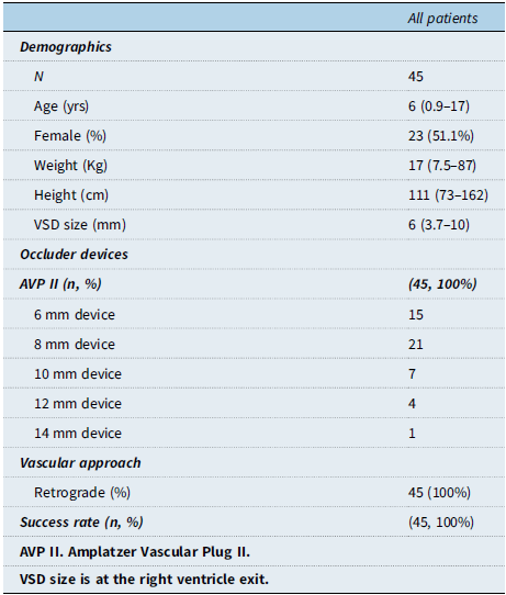
All participants who met the inclusion criteria were sent to the catheterisation laboratory with the intention to treat the defect percutaneously. A total of 45 patients underwent AmplatzerTM Vascular Plug-II (Abbott Cardiovascular; Plymouth, MN, USA) transcatheter closure of their perimembranous ventricular septal defect. The median age was 6 years (range: 0.9–17). Their median weight was 17 kg (range: 7.5–87). The perimembranous ventricular septal defect median defect size at the right ventricle side was 6 mm (range: 3.7–12).
The retrograde approach was used in all the patients. This group included 23 female patients (51.1%) with a group median age of 4 years (range from 2 to 17 years) and weight of 16 kg (12.5–63). Demographic data is shown in Table 1.
Demographic characteristics of the patients

The mean systolic pulmonary arterial pressure was 30.98 ± 6.5 mmHg. The mean of the pulmonary and systemic flow (Qp/Qs) was 1.5 ± 0.2.
The AVP II 6 mm was used in 15 patients each; the AVP II 8 mm was used in 21 patients; the AVP II 10 mm was used in 7 patients; the AVP II 12 mm was used in 4 patients; and the AVP II 14 mm was used in one patient only. Fenestrated ventricular septal aneurysm was also seen. In three patients, there was fenestrated ventricular septum aneurysm with two right ventricle outlet defects during left ventricle angiography, and in one patient, two AVP II size 6 mm were used to close both defects; also another patient has two right ventricle outlet ventricular septal defect during left ventricle angiography where AVP II size 8 mm and 10 mm were selected and finally, a third patient has two right ventricle outlet ventricular septal defect where AVP II 10 mm and 14 mm were used for closure of the defects.
Previous to the procedure, a total of four patients had prolapse of aortic cusp with no aortic regurgitation; one patient had aortic insufficiency and six patients mild mitral insufficiency as well. After the devices were implanted, we found 10 patients (22.2%) with trivial residual shunt across the device evidenced by the angiography during the procedure. In one patient, we used two different devices in the same aneurysmatic defect. Two AVP II 6 mm and the last device was embolised to the right pulmonary artery. The device was capture with a snare, and the residual defect was closed with another AVP II 8 mm device. In some of our patients, we found association with other cardiac defects so we performed successfully during the same procedure, balloon aortoplasty in one patient, patent ductus arteriosus closure in two patients, and atrial septal defect closure in one patient. No increase of previous aortic or mitral valve regurgitation was evidenced by angiography or by transthoracic echo 24 hours after the procedure.
All patients were discharged 24 hours after the procedure. At a mean follow-up of 48 ± 25.7 months, 42 patients showed complete closure of the defect and 3 patients had trivial residual shunt across the device. It is important to mention that we found a good device position at the 1-month follow-up transthoracic echo. No atrioventricular block was found in electrocardiogram, and no increase in previous aortic or mitral regurgitation was found by transthoracic echo during the follow-up.
Discussion
The ventricular septal defect is the most common CHD, comprising 10% of all forms of congenital cardiac defects. Reference Anderson, Becker and Tynan13 The percutaneous closure of the ventricular septal defect has emerged as an important alternative to surgical repair. Initially, the success rates were not high and the complication rate (complete heart block) was a very important limitation.
Perimembranous ventricular septal defect closure using the AmplatzerTM perimembranous ventricular septal defect occluder device has largely been abandoned because of the risk of heart block. In a review published in 2005 by Carminati et al. Reference Carminati, Butera, Chessa, Drago, Negura and Piazza14 where 122 patients were catheterised, there was a 2.5% risk of heart block. In the follow-up, delayed complete heart block occurred in two patients with perimembranous ventricular septal defect, for which a permanent pacemaker was inserted.
Since this early experience, some authors have published case series data on ventricular septal defect closure using different generations of devices in off-label application. AVP II device received FDA approval for use in September 2007 for a various procedure with numerous published reports describing their utility for vascular occlusion procedures. Ebeid et al. Reference Ebeid, Batlivala and Salazar15 in a retrospective review and data reported a total of 20 patients using the Amplatzer vascular plug II in 9 patients and Amplatzer duct occluder II in 11 patients (10 delivered retrograde and 10 prograde). Hua et al. Reference Hua, Aquino and Owada16 reported in a 4-year retrospective chart review of patients with perimembranous ventricular septal defect associated with ventricular septal aneurysm the use of AVP II in 16 patients. Shokka et al. Reference Shokka, Diaa, Gebaly and Hossam17 reported the use in an off-label manner of the AVP II in 12 patient with aneurysmal pmVSD with a complete closure of 92% at the last follow-up. Our series reported on perimembranous ventricular septal defect closure in 45 patients using AVP II devices with a high success rate of 100%. There are reports of use of the AVP in the paediatric cardiac literature for treatment of aortopulmonary collaterals, pulmonary arteriovenous malformations, venovenous collaterals, shunts, coronary fistulas, and certain types of patent ductus arteriosus. Reference Diehm, Kickuth and Silvestro18–Reference Rossi, Rebonato and Greco20 We believe that the AVP II device could be used in all types of perimembranous ventricular septal defect.
For an appropriate selection of these percutaneous devices, Ebeid et al. Reference Ebeid, Batlivala and Salazar15 suggested when the AVP II was used, it was chosen to be 6–7 mm larger than the ventricular septal defect size by colour Doppler, which would be equivalent to the AmplatzerTM duct occluder II (ADO II) retention disc size. El Said et al. Reference El Said, Bratincsak, Gordon and Moore9 proposed three different locations for measurement of the defect: at the left ventricular opening of the ventricular septal defect; at the largest diameter of the aneurysm, usually in the midsegment of the aneurysm; and at the right ventricular opening of the ventricular septal defect aneurysm, usually the smallest diameter of the defect, where they proposed the use of a device 2 mm larger than this last measurement. As mentioned previously, we measured the narrowest portion of the ventricular septal defect on the right ventricular side during the diastolic left ventricular angiogram, and the devices were chosen not to exceed this diameter by 2 mm.
Percutaneous closure of perimembranous ventricular septal defect remains a real challenge; however, with the use of softer and smaller devices, we think that ventricular septal defect closure can be more safe and effective. Besides, the possibility of performing it retrograde stands out, reducing time and complications and obtaining a good success rate with few complications. Hua et al. Reference Hua, Aquino and Owada16 published a 100% successful closure of perimembranous ventricular septal defect in 16 patients using AVP II. We achieved a high complete closure rate of 93.3% using these smaller devices (3 patients with residual shunt across the device at follow-up).
Using AmplatzerTM ventricular septal defect devices, serious complications during and post-procedure have been presented. However, using this softer AmplatzerTM device, only eventual and minor complications have been reported in regard to aortic, tricuspid or mitral regurgitation. In our series, only one patient presented embolisation of the device discovered by transthoracic echo; and in the follow-up, there was no complete heart block or increase in aortic or mitral regurgitation. At follow-up, only 3 patients (6.6%) presented with residual shunt but it was across the device. Hua et al. Reference Hua, Aquino and Owada16 informed by 12 months of follow-up, 83% (10 of 12) of the patients had complete closure, and the other two patients continued to have a residual shunt around the device with complete closure at the 24-month follow-up.
This study demonstrates outstanding results in ventricular septal defect closure referring to the use of softer devices with acceptable complication rate. However, there were certain limitations. Being a retrospective study, some of the obtained data were missing which reduced the impact of this work. In regard to the complications at follow-up, a complete heart block was not observed in any patient; furthermore, it is important to know that all the patients were evaluated only by conventional electrocardiogram. So, we know that it is very important to have holter data for the correct exclusion of complete atrioventricular heart block. This study probably opens gates to future directions in the evaluation of the conductance disturbances after percutaneous PmVSD closure.
Conclusion
The results of this study suggest that the retrograde approach may be implemented successfully in perimembranous ventricular septal defect closure without creating AV loop in children patients. The second generation of the AmpatzerTM vascular plug II seems to offer a safe and attractive alternative for percutaneous closure of the perimembranous ventricular septal defect. AmpatzerTM vascular plug II can be used as an off-label therapy for perimembranous ventricular septal defect closure with good mid-term outcomes.
Acknowledgements
None.
Financial support
This research received no specific grant from any funding agency, commercial, or not-for-profit sectors.
Competing interests
None.