Introduction
The highest diversity of freshwater fish is found in the Neotropics, which contains more than 4400 described species. Within the Characiform order, the largest family is the Characidae with more than 1350 described species (Reis et al., Reference Reis, Kullander and Ferraris Junior2003). The Brycon genus has over 60 species of fish, of which about 40 occur in Central America and South America (Howes, Reference Howes1982).
Within this genre, Brycon cephalus, popularly known as matrinchã, is a rheophilic freshwater fish, found in the Amazon Basin. It reaches sexual maturity between 2 and 3 years of life, and its reproductive period extends from October to January. It has varying environmental abiotic factors (current speed, pH, temperature, pluviosity, light, etc.), and annual maturation and total spawning (Ninhaus-Silveira et al., Reference Ninhaus-Silveira, Foresti, Veríssimo-Silveira and Senhorini2006).
Rheophilic fish require a variety of abiotic environmental stimuli (flowing velocity, pH, temperature, pluviosity, luminosity, etc.) and reproductive migration (‘Piracema’) to ensure gonadal development and effective reproduction. However, when hatchery reared, these species are deprived of their migratory movements and all water abiotic parameters are controlled, not initiating spawning. So, it is necessary to apply hormonal induction techniques to reproduction. These will have the function of inducing the final gonadal maturation and gametes liberation. These artificial reproductive techniques will ensure the breeding and spawning of these migratory species in captivity (Carneiro & Mikos, Reference Carneiro and Mikos2008; Mylonas et al., Reference Mylonas, Fostier and Zanuy2010). In the literature, studies can be found on the induction to reproduction of Brycon cephalus, both males and females, using only crude extract of carp pituitary as inducing agent (Romagosa et al., Reference Romagosa, Narahara, Borella and Fenerich-Verani2001; Cruz-Casallas et al., Reference Cruz-Casallas, Medina-Robles and Velasco-Santamaria2006; Ninhaus-Silveira et al., Reference Ninhaus-Silveira, Foresti, Veríssimo-Silveira and Senhorini2006; Pardo-Carrasco et al., Reference Pardo-Carrasco, Zaniboni-Filho, Arias-Castellanos, Suárez-Mahecha, Atencio-García and Cruz-Casallas2006; Alexandre et al., Reference Alexandre, Ninhaus-Silveira, Veríssimo-Silveira, Buzollo, Senhorini and Chaguri2009).
In males, hormonal induction application leads to an increase in the seminal volume, which is important because it simplifies techniques such as semen cryopreservation, as well as reducing the need for a large number of males in the artificial reproduction process. For fish farms, the advantage of breeding stock reduction is concomitantly linked to food, manual labour and quantity of tanks (Carneiro & Mikos, Reference Carneiro and Mikos2008).
Hormonal induction works as a ‘trigger’ from which a series of cascade neuroendocrine reactions in the reproductive axis (hypothalamus–pituitary–gonad) initiate, taking the stimulus to the sites of production and action of the hormones linked to reproduction. These are gonadotropin-releasing hormone (GnRH) and gonadotrophic hormones, such as follicle stimulating hormone (FSH or GtHI) and luteinizing hormone (LH or GtHII) (Nakayama, Reference Nakayama2011). Generally, these exogenous inductor hormones, besides having the same purpose, stimulate gonadal development, which differs in relation to the target organ of each hormone (GnRH–pituitary; LH and FSH–gonad) (Mylonas et al., Reference Mylonas, Fostier and Zanuy2010).
The most commonly used hormonal inducers for stimulating rheophilic species spermiation are:
-
• Carp pituitary extract (CPE); among treated species we can cite: Cyprinus carpio (Godinho & Godinho, Reference Godinho and Godinho1986), Pseudoplatystoma corruscans, Leporinus elongatus, Piaractus mesopotamicus, Prochilodus lineatus (Carolsfeld et al., Reference Carolsfeld, Godinho, Zaniboni-Filho and Harvey2003), Brycon orbignyanus (Murgas et al., Reference Murgas, Franciscatto and Santos2004; Maria et al., Reference Maria, Viveiros, Freitas and Oliveira2006), Brycon cephalus (Bernardino et al., Reference Bernardino, Senhorini, Fontes, Bock and Mendonça1993; Ninhaus-Silveira et al., Reference Ninhaus-Silveira, Foresti, Veríssimo-Silveira and Senhorini2006), Pseudoplatystoma spp. (Crepaldi et al., Reference Crepaldi, Faria, Teixeira, Ribeiro, Costa, Melo, Cintra, Prado, Costa, Drumond, Lopes and Moraes2006; Ramirez-Merlano et al., Reference Ramirez-Merlano, Medina-Robles and Cruz-Casallas2011; Streit Junior et al., Reference Streit Junior, Sirol, Ribeiro, Digmayer, Gallo, Moraes and Povh2012), Astyanax bimaculatus, Tetragonopterus chalceus (Sato et al., Reference Sato, Sampaio, Ferenich-Verani and Verani2006), Brycon nattereri (Oliveira et al., Reference Oliveira, Viveiros, Maria, Freitas and Izaú2007), Colossoma macropomum (Maria et al., Reference Maria, Azevedo, Santos, Silva and Carneiro2010), Brycon insignis (Shimoda et al., Reference Shimoda, Andrade, Vidal Júnior, Yasui, Silva, Godinho and Souza2007), Salminus hilarii (Honji et al., Reference Honji, Mello, Araújo, Rodrigues-Filho, Hilsdorf and Moreira2011) and Brycon opalinus (Viveiros et al., Reference Viveiros, Maria, Orfão, Nascimento, Corrêa and Caneppele2012).
-
• Analogues of mammalian gonadotropin-releasing hormones (D-Ala6, Pro9 Net-mGnRH) associated with a dopamine inhibitor – highlighting species such as Rhamdia quelen (Carneiro & Mikos, Reference Carneiro and Mikos2008), Brycon henni (Lenis et al., Reference Lenis, Restrepo, Rivera, Monsalve and Cruz-Casallas2009), Leuciscus idus (Jamróz et al., Reference Jamróz, Kucharczyk, Hakuc-Btazowska, Krejszeff, Kujawa, Kupren, Kwiatkowski, Targonska, Jarski, Cejko and Glogowski2008; Cejko et al., Reference Cejko, Targonska, Kowalski, Zarski, Sarosiek, Kucharczyk and Glogowski2012a), Cyprinus carpio (Kayim et al., Reference Kayim, Bozkurt and Ogretmen2010) and Barbus barbus (Targońska et al., Reference Targońska, Kucharczyk, Żarski, Cejko, Krejszeff, Kupren, Król, Dryl, Kowalski and Glogowski2011).
Use of CPE to induce breeding in rheophilic species, despite being the most widespread method in aquaculture, presents some disadvantages, including that: there is no standardization of gonadotrophic hormonal concentration; they not only possess gonadotrophic hormones (FSH and LH), but also growth hormones and osmoregulators, among others, which can cause irregular results, breeding stress and reduce lifespan (Mylonas et al., Reference Mylonas, Fostier and Zanuy2010).
The possibility of the pituitary transmitting diseased animal receptor still exists, because of low sanitary control in production and origin (Donaldson & Hunter, Reference Donaldson and Hunter1983). Furthermore, the commercialization and utilization of pituitary crude extracts are prohibited in some Brazilian regions, due to sanitary reasons.
Therefore, this study aimed at the standardization of a protocol to induce seminal production for Brycon cephalus males by mGnRH (Ovopel®), in order to increase seminal volume while maintaining the quality of semen.
Materials and methods
Two-year-old adult specimens of Brycon cephalus, weighing 0.37 ± 0.06 kg and with an average size of 30.4 ± 3.2, belonging to the Centro Nacional de Pesquisa e Conservação de Peixes Continentais – CEPTA/ICMBIO, Pirassununga, São Paulo, Brazil, were used.
After selection, the males were separated into four concrete tanks, each one with five samples (n = 5). Ovopel® product (Interfish, Hungary) was used as hormonal inductor, it is composed of pellets weighing approximately 3 mg and containing 18–20 μg of [(D-Ala6, Pro9Net)-mGnRH] mammalian analogue + 8–10 mg of a dopamine inhibitor, metoclopramide (Cejko et al., Reference Cejko, Targonska, Kowalski, Zarski, Sarosiek, Kucharczyk and Glogowski2012a). Three different hormonal doses were established as treatments: ⅓ pellet/kg (T1), ⅔ pellet/kg (T2) and 1⅓ pellet/kg (T3). There was also a control group (CO), in which only a physiological solution was applied (0.9% NaCl) as a placebo. To dilute the hormonal doses, 0.5 ml/kg of physiological solution was used (0.9% NaCl). Every treatment was applied in a single dose.
Semen sample
After hormonal application, the temperature was measured every hour and until the time of spawning. After approximately 165 hour-degrees (multiplying the elapsed time between the last hormone injection and the spawning by the mean water temperature this period: t = 6 h; T °C medium = 27°C) from the induction moment, a semen sample was obtained by gentle abdominal massage. The first drop was discarded to avoid any possibility of urine contamination and the rest of the semen was sampled in a 10 ml graduated plastic tube to verify of the total volume released by each male. These data were used to perform the calculation of the ratio of body weight per semen volume (already measured previously). Hereafter, in each sample other seminal parameters measured were for example subjective spermatic motility, duration of sperm motility, pH, osmolality and spermatic concentration.
Seminal parameter evaluation
To estimate the subjective spermatic motility the method of Ninhaus-Silveira et al. (Reference Ninhaus-Silveira, Foresti, Veríssimo-Silveira and Senhorini2006) was used, which set up an arbitrary value scale that varied from 0 to 5, where 0 (no motility), 1 (1–20%), 2 (21–40%), 3 (41–60%), 4 (61–80%) and 5 (81–100%). Thus, one semen drop was placed on a histology slide and activated with the addition of distilled water in the ratio 1:10 (semen:water). Sperm motility duration was measured in seconds from the activation time until observation to be only 10% of motile cells.
To evaluate the seminal pH, semen were placed in plastic tubes and measured with a pH meter (CHECKER by HANNA Instruments®, Porto Alegre, Brazil). The osmolality was assessed by centrifuging the semen at 3000 rpm for 20 min (Quimis®, model 222TM208, Brazil). The supernatant was collected and analyzed using a cryoscopic osmometer (OSMOMAT® model 030, Berlin, Germany).
To determine the spermatic concentration, semen was diluted in a formal-saline solution in a proportion of 1:1000 (semen:solution), which was the count made in a Neubauer hematimetric chamber to determine the average spermatozoa per ml (Sptz/mL):
 \begin{eqnarray*}
{\rm Sptz/mL } &=& {\rm \Big[\!\Big(}\sum\nolimits_{\rm n} {{\rm \Big(\!\Big(}\sum\nolimits_{\rm 2} {{\rm grid \ cells{:}2}} } {\rm \Big)}\sum\nolimits_{\rm 3} \!{{\rm repetition \ p/ }}\nonumber\\
&&{{\rm sample: 3\Big)\Big): n^{*} 50}{\rm .000\Big]^{*}1000}} {\rm }
\end{eqnarray*}
where *n= male quantity.
Statistical analysis
The repetition of the treatments was the number of fish, five replicates for each treatment and control. For data, normality (Shapiro–Wilk) and homogeneity (Bartlett) tests were applied. The Kruskal-Wallis test was applied to non-parametric data, and the same to the analysis of the seminal volume with corporal weight ratio. To results that showed significant differences (P < 0.05), ANOVA and Tukey tests in the R statistic program were applied. Each male was considered as a repetition (n = 5).
Ethics statement
The authors assert that all procedures contributing to this work comply with the Ethical Principles of Animal Experimentation, protocol no. 03/2013/CEUA, adopted by the Brazilian College of Animal Experimentation (COBEA), and were approved by the Ethics Committee on Animal Use (CEUA), Faculty of Engineering, UNESP/Ilha Solteira, São Paulo, Brazil.
Results
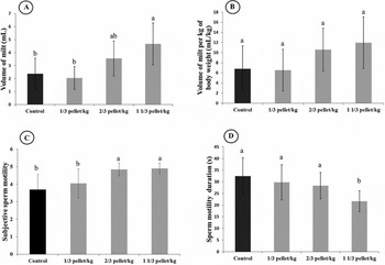
Treatment 3 (T3) resulted a higher production of semen (4.66 ± 1.52 ml) and was significantly different in comparison with the volume produced by the animals submitted to treatments T1 (2 ± 0.9 ml) and T2 (3.5 ± 1.3 ml), and by CO (2.3 ± 1.2 ml) (Fig. 1A ). In relation to the spermatic motility, T2 and T3 showed significantly higher levels [5, (81–100%)] than T1 and CO [4, (61–80%)] (Fig. 1C ). T3 showed a significantly lower average duration of sperm motility (22 ± 5 s) than T1, T2 and CO (30 ± 7 s; 28 ± 6 s; 32 ± 8 s, respectively) (Fig. 1D ). In relation to the seminal parameters of spermatic concentration, pH and osmolality, no statistical difference was observed among the treatments (Table 1). In relation to the analysis of the volume/body weight there was no difference among the treatments and control (Fig. 1B ).
Seminal volume (A), volume of milt per kilo of body weight (B), subjective sperm motility scale (Fribourgh, 1966), levels 0–5 (C), duration of sperm motility (D), semen of Brycon cephalus after hormonal stimulation with three different hormone levels of D-Ala6, Pro9 Net-mGnRH + metoclopramide (Ovopel®): T1 (⅓ pellet/kg), T2 (⅔ pellets/kg), T3 (1⅓ pellet/kg) and control group (physiological solution 0.9%; placebo). The columns represent the mean values found and the bars indicate the standard deviation. Boxes labeled with different letters are statistically different from each other (P < 0.05).

Evaluated pH values, osmolarity and sperm concentration of Brycon cephalus semen after hormonal stimulation with D-Ala6, Pro9 Net-mGnRH + metoclopramide (Ovopel®). Three different hormone levels were tested [T1 (⅓ pellet/kg), T2 (⅔ pellet/kg) and T3 (1⅓ pellet/kg)] with a control group (physiological solution 0.9%; placebo)

a The same letter indicates no significant difference (P > 0.05).
Discussion
Males of rheophilic species, even when rearing in a hatchery, can spermiate; however, to perform the artificial fertilization process, semen obligatorily must be removed by abdominal massage, a process that considerably decreased the quality obtained. Thus, in order to stimulate spermatogenesis and increase seminal liquid, a hormonal induction process is used (Mylonas et al., Reference Mylonas, Fostier and Zanuy2010). As demonstrated in our experiment with B. cephalus, it was verified that after the hormonal induction there was an increase in semen volume production, which will allow a better use of the reproducers.
Although hormonal induction of rheophilic fish is a usual technique for breeders, there is no standardization of hormone concentration for either males or for females. The hormonal doses vary greatly, mainly in males, taking into account factors such as species, number application of latency time and the hormone used. Thus, we can find, in the literature, concentrations used from 1 to 6 mg/kg of pituitary brute extract of Cyprinus carpio (Maria et al., Reference Maria, Viveiros, Freitas and Oliveira2006; Oliveira et al., Reference Oliveira, Viveiros, Maria, Freitas and Izaú2007; Sanches et al., Reference Sanches, Marcos, Baggio, Tessaro, Balen and Bombardelli2011; Viveiros et al., Reference Viveiros, Amaral, Orfão, Isaú, Caneppele and Leal2011, Reference Viveiros, Maria, Orfão, Nascimento, Corrêa and Caneppele2012), 3–5 UI/g of fish of human chorionic gonadotropin (HCG) (Fogli da Silveira et al., Reference Fogli da Silveira, Kavamoto, Cestarolli, Godinho, Ramos and Silveira1990; Kavamoto et al., Reference Kavamoto, Ferraz, Andrade-Talmelli, Meinardes-Pinto, Romagosa, Narahara, Barnabe and Campos1996; Andrade-Talmelli et al., Reference Andrade-Talmelli, Kavamoto and Fenerich-Verani2001; Caneppele et al., Reference Caneppele, Honji, Hilsdorf and Moreira2009) and in the case of GnRHm analogues, approximately 0.1–1.0 pellet/fish kg (Kucharczyk et al., Reference Kucharczyk, Szczerbowski, Luczynski, Kujawa, Mamcarz, Wyszomirska, Szabó and Ratajski2001; Pardo-Carrasco et al., Reference Pardo-Carrasco, Zaniboni-Filho, Arias-Castellanos, Suárez-Mahecha, Atencio-García and Cruz-Casallas2006; Carneiro & Mikos, Reference Carneiro and Mikos2008; Cejko et al., Reference Cejko, Kowalski, Kucharczyk, Targonska, Krejszeff, Zarski and Glogowski2010; Targońska et al., Reference Targońska, Kucharczyk, Żarski, Cejko, Krejszeff, Kupren, Król, Dryl, Kowalski and Glogowski2011).
D-Ala6, Pro9Net-mGnRH has already been tested by many researchers (i.e. Jamróz et al., Reference Jamróz, Kucharczyk, Hakuc-Btazowska, Krejszeff, Kujawa, Kupren, Kwiatkowski, Targonska, Jarski, Cejko and Glogowski2008; Zarski et al., Reference Zarski, Kucharczyk, Targońska, Jamróz, Krejszeff and Mamcarz2009; Kayim et al., Reference Kayim, Bozkurt and Ogretmen2010; Krejszeff et al., Reference Krejszeff, Targonska, Zarski and Kucharczyk2010) with temperate climate species. However, most research performed with this analogue has been focused on female reproduction, as oocyte induction is more complex. Among the studies done with this analogue in males we should mention that of Carneiro & Mikos (Reference Carneiro and Mikos2008) (Rhamdia quelen), Cejko et al. (Reference Cejko, Kowalski, Kucharczyk, Targonska, Krejszeff, Zarski and Glogowski2010) (Leuciscus idus), Targońska et al. (Reference Targońska, Kucharczyk, Żarski, Cejko, Krejszeff, Kupren, Król, Dryl, Kowalski and Glogowski2011) (Barbus barbus), Cejko et al. (Reference Cejko, Targonska, Kowalski, Zarski, Sarosiek, Kucharczyk and Glogowski2012a) (Leuciscus leuciscus) and Cejko et al. (Reference Cejko, Kowalski, Zarski, Dryl, Targonska, Chwaluczyk, Kucharczyk and Glogowski2012b, Reference Cejko, Zarski, Judycka, Kucharczyk, Sarosiek and Kowalski2014) (Barbus barbus).
Cejko et al. (Reference Cejko, Kowalski, Zarski, Dryl, Targonska, Chwaluczyk, Kucharczyk and Glogowski2012b), using D-Ala6, Pro9Net-mGnRH in a 1 pellet/kg dosage in Barbus barbus male induction, obtained the highest semen volume (3 ml) with little pH variation and osmolality in the seminal fluid. For Carneiro & Mikos (Reference Carneiro and Mikos2008), the use of Ovopel® provided the smallest seminal volume in Rhamdia quelen male induction. This result may have occurred because the dosage used was quite low (0.1 pellet/kg). Viveiros & Godinho (Reference Viveiros and Godinho2009) demonstrated in their review that there is a wide variation in existing data with respect to the volume produced by species of fish, and this variation may be related, in addition to the species and the inductor, to the collection method in that it does not permit full withdrawal of semen from the gonad. Ninhaus-Silveira et al. (Reference Ninhaus-Silveira, Foresti, Veríssimo-Silveira and Senhorini2006), using 1 mg/fish kg of CPE in Brycon cephalus male induction, obtained on average 4.0 ml of seminal volume. Similar values were produced by T3 in our study, which allowed the achievement of a higher semen quantity among the tested hormonal concentrations. These similar results may indicate that this methodology is the best way to standardize the Brycon cephalus induction method.
Another factor that may affect seminal production is the time of latency, that is the period after the application of the inducing hormone. For neotropical fish the technique of degree/hour proposed by Woynarovich & Horváth (Reference Woynarovich and Horváth1983) was used to calculate the time when the female would spawn. This consists of multiplying the elapsed time between the last hormone injection and the spawning by the mean water temperature during this period. In our experiment, the latency period of 6 h (162 degree–hours) was adequate because it allowed the production of semen with characteristics (pH, osmolarity, motility and period of motility) of good seminal quality, similar to the control, in addition to increasing the volume of semen produced.
According to Cejko et al. (Reference Cejko, Targonska, Kowalski, Zarski, Sarosiek, Kucharczyk and Glogowski2012a), hormone induction with D-Ala6, Pro9Net-mGnRH provides an increased sperm concentration in the seminal volume. Streit Junior et al. (Reference Streit Junior, Sirol, Ribeiro, Digmayer, Gallo, Moraes and Povh2012) observed that in Pseudoplatystoma reticulatum after hormonal induction with CPE (2.5 mg/kg) spermatic concentration reduction by about 50%, a fact also observed for Salminus maxillosus (Streit Junior et al., Reference Streit Junior, Sirol, Ribeiro, Moraes, Vargas and Watanabe2008) in which a reduction of around 18% was detected in the concentration after induction with CPE (2.5 mg/kg). In contrast with the studies above, our data showed no variation in the spermatic concentration in any of the hormonal doses tested, implying that the hormone used acted in the spermatogenesis stimuli, maintaining the seminal quality. Pardo-Carrasco et al. (Reference Pardo-Carrasco, Zaniboni-Filho, Arias-Castellanos, Suárez-Mahecha, Atencio-García and Cruz-Casallas2006) induced Brycon amazonicus males with CPE and differentiated concentrations of GnRH-a, showing that pituitary extract utilization produces a considerable increase in the seminal fluid, however with a decrease in sperm concentration. However, the GnRH analogue stimulated semen production and maintains seminal characteristics, a report that corroborates the data produced in our experiment.
Spermatic motility is one of the most important seminal parameters for demonstrating spermatozoon viability. Thus, it is desirable that after the hormonal induction the seminal fluid values are maintained or increase, as shown in our study. From preliminary evaluation of male maturity, hormonal induction with GnRH or CPE stimulated spermatogenesis, producing semen with high motility, a fact corroborated by our data and results shown in the studies with Rhamdia quelen (Ferreira et al., Reference Ferreira, Nuner, Luz, Tataje, Esquivel and Restrepo2001), Brycon amazonicus (Pardo-Carrasco et al., Reference Pardo-Carrasco, Zaniboni-Filho, Arias-Castellanos, Suárez-Mahecha, Atencio-García and Cruz-Casallas2006), Piaractus mesopotamicus (Streit Junior et al., Reference Streit Junior, Ribeiro, Moraes, Mendez, Gallo, Digmayer and Povh2006) and Barbus barbus (Cejko et al., Reference Cejko, Kowalski, Zarski, Dryl, Targonska, Chwaluczyk, Kucharczyk and Glogowski2012b). As spermatic motility levels, duration of motility is a preeminent subjective measure among the parameters to determine spermatic viability, because it allows the control of the time required to perform oocyte fertilization by artificial reproduction (Sanches et al., Reference Sanches, Bombardelli, Baggio and Souza2009), as also can suggest data about seminal quality.
The value of the ratio between semen volume and the fish body weight indicates that the two variables are independent. Thus, when compared between treatments, as there was no statistical difference, we can infer that the increase in semen volume obtained after induction is not related to the body weight of the fish, but the effect of the hormone dosage applied. Analyzing Cejko et al. (Reference Cejko, Targonska, Kowalski, Zarski, Sarosiek, Kucharczyk and Glogowski2012a, Reference Cejko, Kowalski, Zarski, Dryl, Targonska, Chwaluczyk, Kucharczyk and Glogowski2012b, Reference Cejko, Zarski, Judycka, Kucharczyk, Sarosiek and Kowalski2014) studies we can infer that the variation in this correlation is related to factors such as hormonal inductor type, latency time and animal physiology state. In conclusion, a dosage of ⅔ pellet/kg of Ovopel® (D-Ala6, Pro9Net-mGnRH + metoclopramide) proved to be the most adequate for hormonal induction of Brycon cephalus males, by increasing the seminal volume produced, together with spermatic motility and duration of sperm motility and maintaining semen quality.
Acknowledgements
I would like to thank the Centro Nacional de Pesquisa Conservação de Peixes Continentais (CEPTA/ICMBio), in the person of engineer José Augusto Senhorini, for allowing the use of Institution facilities and equipment and the animals for this study. I am also grateful to the support (non-financial) from the Laboratório de Ictiologia Neotropical (LINEO), UNESP/Ilha Solteira.
Financial support
This work was supported by the project grant no. 2012/11143–4, São Paulo Research Foundation (FAPESP).