The prepubertal development of the mammary gland influences milk production potential in dairy ruminants. Prepuberty is often regarded as a quiescent phase in the growth of the mammary gland, whereas puberty is known to bring about a phase of active mammary growth with a rapid extension and branching of the duct system. However, Sinha & Tucker (Reference Sinha and Tucker1969) reported a rapid period of allometric growth of mammary parenchyma occurring before puberty that persisted through the first oestrous cycles. During prepuberty the mammary parenchyma develops a complex tree structure of ducts through the mammary fat pad. This prepubertal period is thus decisive for the next step of lobulo-alveolar mammogenesis, which takes place during gestation and continues during future milk production (Akers, Reference Akers1985). During puberty the mammary ductal tree is formed by mammary cell proliferation and extracellular matrix remodelling under the influence of endocrine system (Wiseman et al. Reference Wiseman, Sternlicht, Lund, Alexander, Mott, Bissell, Soloway, Itohara and Werb2003).
Wallace (Reference Wallace1953) was the first to suggest that the ovaries played a critical role during mammogenesis in cattle, reporting a strong decrease in mammary parenchyma in the mammary glands from ovariectomized heifers. Thus the effects of ovarian hormones on mammary development were thought to become evident early in the animal's life. Purup et al. (Reference Purup, Sejrsen, Foldager and Akers1993) reported that parenchyma weight and mammary DNA content were 5-times lower in ovariectomized heifers than in controls. Several studies have shown that ovariectomy inhibits the development of mammary glands in prepubertal heifers, leading to a 90% decrease in development of mammary parenchyma (Purup et al. Reference Purup, Sejrsen and Akers1995). Heifers ovariectomized before 2·5 months of age presented impaired mammary development (Berry et al. Reference Berry, Jobst, Ellis, Howard, Capuco and Akers2003) whereas heifers ovariectomized between 2·5 and 3 months of age showed altered but not totally inhibited mammogenesis (Purup et al. Reference Purup, Sejrsen, Foldager and Akers1993, Reference Purup, Sejrsen and Akers1995). In addition, ovariectomy performed on goats before puberty impacted mammogenesis by limiting parenchyma proliferation and morphology (Dessauge et al. Reference Dessauge, Finot, Wiart, Aubry and Ellis2009). In prepubertal ovariectomized goats, mammary cells showed significantly less proliferation, and the disorganization of mammary tissue was linked to a severe decrease in the level of cell adhesion agents. By contrast, prepubertal ovine mammary development was unaffected by ovariectomy (Ellis et al. Reference Ellis, McFadden and Akers1998). This study demonstrated that prepubertal mammary growth in the ewe lamb did not require the presence of the ovary. The molecular mechanisms underlying mammogenesis before puberty are not well characterized in small ruminants. However, improving prepubertal mammary development is important for enhancing subsequent lactation.
Oestrogens and progesterone are known to be the main ovarian hormones, and it is now well established that both are essential for mammary epithelial cell (MEC) proliferation and differentiation (Erb, Reference Erb1977; Romagnolo et al. Reference Romagnolo, Akers, Wong, Boyle, McFadden, Byatt and Turner1993). In rodents, data obtained with oestrogen receptor (ER) and progesterone receptor (PR) knock-out models confirm that oestradiol is required for ductal morphogenesis, while progesterone signalling is critical for lobulo-alveolar development (Korach, Reference Korach1994). In an ovarian steroid exclusion experiment, the administration of oestrogen alone or in combination with progesterone in the intact prepubertal heifer was shown to stimulate MEC proliferation, whereas progesterone treatment alone had no effect on prepubertal mammary cell proliferation (Woodward et al. Reference Woodward, Beal and Akers1993, Connor et al. Reference Connor, Meyer, Li, Van Amburgh, Boisclair and Capuco2007). It is generally accepted that ovarian and growth hormones exert an influence on the mammary gland, and that the transcriptional biological responses are mainly mediated by receptors. For oestradiol and progesterone, the biological responses are predominantly mediated by oestrogen receptor alpha (ERα) and progesterone receptor (PR), respectively. Capuco et al. (Reference Capuco, Ellis, Wood, Akers and Garrett2002) showed that oestrogen and progesterone receptors were co-localized in MEC but not in stromal cells. They reported that 99% of the proliferating cells in the mammary tissue of pre-pubertal heifers do not express oestrogen receptors.
The activity of matrix metalloproteinases (MMP) and the degradation of the extracellular matrix also play a major role in the extensive remodelling that occurs in the mammary gland during development. MMP are expressed in the bovine mammary gland, and expression has been linked to mammary development (Rabot et al. Reference Rabot, Sinowatz, Berisha, Meyer and Schams2007). However, no data are available on the effect of ovarian steroids on MMP activity or the involvement of MMP in extracellular matrix degradation during mammogenesis.
Discovery of the ovary-mediated mammogenesis mechanisms will permit novel goat management strategies to optimize mammary growth and ultimately improve subsequent lactation efficiency in dairy animals. Studies of the systemic effects of ovarian hormones on prepubertal mammary development in goats are limited, and the molecular mechanisms underlying the influence of ovarian steroids on mammogenesis are not yet fully understood. In addition, the control of allometric mammary development seems to differ among dairy species, and so novel information using a caprine model will add to overall understanding of mammogenesis in ruminants. We had previously demonstrated that ovaries were required for pubertal mammogenesis in goats. The aim of the present study was thus twofold: (1) to improve knowledge of the molecular pathways involved in prepubertal caprine mammogenesis, especially regarding tissue remodelling and (2) to determine the critical period for the requirement of ovarian secretions in prepubertal caprine mammogenesis, using a serial ovariectomy approach.
Materials and Methods
All of the animal procedures were approved by the French Ministry of Agriculture's animal ethics committee in compliance with French regulations (Decree No. 2001-464, 29 May 2001).
Animals and issue preparation
The experiment was conducted on 30 Alpine goats from the experimental farm herd kept by the French National Institute for Agricultural Research at Le Rheu. Birth weights were between 4·3 and 6 kg (mean=5±0·3 kg). No twin goats were included, to maximize growth and survival. All the goats were born between 8 February and 17 February 2008. From birth to weaning, the goats were reared by feeding collectively with milk replacer and housed in a nursery. From weaning to the end of the experiment, they were fed collectively with straw, hay and concentrate (Fluvia Junior, Sanders, Bruz, France) to optimize average daily weight gain (125–150 g/d).
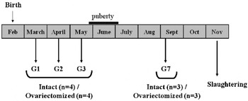
A serial ovariectomy approach was used to test our hypothesis. Goats in prepubertal period were either ovariectomized at 1 (Ovx-1, n=4), 2 (Ovx-2, n=4) and 3 (Ovx-3, n=4) months of age, or sham-operated (Int-1, n=4; Int-2, n=4; Int-3, n=4). Six additional goats were ovariectomized (Ovx-7, n=3) or sham-operated (Int-7, n=3) at 7 months of age, in the post-pubertal period (Fig. 1). Goats are considered to be pubescent at around 4 and 5 months of age and are usually inseminated after they have reached 32 kg of body weight. In this study, all of the goats reached this body weight between 5 and 6 months of age. At 9 months of age, the goats were slaughtered under general anaesthesia with subsequent euthanasia (Rompun i.v. 1 ml; Dolethal, i.v. 25 ml). Entire udders were removed and weighed (with teats and skin) and then mammary glands were separated into right and left halves along the medial suspensory ligament. At slaughter, ovariectomy and presence of ovaries (with corpus luteum) in Ovx and Int goats respectively were confirmed by inspection of genital tracts.
Experimental design. At 1 (G1), 2 (G2), 3 (G3) and 7 (G7) months of age, goats were either ovariectomized (Ovx) or sham-operated (Int). Experimental design included 8 goats in each group (G1, G2 and G3) with 4 animals per treatment except for G7 in which 6 animals were used (3 animals per treatment). At 9 months of age, goats were sacrificed to remove the mammary gland.

Blood sampling and progesterone assays
Blood samples were taken 15 min before slaughter to determine plasma hormone concentrations. Sampling was performed using Monovette syringes coated with sodium heparin (Sarstedt, Nümbrecht, Germany). Plasma was separated by centrifuging at 3000 g at 4°C for 15 min and stored at −20°C until analysis. Plasma progesterone concentrations were measured using an ELISA. The sensitivity of the assay was 0·1 ng/ml. The intra-assay and inter-assay coefficients of variation were 12·3 and 7·5% respectively. Progesterone assay was performed at the Reproductive and Behavioural Physiology Laboratory (INRA, Tours, France).
Tissue sampling
The right mammary gland was skinned and dissected to collect several samples of mammary tissue. In the young goat, the mammary parenchyma develops into the stroma in a diffuse and generally homogenous manner. Based on appearance we could not distinguish parenchymal tissue from the associated stromal tissue. Therefore mammary tissue samples were taken from a region located superior to the teat, approximately mid-way to the distal margin of the mammary gland which were equally composed of parenchymal and stromal tissue. A fraction of the freshly dissected tissue segments was cut into small pieces (1 g), immediately frozen in liquid nitrogen, and stored at −80°C for RNA, DNA, and protein analyses. The remainder was rinsed in phosphate buffer saline (PBS) before fixation for immunohistochemistry analysis.
Histological analysis
Mammary tissue samples used for histological analysis were fixed in 4% paraformaldehyde (pH 7·4) for 2 h and paraffin-embedded using standard protocols. Tissue sections (thickness 5 μm) mounted on SuperFrost Plus slides (Labo-Moderne, Paris, France) were deparaffinized in three changes of xylene and rehydrated in a graded ethanol-water series. After rehydration, tissue sections were stained with haematoxylin and eosin or processed for immunohistochemical detection of proliferating cells (using Ki67 antigen, Ab 15580, AbCAM, Paris, France, dilution 1:1000 in PBS-BSA 1%) or ERα positive cells (sc-543, Santa Cruz Biotechnology, Inc., Heidelberg, Germany, dilution 1:100 in PBS-Triton X100 0·2%–BSA 1%). Tissue sections were then quenched in Peroxoblock (00-2015, Invitrogen Life Technologies, Berlin, Germany) for 2 min to eliminate endogenous peroxidase activity. Slides were washed in PBS.
Slides were scanned using NanoZoomer Digital Pathology technology (Hamamatsu Photonics, Massy, France) with automatic detection of the whole tissue sections. Two tissue sections per animal (about 1 cm2 per section) were analysed for epithelial area measurement and Ki67 and ERα positive cell counting. All histological analyses were performed with JMicroVision v1·27 freeware, using specific macro based on colour threshold for parenchymal area measurement, and on both colour threshold and particle size for Ki67 and ERα positive-cell counting. Parenchymal area was expressed as a percentage of total measurement area (i.e. about 2 cm2). Ki67 and ERα positive-cell quantifications were expressed as a number of positive-cells relative to mammary tissue area.
DNA quantification of mammary tissue
Mammary tissue samples (100 mg) were first weighed and homogenized in 2·5 ml of sodium phosphate extraction buffer (0·05 m Na2HPO4, 0·05 m NaH2PO4, 2 mm EDTA and 2 m NaCl, pH 7·4). The suspension was then homogenized using an Ultra-Turrax homogenizer for 30 s (twice), sonicated for 1 min and centrifuged at 4000 g for 1 h. The supernatant was transferred to a new tube, and the pellet was resuspended in 2·5 ml of the extraction buffer and subjected to three successive extraction steps of homogenization, sonication and centrifugation as described above. At the end of each centrifugation, the supernatant was recovered and added to the first. The DNA mixture was then analysed by fluorescence using Hoechst 33258 dye (Sigma Aldrich Chimie, Lyon, France). Aliquots from the DNA mixture were mixed with sodium phosphate buffer stained with Hoechst dye to a final dye concentration of 1 μg/ml.
Protein extraction
Total proteins were extracted from frozen mammary tissue samples using the tissue protein extraction reagent (78510, Thermo Fisher Scientific, Brebières, France). After homogenization, the mixture was centrifuged at 10 000 g at 4°C for 5 min and the supernatant containing total proteins was recovered. Protein concentration was determined by the Bradford method using the Bradford protein assay (Sigma Aldrich Chimie, Lyon, France). Lysates were then combined with sample buffer (50 mm Tris–HCl, pH 6·8, 2% SDS, 0·1% bromophenol blue, 20% glycerol and 0·1 m dithiothreitol) and boiled for 5 min at 95°C for resolution by SDS-PAGE.
Western blot analysis
Proteins (30 μg per lane) were separated on 10% SDS-polyacrylamide gels, blotted to polyvinylidene difluoride membranes (GE Healthcare Bio-Sciences AB, Uppsala, Sweden) and incubated with blocking solution (5% dry skimmed milk dissolved in Tris buffer saline Tween (TBS-T) buffer, 50 mm Tris–HCl, pH 8·6, 150 m NaCl and 0·1% Tween) for 30 min. Membranes were incubated overnight at 4°C with the appropriate dilution of the following primary antibodies (all antibodies were used at 1:1000 dilution): a mouse monoclonal anti-E-cadherin (M3612, Ozyme, Saint-Quentin-en-Yvelines, France), anti-proliferating cell nuclear antigen (PCNA) (M0879, Dako, Trappes, France), anti-actin (A5441, Sigma Aldrich Chimie, Lyon, France) and a rabbit polyclonal anti-fatty acid binding protein 4 (FABP4) (sc-18661, Santa Cruz Biotechnology, Inc., Heidelberg, Germany). Membranes were then washed with TBS-T and incubated with horseradish peroxidase (HRP)-conjugated anti-mouse or anti-rabbit secondary antibody (31450, 31464, Thermo Fisher Scientific, Brebières, France) used at 1:500 for 1 h at room temperature. Images were scanned at 16-bit/600-dpi resolution on a Canoscan D1250 U2 flatbed scanner (Canon France, Courbevoie, France), saved as TIFF files and calibrated on an optical density scale. The integrated optical densities of the bands were quantified using ImageQuant software (GE Healthcare Bio-Sciences AB, Uppsala, Sweden). Each sample was normalized for actin content.
Gelatin zymography for MMP2
Gelatin zymography was used to quantify both activated and latent forms of MMP2 in tissue extracts. Proteins (5 μg per lane) underwent electrophoresis in 10% SDS-PAGE gels co-polymerized with 1 mg/ml of gelatin (Sigma Aldrich Chimie, Lyon, France).
Gels were scanned at 8-bit/500-dpi resolution on a Canoscan D1250 U2 flatbed scanner (Canon France, Courbevoie, France). Pro-MMP2 (66 kDa) and active MMP2 (57 kDa) activities were measured by quantifying the integrated optical density of the clear bands using ImageQuant software.
RNA extraction and cDNA synthesis
Total RNA was extracted from mammary tissue samples (50 mg) using Trizol (Invitrogen Life Technologies, Berlin, Germany) and the RNeasy Mini kit (Qiagen, Courtaboeuf, France) according to the manufacturer's instructions. RNA quality was determined on an Agilent 2100 Bioanalyzer (Agilent Technologies, Massy, France) by RNA profiling and measurement of RNA integrity number using Agilent 2100 Expert Software, version B.02 (Agilent Technologies, Massy, France). RNA (1 μg) was then reverse-transcribed at 42°C for 1 h using a ThermoScript RT-PCR System (Invitrogen Life Technologies, Berlin, Germany) according to the manufacturer's protocol for oligo(dT)15-primed cDNA-synthesis. Before use in RT-qPCR, cDNA was diluted in sterile water (1:50).
Primer design and testing
Gene sequences for primer design were based on bovine mRNA sequences and were obtained from the National Center for Biotechnology Information's Genebank. Only primers for 60S acidic ribosomal protein P0 (RPLP0) were taken from the literature (Robinson et al. Reference Robinson, Sutherland and Sutherland2007). Sequence data for the primers used for real-time PCR were: RPLP0 forward 5′-ACTCTGCATTCCCGCTTCCT-3′, reverse 5′-GGTACAGATGCGACGGTTGG-3′; 18S rRNA forward 5′-CAAATTACCCACTCCCGACCC-3′, reverse 5′-AATGGATCCTCGCGGAAGG-3′; β-actin B forward 5′-TCATCACCATCGGCAATGAG-3′, reverse 5′-GGTAGTTTCGTGAATGCCGC-3′; GAPDH forward 5′-GTCTTCACTACCATGGAGAAGG-3′, reverse 5′-TCATGGATGACCTTGGCCAG-3′; Cyclophilin A forward 5′-GGATTTATGTGCCAGGGTGGTGA-3′, reverse 5′-CAAGATGCCAGGACCTGTATG-3′; ERα forward 5′-CAGGAGGAAGAGCTGTCAGG-3′, reverse 5′-ATCATCTCTCTGGCGCTTGT-3′; PR forward 5′-TGCAGGACATGACAACAGCA-3′, reverse 5′-TTCCGAAAACCTGGCAGTG-3′.
Quantitative PCR
Quantitative PCR analysis was performed on an ABI Prism 7000 sequence detection system (Applied Biosystems, Courtaboeuf, France) using SYBR Green PCR master mix. Reactions were carried out in 96-well optical reaction plates with 200 nm of each specific primer and 2·5 μl of diluted cDNA. Each sample was run in triplicate with a non-template control included.
Data obtained from ABI Prism 7000 Software, version 1.1 (Applied Biosystems, Courtaboeuf, France) on raw cycle threshold (Ct) values were used for the comparative Ct method (delta delta cycle threshold (ΔΔCt)) using specific efficiencies (Pfaffl, Reference Pfaffl2001). R value has been calculated as above:

E is the specific efficiency of each gene performed. The 4 intact groups (Int-1, Int-2, Int-3 and Int-7) were used as control (biological replicates).
Evaluation of reference gene expression stability
A reference gene is required to normalize qPCR data. Candidate reference genes were chosen and evaluated in this study: β-actin, GAPDH, cyclophilin A, 18S ribosomal RNA, and RPLP0. GeNorm version 3.4 software (Visual Basic application tool for Microsoft Excel) was used to calculate the stability of the candidate reference genes. T he GeNorm application generates a stability measure value (M) for the candidate reference gene, which can be used to rank the reference genes (Vandesompele et al. Reference Vandesompele, De Preter, Pattyn, Poppe, Van Roy, De Paepe and Speleman2002).
The pairwise RPLP0-cyclophilin A was determined by GeNorm as the optimal internal control for caprine mammary gland gene expression profiling and used to normalize qPCR data.
Statistical analysis
Data on mammary gland weight and DNA concentration, parenchyma concentration, parenchymal area, pixel intensities of Western blot and zymography analysis, and quantitative PCR (ΔΔCT) are expressed as means±sem. Data underwent analysis of variance (ANOVA) with Student's t test to compare the mean values resulting from the both treatments (Int and Ovx) and interactions between treatment and age. We considered effects to be statistically significant at P<0·01. Statistical analyses were carried out using the R freeware version 2.12·2.
Results
Validation of ovariectomy
Plasma progesterone concentrations measured at 9 months of age were significantly lower in ovariectomized goats whatever the age of ovariectomy (Table 1). At slaughter, the presence or the absence of ovaries (in Int and Ovx goats, respectively) was verified in order to validate the experimental model of ovariectomy. We observed the absence of ovaries in Ovx goats and that Int goats presented multiples corpus luteum in their ovaries (data not shown).
Effects of ovariectomy on udder weight, fatty acid binding protein (FABP4) level, ERα-positive cells number and progesterone receptor (PR) expression in mammary tissue and plasma concentration of progesterone

Int=Intact goat; Ovx=Ovariectomized goats
T=Treatment; A=Age; T×A=interaction of treatment and age; n=number
NS=not significant (P⩾0·1); †0·05<P<0·1 (tendency); *P<0·05; **P<0·01; ***P<0·001
sem=standard error of the mean
Early ovariectomy had no effect on mammary gland weight but strongly affected development of mammary parenchyma
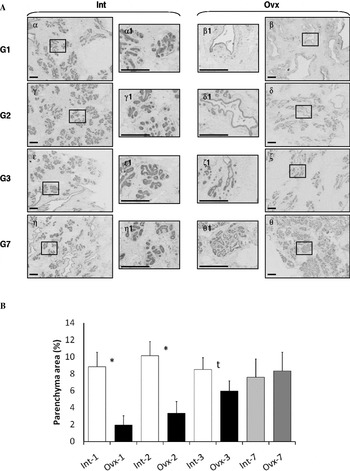
The mammary gland weights of Ovx and Int goats were not significantly different whatever the ovariectomy period (Table 1). However, histological analysis revealed major differences in the appearance of epithelial structures in mammary tissue of Ovx v. Int goats (Fig. 2a). The mammary glands of intact goats showed classical mammary epithelial structures with round, developed, and very complex ducts. By contrast, ovariectomized goats (Ovx-1, Ovx-2 and Ovx-3) showed strongly impaired mammary gland development. The reduced number of epithelial structures that were observed in tissue sections from Ovx-1, 2 and 3 goats showed minimal development characterized by reduced branching and bud-like formation. Also, we determined the impact of ovariectomy on the adipose tissue in mammary parenchyma-stroma border samples, using the quantification of FABP4. We observed a significant interaction between age and treatment in FABP4 levels (Table 1).
Effect of ovariectomy on mammary parenchyma development. (a) Histological analysis was performed on paraffin-embedded mammary gland parenchyma after haematoxylin and eosin staining, from Int (left) and Ovx (right) goats at 1 (α and β), 2 (γ and δ), 3 (ε and ζ) or 7 (η and θ) months of age. Central panels (α1 to θ1) represent high magnifications from lateral panels (α to θ). Note the presence of organized ductal network in the Int goats compared with a single ductal rudiment in the Ovx goats. These organizational differences were more marked when ovariectomy was performed early. Bars represent 100 μm. (b) Epithelial area was evaluated from histological sections using JMicroVision freeware. Data are expressed as means±sem. An effect was considered significant at P<0·01 (*) and tended to be significant at P<0·05 (t). Note a significant decrease in epithelial area of Ovx goats at Ovx-1 and Ovx-2 and a tendency at Ovx-3.

From histological scans, we measured the areas of mammary epithelium (Fig. 2b). We found a relationship between development of epithelial structures and age at ovariectomy. Goats ovariectomized at 1 and 2 months of age (Ovx-1 and Ovx-2) had significantly (5-times) less epithelium than Int animals (P<0·001). Ovariectomy at 3 months of age (Ovx-3) tended to decrease the percentage of epithelial area (P=0·11). After puberty (G7), ovariectomy had no effect on parenchymal area.
Early ovariectomy affected mammary DNA concentration
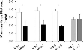
We focused on the DNA concentration to evaluate the number of cells in the samples of mammary tissue. Results presented in Fig. 3 show that the ovariectomy performed at 1, 2 and 3 months of age (Ovx-1, Ovx-2, Ovx-3) caused a 50% reduction in mammary tissue DNA concentrations (P<0·001). Ovariectomies performed after puberty (Ovx-7) did not influence DNA concentrations compared with the Int-7 group (P=0·90).
Effect of ovariectomy on mammary tissue DNA concentration. Mammary tissue DNA concentration was determined by Hoechst 33258 staining in Ovx and Int goats. Data given in mg of DNA content per g of mammary tissue are expressed as means±sem. Effect is considered significant at P<0·01 (*).

Early ovariectomy affected proliferation of mammary cells
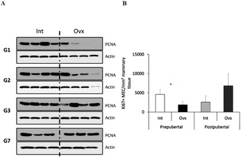
Goats ovariectomized before 2 months of age (Ovx-1 and Ovx-2) showed a significant, 2·5-fold decrease in PCNA expression (P<0·01). PCNA levels in Ovx-3 and Ovx-7 groups were not different compared with Int-3 and Int-7 groups respectively (Fig. 4a). Additionally, we evaluated mammary cell proliferation directly on mammary sections using Ki67 immunodetection. We observed that the number of Ki67-positive cells per mm2 of mammary tissue decreased when goats were ovariectomized before puberty (Fig. 4b, P<0·01), and was equivalent between Ovx-7 and Int-7.
Effect of ovariectomy on mammary cell proliferation analysed by Western blot (PCNA) and immunodetection (Ki67) in mammary gland samples. (a) Proliferating cell nuclear antigen (PCNA) was detected on gels at 36 kDa using Western blot analysis. Each band corresponds to one goat for each period. Βeta-actin was used to normalize PCNA levels. (b) Ki67-positive cell number per mm2 of tissue in mammary gland of Ovx and Int prepubertal goats (G1, G2 and G3) and postpubertal goats (G7) was determined by immunodetection. Data are expressed as means±sem. Effect is considered significant at P <0·01 (*). Note that ovariectomy performed before puberty decreased cell proliferation in goat mammary tissue.

Early ovariectomy inhibited mammary gland remodelling
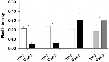
Western blot results showed that E-cadherin was strongly decreased in goats ovariectomized before 2 months of age (Fig. 5, Ovx-1: 80% and Ovx-2: 85%) but was not different in goats ovariectomized at 3 months and 7 months, relative to respective Int groups. The relative abundance of E-cadherin was increased in the mammary extracts from goats ovariectomized after puberty (Ovx-7: 45%, P=0·09).
Effect of ovariectomy on the abundance of E-cadherin in mammary extracts. Bands corresponding to E-cadherin were quantified from WB analysis using ImageQuant software. Pixel intensity data are expressed as means±sem. Effect was considered significant at P<0·01 (*). Note that E-cadherin levels in mammary tissue were decreased in Ovx-1 and Ovx-2 goats and increased in Ovx-7 goats.

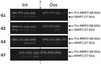
Gelatinase MMP2 activity in mammary gland extracts from all groups was studied by gelatin zymography (Fig. 6). MMP2 activity was decreased in mammary glands of goats ovariectomized at 1 and 2 months of age (P=0·01 and P=0·02, respectively), whereas there was no difference at 3 and 7 months of age relative to respective Int groups.
Effect of ovariectomy on matrix metalloproteinase 2 (MMP2) activity in caprine mammary tissue. Mammary explants of Ovx and Int goats were subjected to gelatin zymography. The zymography bands corresponding to pro-MMP2 and MMP2 gelatinase activity were detected at 66 kDa and 57 kDa respectively. Each band corresponds to one goat for each period. Note that ovariectomy significantly decreased MMP2 activity in G1 and G2 goats.

Ovariectomy modified expression of ovarian steroid receptors
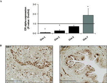
Goats ovariectomized before puberty (Ovx-1, Ovx-2 and Ovx-3) presented a significant decrease in ERα transcripts in mammary glands (Fig. 7a). However, qPCR results revealed an over-expression of ERα transcripts in Ovx-7 group compared with the Int-7 group. Immunolocalization of ERα was performed on mammary gland tissue sections. We observed that ERα was predominantly confined to parenchyma (Fig. 7b) and the proportion of ERα-positive cells was lower in Ovx than in Int animals (Table 1).
Expression of oestrogen receptor alpha (ERα) in the mammary gland determined by real-time qPCR and. Immunodetection. (a) Quantitative PCR data given R value (see Materials and Methods) are expressed as means±sem. Intact goats were used calibrator and the dashed line indicates gene upregulation and downregulation, in the mammary gland samples from Ovx goats compared with Intact goats. Effect is considered significant at P<0·01 (*). Note that ovariectomy significantly decreased ERα expression in G1 and G2 goats but increased ERα expression in G7 goats. (b) Histological analysis was performed on paraffin-embedded mammary gland parenchyma after ERα immunodetection, from Int (left) and Ovx (right) goats at 1 month of age and slaughtered at 9 months of age. Bars represent 100 μm.

Ovariectomy caused a severe decrease in PR expression whatever the period (Table 1, P<0·001), with undetectable PR transcription levels. PR expression was reduced 16-fold in Ovx-1 goats and 17-fold in Ovx-2, Ovx-3 and Ovx-7 goats compared with the corresponding intact groups.
Discussion
Our surgical model was validated in that ovariectomy led to a total reduction of systemic progesterone concentration and a significant decrease in uterus size from Ovx animals (−57%, P<0·001, data not shown), supporting a drop in systemic oestrogen concentration (Berry et al. Reference Berry, Jobst, Ellis, Howard, Capuco and Akers2003). In a previous study we demonstrated that oestrogen concentrations were significantly decreased in ovariectomized prepubertal goats (Dessauge et al. Reference Dessauge, Finot, Wiart, Aubry and Ellis2009). Taken together, our results suggest that our observations were linked to changes in systemic oestrogen and progesterone concentrations, although we cannot rule out the involvement of other ovarian factors in prepubertal goat mammary gland development.
Mammary tissue organization presented a strong developmental failure following ovariectomy, while total udder weight remained unaffected. Histological analysis revealed that in early-ovariectomized animals, mammary parenchymal tissue was rudimentary and undeveloped, whereas in intact animals the mammary gland was already well-structured and organized in lobules with epithelial ducts. This impaired mammary gland organization was associated with a significantly reduced mammary epithelial tissue area. In addition, we observed that the abundance of FABP4 protein, a specific adipose tissue marker (Samulin et al. Reference Samulin, Berget, Lien and Sundvold2008), was increased in ovariectomized goats. Thus taken together, these results suggest that ovariectomy performed before puberty would promote adipose mammary tissue development at the expense of mammary epithelium. These observations are in agreement with previous studies conducted on heifers and goats (Berry et al. Reference Berry, Jobst, Ellis, Howard, Capuco and Akers2003, Dessauge et al. Reference Dessauge, Finot, Wiart, Aubry and Ellis2009). Berry et al. (Reference Berry, Jobst, Ellis, Howard, Capuco and Akers2003) showed that heifers ovariectomized at 2·5 months of age and slaughtered at 9 months presented significantly less mammary epithelium and lumen areas, associated with an increase in stromal area.
Our study demonstrated that the removal of the main source of oestradiol and progesterone before puberty resulted in a decrease in the mammary DNA concentration as well as MEC proliferation. This decrease in percent of cells proliferating following ovariectomy was found in several studies on heifers. Cell proliferation in the mammary glands of heifers ovariectomized before puberty and slaughtered at 6 months of age was 10-times lower than in intact animals. In addition, in these ovariectomized heifers, less than 1% of MEC were in proliferation at 6 months of age, against 2·5% in the intact animals (Berry et al. Reference Berry, Jobst, Ellis, Howard, Capuco and Akers2003). In goats, we also confirmed that Ki67-positive cell number and PCNA levels were negatively affected by ovariectomy. These results show that ovarian secretions control mammary cell proliferation before puberty.
Mammary epithelial cells are surrounded by stromal tissue composed of adipocytes, fibroblasts, capillaries and extra-cellular matrix. For the epithelial ducts to grow into the stromal tissue, it must be remodelled. Dessauge et al. (Reference Dessauge, Finot, Wiart, Aubry and Ellis2009) demonstrated that the removal of ovaries at the time of puberty induced a down-regulation of several cellular adhesion molecules (β-catenin, α-E-catenin, N-cadherin, E-cadherin, P-cadherin and Pan-cadherin). Here, we found a decrease in E-cadherin protein relative abundance following ovariectomy, but only in animals ovariectomized at 1 and 2 months of age. Thus the rudimentary organization of the mammary gland that we observed in goats ovariectomized before puberty, and more particularly before 3 months of age, may be partly due to a reduction either in cell number as discussed before, or in cell-cell adhesion, or both. A decrease in metalloprotease activity may also occur, preventing the gland from undergoing tissue remodelling. MMP play a predominant role in the different stages of mammogenesis, at puberty and during gestation, and in tissue regression during mammary involution. In the mammary gland, metalloproteinase 2, or gelatinase A, is mainly expressed by myoepthelial cells (Monteagudo et al. Reference Monteagudo, Merino, San-Juan, Liotta and Stetler-Stevenson1990). During mammogenesis, its expression increases after the initiation of puberty during the development and ramification of the mammary ducts. This suggests a role of MMP in extracellular matrix remodelling to enable ductal development (Rudolph-Owen & Matrisian, Reference Rudolph-Owen and Matrisian1998). MMP may also influence MEC proliferation/apoptosis balance. In mice, the over-expression of MMP3 in the mammary gland induced an increase in the proliferation rate of epithelial cells both at the time of puberty and after puberty in non-pregnant mice (Sympson et al. Reference Sympson, Talhouk, Alexander, Chin, Clift, Bissell and Werb1994; Witty et al. Reference Witty, Wright and Matrisian1995). In our study, metalloprotease activity measured in goats ovariectomized at 1 and 2 months of age (Ovx-1 and Ovx-2) decreased. We can thus hypothesize that ovarian secretions are closely associated with the expression or activation of MMPs during early puberty. Consequently, the decrease in metalloprotease activity may have contributed to the decrease in MEC proliferation and mammary gland remodelling mediated by the extracellular matrix proteins.
In this experiment, ovariectomy negatively affected the development of the mammary gland when it was performed before puberty. At both structural and molecular levels, effects were more marked when the ovariectomy was performed at the early prepubertal stage (Ovx-1 and Ovx-2). These alterations of mammary development were associated with a significant decrease in ERα expression in 1-month-old and 2-month-old ovariectomized goats, and in PR expression whatever the age at ovariectomy. The expression of genes coding for progesterone receptors is known to be under the control of oestrogens. The promoter region of PR genes contains an oestrogen response element half-site (Kastner et al. Reference Kastner, Krust, Turcotte, Stropp, Tora, Gronemeyer and Chambon1990). The oestrogen response element allows ERα to bind the PR promoter and thus ERα can activate PR gene transcription (Petz et al. Reference Petz, Ziegler, Schultz, Kim, Kemper and Nardulli2004). Ovaries are known to be the main source of oestradiol and progesterone. Here, we inhibited ovarian secretions by ovariectomy, thus ERα binding to PR genes would be partially inhibited and consequently PR transcription would be inactivated. This would explain the marked decrease in PR expression observed in all the ovariectomized groups. In the heifer mammary gland, ERα is mainly expressed by MEC (Capuco et al. Reference Capuco, Ellis, Wood, Akers and Garrett2002). In our study, immunohistochemical data confirm that ERα is predominantly expressed in mammary parenchyma. Also, in ruminants, early ovariectomy not only affected parenchyma development, but also resulted in a lower proliferation rate (Ki67-positive cell number) and oestrogen receptivity (ERα-positive cell number). Hence the reduction of ERα expression in goats ovariectomized at 1 and 2 months of age may be due to the significant decrease in proliferation, mainly concerning MEC in these animals. This was probably due to inhibition of the negative feedback that oestradiol usually exerts on its receptor expression. Similar results had already been obtained in the prepubertal goats (Dessauge et al. Reference Dessauge, Finot, Wiart, Aubry and Ellis2009) but not in heifers (Berry et al. Reference Berry, McFadden, Pearson and Akers2001, Reference Berry, Jobst, Ellis, Howard, Capuco and Akers2003), in which the ovariectomy induced an increase in the ERα expression whatever the age of ovariectomy. The high negative feedback of oestradiol in the promoter region of several genes could explain the increase of E-cadherin levels in goats ovariectomized after puberty. In addition, this drop in ERα expression was not found in animals ovariectomized at 3 months of age, which did not present a significant decrease in epithelial cell proliferation. Unfortunately, we were unable to develop an immunohistochemical protocol for Ki67-ERα antigen double-labelling in goats. Capuco et al. (Reference Capuco, Ellis, Wood, Akers and Garrett2002), using double-labelling Ki67-ERα in oestrogen-treated heifers, demonstrated that 99% of proliferating cells did not express ERα. This suggests that mammary gland cell proliferation in response to oestrogen may be stimulated by a paracrine route via the mammary fat pad as discussed previously. Interestingly, our data show that ovariectomy negatively affected ERα expression in goat mammary gland when performed before puberty. It had been shown that ovariectomy on prepubertal heifers had no effect (Meyer et al. Reference Meyer, Capuco, Boisclair and Van Amburgh2006) or positively affected ERα expression in mammary gland, whatever the age at ovariectomy (Berry et al. Reference Berry, McFadden, Pearson and Akers2001, Reference Berry, Jobst, Ellis, Howard, Capuco and Akers2003). In addition, a study conducted on ovariectomized prepubertal ewe lambs showed that prepubertal mammary development did not require the presence of the ovary in this species (Ellis et al. Reference Ellis, McFadden and Akers1998), unlike in heifers and goats. Despite the fact that the slaughtering ages in Berry's studies (6 months, Berry et al. Reference Berry, McFadden, Pearson and Akers2001, Reference Berry, Jobst, Ellis, Howard, Capuco and Akers2003) were different from the slaughtering age in our study (9 months), these conflicting results suggest that ewes, goats and heifers present differences in ovarian steroid receptivity during pubertal mammogenesis. The existence of other endocrine pathways involved in prepubertal mammary development such as growth hormone (GH)-IGF-1 axis is highlighted in studies on heifers (reviewed in Akers et al. Reference Akers, Ellis and Berry2005).
It is known that the GH-IGF-1 axis plays a key role in mammogenesis, and that these factors are closely related to oestrogenic axis signalling. Several studies have shown that ovariectomizing heifers before puberty induces a decrease in cellular proliferation in the mammary gland, associated with a reduction in the stromal IGF-1 production and IGF-1 receptor expression (Berry et al. Reference Berry, Jobst, Ellis, Howard, Capuco and Akers2003; Akers et al. Reference Akers, Ellis and Berry2005). Conversely, heifers treated with oestrogens presented an increase in stromal IGF-1 and a decrease in genes encoding for IGF-1-binding protein (IGFBP-3) (Berry et al. Reference Berry, McFadden, Pearson and Akers2001; Akers et al. Reference Akers, Ellis and Berry2005). Secretion of prolactin could also play a part in the prepubertal development of the mammary gland. It has been shown in heifers that the secretion of pituitary prolactin increased in the main stages of mammogenesis, i.e. between 2 months and 3 months after birth, and at 9 months (Sinha & Tucker, Reference Sinha and Tucker1969). Thus it will be of interest to focus specific studies on the role of prolactin and GH-IGF-1 axis in prepubertal goat mammogenesis.
Conclusions
In conclusion, this study suggests that ovarian secretions play an important role in mammary gland development in the prepubertal goat, especially before 3 months of age. Removal of the main source of oestradiol and progesterone before puberty would induce a deregulation of ERα and PR expression. These results showed a severe developmental failure of mammary epithelium, in mammary cell proliferation and in the ability of tissues to support remodelling. This study highlights the existence of a critical period (before 3 months of age) for ovarian hormone action on mammary gland development. It thus sheds new light on the molecular mechanisms involved in mammary prepubertal tissue remodelling and defines more precisely the critical period for ovarian hormone effects on prepubertal allometric mammary development in goats.
The authors thank Michel Chorho, Eric Siroux and Jean-Marc Aubry for animal assistance and knowledge on goat rearing, Vanessa Lollivier for critical reading of the manuscript, Remy Le Guével from ImPACcell and Alain Fautrel from H2P2 laboratory for help in histological analysis, and Auvergne Traduction Technique (Clermont-Ferrand, France) for language editing. This research was supported by the French National Institute of Agricultural Science (INRA), the PHASE department.