Introduction
Fungus gnats are one of the most species-rich dipteran families, Mycetophilidae (Diptera), in the Neotropical Region. Although the monophyly of Mycetophilidae is a consensus (Søli Reference Søli1997; Amorim and Rindal Reference Amorim and Rindal2007; Rindal et al. Reference Rindal, Søli and Bachmann2009), a robust phylogeny of this family is still wanting. Seven subfamilies are recognized for Mycetophilidae s.s.: Sciophilinae, Gnoristinae, Mycomyinae, Leiinae, Manotinae, Allactoneurinae, and Mycetophilinae (e.g., Väisänen Reference Väisänen1984; Matile Reference Matile and Evenhuis1989; Rindal et al. Reference Rindal, Søli and Bachmann2009). However, the monophyly of Sciophilinae, Gnoristinae, and Leiinae is still questionable. Subfamilial rank has been given to the tribe Metanepsiini (Väisänen Reference Väisänen1984; Ševčík and Hippa Reference Ševčík and Hippa2010) of Gnoristinae, but this clade would almost certainly make the rest of Gnoristinae paraphyletic (Kallweit Reference Kallweit1998).
The present diversity of Leiinae comprises 32 recent genera and almost 550 recent species worldwide. Leiinae are well represented in the fossil record (Evenhuis Reference Garrett1994; Blagoderov and Grimaldi Reference Blagoderov and Grimaldi2004), with 54 species. Particularly noteworthy are 8 genera currently assigned to Leiinae in Cretaceous amber (some of these genera may belong elsewhere). The recurrent discussion about the monophyly of Leiinae (e.g., Søli et al. Reference Søli, Vockeroth, Matile, Papp and Darvas2000; Hippa et al. Reference Hippa, Jaschhof and Vilkamaa2005; Jaschhof and Kallweit Reference Jaschhof and Kallweit2009; Rindal et al. Reference Rindal, Søli and Bachmann2009) reflects the problems in establishing subfamily limits. Two characters (short R1 and longitudinal r-m) were used by Edwards (Reference Edwards1925) to define Leiinae. These features are indeed met by most leiine genera, but there are a number of exceptions (Jaschhof and Kallweit Reference Jaschhof and Kallweit2009). The sinuous vein CuA has more recently been considered a synapomorphy for Leiinae (Baxter and Poinar Reference Baxter and Poinar1994; Jaschhof and Kallweit Reference Jaschhof and Kallweit2009), but this actually delimits only a smaller clade, without solving the problem of placing various genera in this clade or Gnoristinae.
Docosia Winnertz is a distinctive genus, possibly close to Tetragoneura Winnertz (Edwards Reference Edwards1925). Väisänen (Reference Väisänen1986) suggested that Tetragoneura plus Ectrepesthoneura Enderlein should be treated as gnoristines. Docosia has CuA more or less straight and, even considering that this condition is plesiomophic, it could fit together with Tetragoneura (whatever its placement). Some other features could suggest the placement of the genus in Leiinae, but molecular analysis (Rindal et al. Reference Rindal, Søli and Bachmann2009) suggests that Docosia should be assigned to Gnoristinae.
Docosia presently comprises 65 described extant species worldwide of which 46 are Palaearctic (e.g., Chandler et al. Reference Chandler, Bechev and Caspers2006; Kurina Reference Kurina2006, Reference Kurina2008; Laštovka and Ševčík Reference Laštovka and Ševčík2006; Ševčík Reference Ševčík2006b; Ševčík and Laštovka Reference Ševčík and Laštovka2008; Xu et al. Reference Xu, Wu and Yu2003), 16 are Nearctic (e.g., Garrett Reference Garrett1925; Van Duzee Reference Van Duzee1928), 1 is Oriental (Reference Ševčík and HippaŠevčík 2010), and 2 are Neotropical (Edwards Reference Edwards1933). Information on the biology of Docosia has been provided mainly through the contributions of Reference ChandlerChandler (2010), Ševčík (Reference Ševčík2006a), and Rulik and Kallweit (Reference Rulik and Kallweit2006).
In this paper we describe a new Neotropical species of Docosia from Colombia and compare features shared by this species with the remaining species of the genus. Comments are made on the implications of the relationship of this species with other species of the genus for understanding the biogeography of the Neotropical Region.
Material and methods
The specimens examined in this study belong to the Diptera collection of the Instituto de Investigación de Recursos Biológicos Alexander von Humboldt (IAvH), Bogotá, Colombia. The holotype and some of the paratypes are deposited in IAvH. One male and one female paratype have been placed in the Diptera collection of the Museu de Zoologia da Universidade de São Paulo (MZUSP).
Head, thorax, wing, and terminalia were drawn after dissection. Soft parts were cleared in 10% KOH at 40 °C for 4–6h, neutralized in acetic acid, and mounted on permanent slides with Canada balsam. Some specimens were mounted temporarily in glycerin or in jelly with phenol (modified from Zandler Reference Zandler2003) on depression slides. Photographs were taken using a Leica DC camera attached to a Leica MZ16 stereomicroscope and a DM2500 transmission microscope. Photographs were prepared using AutoMontage software and edited with Adobe Photoshop CS. Drawings were prepared using a camera lucida and redrawn using Adobe Illustrator 11.0. Morphological terms follow Søli (1997), except for wing venation, which follows Amorim and Rindal (2007).
Docosia Winnertz
Docosia Winnertz, Reference Winnertz1863: 802.
Type species:Mycetophila sciarina Meigen (Johannsen Reference Johannsen and Wytsman1909: 92).
Diagnosis
Three ocelli, lateral ones close to eye margin. Sc ending free or in R; R1 obviously longer than r-m; point of furcation of M very basal in the wing, r-m nearly horizontal, M1+2 shorter than r-m. Wing may be darkened, but without markings and without macrotrichia on the membrane. Male cerci with combs of blackish spines. Long apical setae on T9.
Docosia adusta sp. nov.
Diagnosis
Lateral ocelli not touching eye margin. Laterotergite with a group of 9–12 setae of different lengths on posterior third. Legs and halteres dark. Wing membrane brownish, mainly along anterior margin; Sc without macrotrichia; strong medial and cubital veins. Gonocoxite setose fused to each other ventrally, with a pair of distal mesal extensions, gonocoxal bridge well produced; gonostylus simple, short, with a single apical long seta and 2 small subapical setae; T9 more or less rectangular, longer than wide.
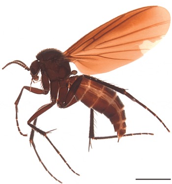
Docosia adusta. Female paratype, habitus. Scale bar =1mm.

Material examined
Holotype ♂,: labelled “COLOMBIA, Cundinamarca, PNN [Parque Nacional Natural] Chingaza Alto de La Bandera, Malaise trap, 04°31′N, 73°45′W, 3,600 m, 15.xi–01.xii.2001, L. Cifluentes leg., M. 2600 [Malaise trap # 2600]”. Paratypes: 2 ♂, 4 ♀, same data as holotype.
Description
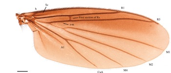
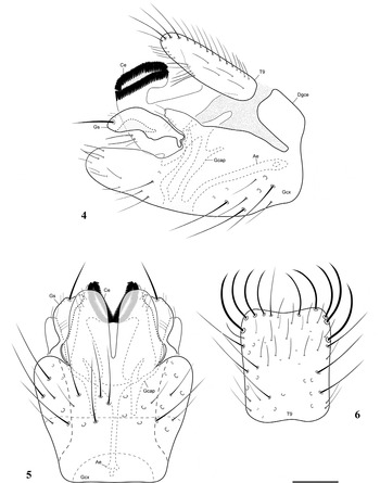
Male (Figs. 2, 4–6). Wing length 4.1mm, wing width 1.7mm. Head (Fig. 2). Vertex dark brown, with scattered setae. Three ocelli approximately aligned, mid-ocellus smaller than lateral ones, lateral ocelli separated from mid-ocellus by about 2.0 times its width and from eye margin by about its own diameter. Occiput dark brown. Eyes setose. Antennal scape and pedicel quite short, brown, with longer setae dorsally along distal margin; 14 brown flagellomeres, first slightly longer than second, flagellomeres 2–14 same length, almost 1.5 times longer than wide, with scattered setae and a short basal neck. Frons and clypeus brown, covered with short setae; labella light brown, proximal article brown; maxillary palpus brownish, lighter towards apex, five palpomeres, first and second palpomere rounded, apical ones increasingly longer, fourth as long as third, last palpomere 1.5 times penultimate one. Thorax (Fig. 2). Scutum, scutellum, and pleural sclerites dark brown. Pleural membrane brown. Scutum moderately arched, covered with small scattered setae and stronger supra-alar, dorsocentral, and acrostichal setae. Scutellum with eight scutellar bristles, four of them twice length of remaining four, plus many additional setulae. Pronotum with 2 strong setae and some additional setae. Anespisternum with many setulae along posterior margin, katepisternum more or less square ventrally. Mesepimeron not reaching ventral margin of thorax, bare. Laterotergite only slightly projecting, with a group of 9–12 setae of different lengths on posterior third. Mediotergite slightly curved in profile, bare. Metepisternum with five setulae posteriorly. Haltere pedicel brownish, knob light brown, setulae on pedicel, knob more densely setulose. Legs brown. First tarsomere more than twice length of second tarsomere; mid and hind tibiae with long erect darker bristles, in midtibia dorsally and ventrally, in hind tibia more conspicuously dorsally. Tibial spurs 1:2:2, brown, almost 3 times tibial width at apex, internal spurs shorter. Tarsal claws with small basal tooth. Wing. Membrane homogeneously light brown, darker along anterior margin; membrane densely covered with microtrichia in all cells. R1, R5, and r-m with macrotrichia dorsally and ventrally; M1, M2, M4, CuA, and A1 with macrotrichia only dorsally. Sc short, ending free, vanishing to apex, without macrotrichia. C ending much before wing apex, extending about one-sixth of distance between R5 and M1. First sector of Rs perfectly transverse, devoid of macrotrichia. R1 long, reaching C on apical third of wing; R5 short, reaching C quite before wing apex, well sclerotized, slightly sinuous at apex; r-m almost perfectly longitudinal, slightly curved at basal end, well sclerotized, more than 4 times length of r-m. M1+2 short, bare, strongly displaced to wing base; medial fork complete, 7 times length of M1+2, M1 and M2 only slightly divergent towards apex; M4 basally well produced, distally reaching wing margin, M4 and CuA only slightly divergent along their length; first sector of CuA slightly shorter than second sector of CuA. A1 incomplete, not produced on apical half. Abdomen. Abdomen brown, setose, lateral margins of tergites and sternites with slender, less sclerotized band. T8 short and wide, S8 longer than wide. Terminalia brown, conspicuous, not particularly elongate. Terminalia (Figs. 4–6). Gonocoxite setose, elongate, fused to each other ventrally, with a pair of distal mesal extensions, gonocoxal bridge well produced; gonostylus short, simple, elongate, slightly curved mesally, with single long apical seta and 2 subapical setae on rounded apex; aedeagus distinctively sclerotized, filiform, trifid basally; T9 more or less rectangular, longer than wide, setose, with four pairs of longer, curved setae on distal margin; cercus very developed, with two rings of distinctively sclerotized spines.
Docosia adusta. Male paratype, thorax, lateral view (anp, anepisternum; cx I, fore coxa; cx II, midcoxa; cx III, hind coxa; ktp, katepisternum; ltg, laterotergite; mes, metepisternum; mep, mesepimeron; mtg, mediotergite; pem, proepimeron; pes, proepisternum; pnt, pronotum; sctl, scutellum). Scale bar=0.3mm.

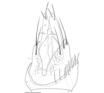
Female (Figs. 1, 3, 7). As in male, except for following features. Wing (Fig. 3). Length 4.4mm, width 1.7mm. Head (Fig. 1). Antennal flagellomeres not as elongate as in male, flagellomeres closer to each other. Terminalia (Fig. 7). Yellowish. S8 weakly sclerotized, with mesal distal projection covered with scattered microtrichia; T8 wide, very short mesally, setae only at lateral margins; S9 (genital fork) weakly sclerotized, wide at anterior end; T9 almost divided in two separate lateral sclerites, with some long setae along distal margin; Ce1 weakly sclerotized, mostly covered by T9, Ce2 more or less cylindrical, weakly sclerotized, devoid of microtrichia, 1 longer seta and 1 shorter seta on basal third and 2 longer subapical setae (one dorsal and one ventral), besides 1–2 additional short setae.
Docosia adusta. Female paratype, wing. Scale bar=0.3mm.

Etymology
The species epithet is feminine, derived from the Latin adustus, for “brown”, and referring to the brown wing membrane (especially along the anterior margin) and the general body color.
Comments
The males of this species have the terminalia rotated to some degree. Edwards (Reference Edwards1941: 71) considered that the pair of dorsal sclerites in the male terminalia of Docosia gilvipes (Walker) correspond to T9 divided into two separate plates by a transverse suture. This actually does not seem to be the case in D. adusta sp. nov. The basal sclerites are entirely bare and clearly correspond to the dorsal fusion of lateral extensions of the gonocoxites, resulting in a complete ring anteriorly in the terminalia. This feature is also present in most other species of the gilvipes group (e.g., D. pseudogilvipes Kurina (Kurina Reference Kurina2008, fig. 11)). In the female terminalia the shape of sternite 8 is unusual in relation to that of other genera of Mycetophilidae, in the sense that this sclerite has a single median distal extension, ending acuminate. This is also seen in other species of the genus, like D. gilvipes (Laštovka and Ševčík Reference Laštovka and Ševčík2006, fig. 17b). Tergite 9 also has a deep incision distally, which almost divides the plate into two separate lateral sclerites. A single conspicuous cercomere is visible, but this is actually Ce2, the basal cercomere being weakly sclerotized and partially covered by T9.
Docosia adusta. Male paratype, terminalia. 4, Lateral view; 5, ventral view, without tergite 9; 6, tergite 9, dorsal view (Ae, aedeagus; Ce, cercus; Dgce, dorsal extension of gonocoxite; Gcap, gonocoxal apodeme; Gcx, gonocoxite; Gs, gonostyle; T, tergite). Scale bar=0.1mm.

Discussion
Docosia adusta is different from the two other Neotropical species of the genus, D. pammela Edwards and D. cuzcoensis Edwards, in many respects. The other Neotropical species are small (3mm long), entirely black, and with the posterior wing veins weakly sclerotized; D. adusta is much larger, dark brown, and with a darkened wing membrane.
A detailed study of the relationships within Docosia is outside the scope of this paper. Nevertheless, it is obvious that D. adusta is not directly connected with the two known Neotropical species. The features shared by D. adusta, D. pammela, and D. cuzcoensis are of wider distribution in Docosia and do not correspond to shared exclusive apomorphies. This includes Sc ending free, anespisternum setose, gonocoxites connected to each other dorsally, aedeagus filiform, T9 with some stronger setae along the distal margin, and cerci with rows of black spines.
Docosia adusta seems to be more closely related to the Palaearctic species D. gilvipes and D. pseudogilvipes, the morphology of whose male terminalia was carefully described and illustrated by Kurina (Reference Kurina2008). A close relationship is suggested by the shape of the cercus with two pairs of rather long rows of spines, the more or less simplified gonostyle, and the distal extension of the syngonocoxite ventrally. From this perspective, D. adusta is considerably different from D. carbonaria Edwards, D. cephaloniae Chandler et al., D. chandleri Ševčík and Laštovka, D. enos Chandler, Bechev, and Caspers, D. expectata Laštovka and Ševčík, D. flavicoxa Strobl, D. fumosa Edwards, D. fuscipes (von Roser), D. helveola Chandler, and D. lastovkai Chandler. Laštovka and Ševčík (Reference Laštovka and Ševčík2006) and Kurina (Reference Kurina2008) suggested that D. gilvipes could have an isolated position within the genus and could be placed in a separate subgenus or genus (along with D. pseudogilvipes (Kurina Reference Kurina2008)). Some undescribed Nearctic species of Docosia also belong to this group (J. Ševčík, personal communication).
With reference to a revision of the Palaearctic species of Docosia, Petr Laštovka has commented that relationships within the Nearctic portion of the genus are complex (P.J. Chandler, personal communication). He recognized about 50 species with “interesting phyletic relations” and that there are Nearctic species from the gilvipes group “more numerous than those with Sc bare and ending in R1 (apart from distinct features of terminalia)”.
Docosia adusta. Female paratype, terminalia, laterodorsal view (ce, cercus; S, sternite; T, tergite). Scale bar=0.1mm.

Independently of the question of the rank of this clade (species group, genus, or subgenus), if the association of D. adusta with this Holarctic group of species within the genus is correct, the presence of this species in Colombia corresponds to a secondary extension of a Nearctic clade to the south, reaching northeastern South America. This pattern is actually not restricted to Docosia within the Mycetophilidae. Two undescribed species of Cordyla Meigen (Mycetophilidae: Mycetophilinae) show this pattern as well, one reported from Costa Rica (Vockeroth Reference Vockeroth, Brown, Borkent, Cumming, Wood and Zumbado2009), the other reaching Colombia (Oliveira et al. Reference Oliveira, Silva and Amorim2007).
The overlap of different biogeographic elements in Colombia is actually even more complex. Besides the presence of elements of Nearctic origin higher in the Andes, close to localities with elements of tropical origin in the Neotropical Region, areas of higher altitude in Colombia also have a number of typical circum-antarctic elements, such as species of Neoaphelomera Miller, Allocotocera Mik, Parvicellula Marshall, Austrosynapha Tonnoir, Paraleia Tonnoir, and Procycloneura Edwards (Oliveira et al. Reference Oliveira, Silva and Amorim2007). Further studies of the diversity of Docosia and its phylogenetic connections would contribute to a better understanding of the biogeographic history of the genus and, hence, the biogeographic evolution of the region.
Acknowledgements
The material used in this study was very kindly made available by Michael Sharkey (National Science Foundation Grant DEB-0205982), to whom we are most grateful. Additional information on the project can be found at the web address of the project (http://www.sharkeylab.org/biodiversity/static.php?app=colombia&page=index). The first author has a graduate fellowship from the Fundação de Amparo à Pesquisa do Estado de São Paulo (Grant 2008/52324-6) and the second author has a fellowship from the Brazilian National Science Research Agency(CNPq). Jan Ševčík helped us with discussions on the Palaearctic Docosia fauna and some advice, and we are grateful to him. Brad Sinclair and two anonymous reviewers helped with an excellent review of the manuscript, with useful suggestions and corrections. The authors sincerely acknowledge J.R. Vockeroth and the general editors of the Manual of Nearctic Diptera for the dedication that resulted in the chapter on the Nearctic Mycetophilidae and the whole manual, respectively. The effort spent in compiling the collective knowledge about Nearctic Diptera has saved countless hours of work by researchers, technicians, and students trying to find scattered primary sources of information worldwide, and prevented an incalculable number of mistakes by those dealing with the complex set of information about each family.