Introduction
Gyrodactylidae van Beneden et Hesse, 1832 is one of the most species-rich groups of monopisthocotylid monogeneans, common in all oceans and freshwater ecosystems of all continents infecting not only fish, like most monogeneans, but also crustaceans, amphibians and cephalopods (Boeger et al., Reference Boeger, Kritsky and Pie2003). Recently, this family has been restricted only to the viviparous gyrodactylids, with the oviparous species placed in a separate family Oogyrodactylidae Harris, 1983 (Boeger et al., Reference Boeger, Kritsky, Patella and Bueno-Silva2021). The defining trait of reproductive biology within Gyrodactylidae sensu stricto is the phenomenon of hyperviviparity, whereby several daughter generations of embryos can gestate one inside another within the parental uterus (Bakke et al., Reference Bakke, Cable, Harris, Baker, Muller and Rollinson2007). This unique reproductive ability combined with a tendency toward progenesis has a strong influence on many aspects of gyrodactylid morphology and their mode of transmission. As gyrodactylids have no specialized larval transmission stage, adult worms have evolved adaptations to host switching reflected in their locomotory behaviour and the structure of their sensory system and attachment organs. Despite a simplified and uniform morphology of internal organs, the gyrodactylids show a striking diversity in mechanisms of host attachment.
In most gyrodactylids, the posterior attachment organ (haptor) is armed with one pair of hook-shaped hamuli connected by ventral and dorsal bars and eight pairs of marginal hooks. Some genera, however, have a more complex haptoral armament containing additional sclerites and their haptor has been modified into a flattened disc. Molecular evidence (Přikrylová et al., Reference Přikrylová, Vanhove, Janssens, Billeter and Huyse2013, Reference Přikrylová, Barson and Shinn2021) indicates that these genera do not form a single monophyletic group and their complex haptors may have evolved independently in several phylogenetic lineages. Two of these genera, Fundulotrema Kritsky & Thatcher, 1977 and Swingleus Rogers, 1969, nest within Gyrodactylus von Nordmann, 1832, and another genus, Mormyrogyrodactylus Luus-Powell et al., Reference Luus-Powell, Mashego and Khalil2003, endemic to Africa, forms a cluster with several other African gyrodactylids that lack supplementary sclerites in the haptor (Přikrylová et al., Reference Přikrylová, Barson and Shinn2021). The phylogenetic position of the remaining gyrodactylids with a disc-shaped haptor is uncertain, either because their placement in the phylogenetic trees is unresolved (Macrogyrodactylus Malmberg, 1957) or because the molecular evidence for these genera is lacking (Accessorius Jara et al., Reference Jara, An and Cone1991 and Polyclithrum Rogers, Reference Rogers1967). Macrogyrodactylus, one of the genera in the latter group, has been especially extensively studied using both electron and confocal microscopy (El-Naggar et al., Reference El-Naggar, Arafa, El-Abbassy and Kearn2001, Reference El-Naggar, Arafa, El-Abbassy, Stewart and Halton2004, Reference El-Naggar, Arafa, El-Abbassy, Stewart and Halton2007; Arafa et al., Reference Arafa, El-Naggar and Kearn2003, Reference Arafa, El-Naggar and Kearn2013; Arafa, Reference Arafa2011), and a good deal of what is currently known about gyrodactylid muscle and nervous systems has been derived from these studies. The most elaborate haptor, however, with at least seven pairs of supplementary sclerites and an array of supporting ribs, has been described in Polyclithrum, which has so far been relatively little studied.
The first species of Polyclithrum, Polyclithrum mugilini Rogers, Reference Rogers1967, has been described by Rogers (Reference Rogers1967) from the flathead grey mullet (Mugil cephalus Linnaeus, 1758) of Lake Seminole (USA). A similar gyrodactylid species was reported from the same host species by Skinner (Reference Skinner1975) and was assigned to a new genus Micropolyclithrum, as Micropolyclithrum parvum Skinner, Reference Skinner1975, based primarily on the presence of the so-called double bar in the haptor. Yet another newly described gyrodactylid genus, Accessorius with a single species Accessorius peruensis Jara et al., Reference Jara, An and Cone1991, was also provisionally placed in the same subfamily as Polyclithrum (Polyclithrininae) (Jara et al., Reference Jara, An and Cone1991), but a shared trait of both genera, an arrangement of marginal hooks in three groups, is also present in Fundulotrema and Swingleus and does not seem to be sufficient to justify this taxonomic decision. Ernst et al. (Reference Ernst, Whittington and Jones2000) have revised the description of P. mugilini and added three new species to the genus Polyclithrum: Polyclithrum alberti and Polyclithrum corallense from Australia and Polyclithrum boegeri from Brazil. These authors also called into question the validity of Micropolyclithrum arguing that the double bar of this species was a misinterpretation of accessory bar 1, one of the haptoral sclerites present in Polyclithrum species. The last species of Polyclithrum, Polyclithrum ponticum Gerasev et al., (Reference Gerasev, Dmitrieva and Gaevskaia2002) from the grey mullets of the Black Sea, has been described by Gerasev et al. (Reference Gerasev, Dmitrieva and Gaevskaia2002) who also showed that the haptor of both P. ponticum and P. mugilini has a pair of large ear-shaped sclerites (ESSs) that had not been reported in earlier descriptions.
Aiming to gain a better understanding of the microanatomy of Polyclithrum, we have examined the neuromusculature and morphology of haptoral sclerites of P. ponticum by confocal microscopy. Particular emphasis in this study was placed on the armament and musculature of the haptor in connection with the mechanisms of attachment to the host.
Material and methods
Specimens of P. ponticum were collected from M. cephalus caught off the Crimean shores of the Black Sea (44°54′23″N, 35°11′00″E) in June 2016. All monogeneans were collected from gills alive and some of them (n = 5) were cleared in a drop of sodium dodecyl sulphate (SDS), mounted in glycerine jelly prepared with 0.5 g of carbolic acid and photographed using an Olympus CX41 microscope (Olympus Corporation, Tokyo, Japan) with phase-contrast optics and CellSens digital image analysis software (Olympus Corporation, Tokyo, Japan). Other worms (n = 11) were fixed with 4% (w/v) paraformaldehyde in 0.01 M phosphate-buffered saline (PBS) at ambient temperature and after 6 h of fixation were transferred to 0.01 M PBS containing 0.05% (w/v) sodium azide (Sigma-Aldrich, St. Louis, Missouri, USA) for storage at 4–6 °C.
All subsequent steps were performed on an orbital shaker at room temperature. The worms were permeabilized for 2–4 h in 0.01M PBS containing 0.25% Triton X-100 (Tr-PBS; Sigma-Aldrich, St. Louis, Missouri, USA) and blocked with Image-iT FX Signal Enhancer (Molecular Probes, Eugene, Oregon, USA) for 1 h. Blocked animals were rinsed 4 × 15 min in Tr-PBS and incubated for 10–12 h in antibodies against serotonin (5HT) (Sigma-Aldrich, St. Louis, Missouri, USA; diluted 1:600) or against FMRFamide (Abcam, Cambridge, Massachusetts, USA; diluted 1:600) to stain for FMRFamide-related peptides (FaRPs). After incubation in primary antibodies, the animals were rinsed again 4 × 15 min in Tr-PBs and incubated for 5 h in Alexa Fluor 488-conjugated secondary antibodies (Life Technologies, Carlsbad, California, USA; diluted 1:300). After that, the monogeneans were rinsed for 15 min in Tr-PBS and stained 2–3 h with Alexa Fluor 543-conjugated phalloidin (Life Technologies, Carlsbad, California, USA; diluted 1:200), then rinsed once in 0.01M PBS and mounted on slides with Vectashield (Vector Laboratories Inc., Burlingame, California, USA). Staining specificity was verified by omitting primary antibodies.
Confocal images were acquired on a Leica TCS SP5 confocal laser scanning microscope (Leica Microsystems GmbH, Wetzlar, Germany) at the ‘Taxon’ Research Resource Centre (Zoological Institute of the Russian Academy of Sciences, St. Petersburg, Russia). Haptoral sclerites and the armament of the male copulatory organ were visualized in the reflection mode of the confocal microscope as described in Petrov et al. (Reference Petrov, Gerasev, Popyuk and Dmitrieva2016): the worms were illuminated with a helium–neon (633 nm) laser and the images were obtained at a detection range of 625–645 nm. All confocal images in the paper were presented as maximum intensity projections of Z-stacks. Three-dimensional schemes were made using ZBrush 4R6 (Pixologic, Los Angeles, California, US).
Results
Musculature of the body wall and cephalic lobes
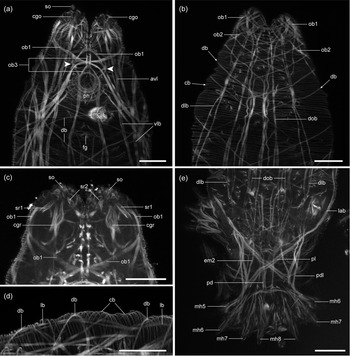
The body is subdivided into a spindle-shaped body proper and a disc-shaped haptor. The anterior end of the body bears a pair of cephalic lobes through which pass the ducts of large cephalic glands located on either side of the body slightly posterior to the pharynx. The glands open ventrally at the tips of the cephalic lobes (cgo, fig. 1a). Cone-shaped sensory spike organs (so, fig. 1a, c) open at the distal extremities of the cephalic lobes slightly dorsal to the openings of the cephalic glands (fig. 1a). Two small muscles (sr1–2, fig. 1c) originate at the base of each cephalic lobe, on their medial and lateral sides, and insert on the spike organs near their openings. A similar set of muscles (cgr, fig. 1c) extends from the openings of the cephalic glands to the lateral body wall just posterior to the cephalic lobes.
Confocal images of phalloidin-stained body-wall musculature of Polyclithrum ponticum: (a) anterior half of the body proper in ventral view. Arrowheads indicate the anterior endings of the ventral bundles of longitudinal body-wall muscles; (b) anterior half of the body proper in dorsal view; (c) anterior end of the body showing the retractor muscles of the spike organs and cephalic glands; (d) anterolateral portion of the body showing the arrangement of the three layers of body-wall muscles. Note that the longitudinal muscles lie internal to the diagonal muscles; and (e) posterior end of the body proper and haptor in dorsal view. Scale bars: 20 μm. Abbreviations: avl, anterior portions of ventrolateral body-wall muscles; cb, circular body-wall muscles; cgo, cephalic gland opening; cgr, cephalic gland retractor muscle; db, diagonal body-wall muscles; dlb, dorsolateral bundles of longitudinal body-wall muscles; dob, dorsal bundles of longitudinal body-wall muscles; em2, extrinsic haptoral muscle 2; fg, female gonopore; lab, lateral bundles of body-wall muscles; lb, longitudinal body-wall muscles; mc, male copulatory organ; mh5–8, marginal hooks 5–8; ob1–3, oblique bundles of body-wall muscles; pd, posterior portions of dorsal longitudinal body-wall muscles; pdl, posterior portions of dorsolateral longitudinal body-wall muscles; ph, pharynx; pl, posterior portions of lateral longitudinal body-wall muscles; so, spike organ; sr1–2, retractor muscles of spike organ; and vlb, ventrolateral bundles of longitudinal body-wall muscles.

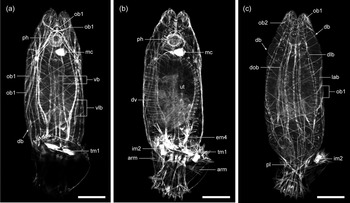
The body-wall musculature comprises outer circular (cb, fig. 1b, d), intermediate diagonal (db, figs 1a, b, d and 2a, c) and inner longitudinal (lb, fig. 1d) layers. The circular layer consists of densely spaced thin muscle fibres that are regularly arranged throughout the body proper and are present dorsally in the haptor. The diagonal layer is composed of two sets of widely spaced criss-crossing muscle fibres that tend to be more densely arranged at the anterior end of the body and are lacking in the haptor. The longitudinal body-wall muscles are grouped into several paired bundles in dorsal (dob, figs 1b, e and 2c), dorsolateral (dlb, figs 1b, e and 2c), lateral (lab, figs 1e and 2c), ventrolateral (vlb, figs 1a and 2a) and ventral (vb, fig. 2a) positions. The ventrolateral bundles are especially well-developed and anchor anteriorly at the tips of the cephalic lobes or curve around them laterally to the dorsal side. The ventral bundles lie submedially flanking the mouth and the female gonopore (fg, fig. 1a) and end anteriorly just in front of the mouth (arrowheads, fig. 1a). Posteriorly, the ventral and ventrolateral bundles provide some extrinsic muscles for sclerites (see below), the lateral bundles curve medially in the haptor to cross the corresponding opposite bundles (pl, figs 1e and 2c), and the dorsal (pd, fig. 1e) and dorsolateral (pdl, fig. 1e) bundles converge to end together at the posterior extremity of the haptor, with one dorsal fibre giving off a branch (em3, figs 5 and 6a, c) attached to haptoral sclerite A5. In the anterior half of the body, there are muscle bundles that are oriented obliquely to the main body axis, but their position internal to other muscle layers suggests that they are derived from the longitudinal muscles. The most prominent of these bundles (ob1, figs 1a–c, 2a, c and 3a) originate posteriorly in the dorsolateral position (fig. 2c), continue diagonally to the ventral side, where they cross the corresponding contralateral muscles in front of the mouth (figs 1a and 2a), and then either anchor to the tip of the cephalic lobe on the opposite side of the body or curve again to the dorsal side and cross the cephalic lobe diagonally to end on its medial side (figs 1b and 2c). Some of the oblique muscles are much shorter and occur only in front of the pharynx curving laterally around the cephalic lobes (ob2, figs 1b and 2c). Another pair of muscle bundles (ob3, fig. 1a) begins posteriorly in the same position as the ventral bundles of longitudinal muscles, but cross each other in front of the mouth and terminate anteriorly on the medial sides of the cephalic lobes.
Confocal images of phalloidin-stained musculature showing the overall view of Polyclithrum ponticum in ventral view (a), in the interior of the body (b) and in dorsal view (c). Scale bars: 50 μm. Abbreviations: arm, separate muscle fibres of anterior haptoral ribs; db, diagonal body-wall muscles; dlb, dorsolateral bundles of longitudinal body-wall muscles; dob, dorsal bundles of longitudinal body-wall muscles; dv, dorso-ventral muscles; em4, extrinsic haptoral muscle 4; im2, inner hamular root muscle 2; lab, lateral bundles of body-wall muscles; mc, male copulatory organ; ob1–2, oblique bundles 1–2 of body-wall muscles; ph, pharynx; pl, posterior portions of lateral longitudinal body-wall muscles; tm1, transverse hamular muscle 1; ut, uterus; vb, ventral bundles of longitudinal body-wall muscles; and vlb, ventrolateral bundles of longitudinal body-wall muscles.

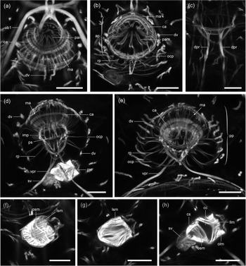
Confocal images of phalloidin-stained musculature of the pharynx and male copulatory organ of Polyclithrum ponticum: (a, b) anterior pharyngeal region; (c) dorsal part of the pharynx showing dorsal pharyngeal retractor muscles; (d) pharynx and male copulatory organ; (e) posterior pharyngeal region and ventral pharyngeal retractor muscles; (f, g) male copulatory organ on the side of the short spinelets; and (h) male copulatory organ on the side of the long spine. Scale bars: 10 μm. Abbreviations: ap, anterior pharyngeal region; ca, circular muscles of anterior pharyngeal region; cc, cavity of copulatory organ; cs, sphincter of copulatory organ; dpr, dorsal pharyngeal retractor muscle; dv, dorso-ventral muscles; imp, meridional muscles in inner wall of posterior pharyngeal region; llm, longitudinal muscles of copulatory organ on side of long spine; lsm, longitudinal muscles of copulatory organ on side of short spines; ma, meridional muscles of anterior pharyngeal region; mc, male copulatory organ; ob1, oblique bundles 1 of body-wall muscles; ocp, circular muscles in outer wall of posterior pharyngeal region; olm, oblique muscle fibers of copulatory organ on side of long spine; osm, oblique muscle fibers of copulatory organ on side of short spine; pam, longitudinal muscles of pharyngeal papillae; pp, posterior pharyngeal region; ps, pharyngeal sphincter; rp, radial muscles of posterior pharyngeal region; sv, seminal vesicle; and vpr, ventral pharyngeal retractor muscle.

Musculature of the digestive system
The pharynx is located ventrally between the cephalic glands and is subdivided into smaller anterior (ap, fig. 3b) and a larger posterior (pp, fig. 3e) regions. Both pharyngeal regions are roughly spherical in shape and are separated by a shallow circular constriction. The anterior region bulges into the pharyngeal pocket and its distal end bears a wreath of eight conical papillae. Both pharyngeal walls in the posterior region contain a single muscle layer consisting of very thin circular fibres in the outer wall (ocp, fig. 3b, d, e) and longitudinal (meridional) fibres in the luminal wall (imp, fig. 3d); the space between the walls is crossed by bundles of radial fibres (rp, fig. 3b, d, e). A few circular fibres (ps, fig. 3d) present within the luminal wall at the posterior end of the pharynx form a sphincter at the junction with the gut. In the anterior region, the musculature of the outer wall is two-layered, with numerous thin meridional muscle fibres (ma, fig. 3a, b, d, e) lying to the outside of 6–7 ribbon-shaped circular muscles (ca, fig. 3a, b, d, e). The luminal wall in the anterior region appears to lack muscles. The movement of each pharyngeal papilla is controlled by two bundles of muscle fibres (pam, fig. 3a, b) attached with one end to the tip of the papilla and with the other to the outer wall of the anterior region about midway along its length. The pharynx is enclosed in a sheath of dorsoventral muscles (dv, fig. 3a, b, d, e) and provided with two pairs of retractor muscles: a ventral pair (vpr, fig. 3d, e) that arcs around the uterus and attaches to the ventrolateral body wall; and a pair of short dorsal retractors (dpr, fig. 3c) that extend posteriorly to their attachment on the dorsal body wall. The gut lacks any distinct musculature.
Musculature of the reproductive system
In the individuals examined in this study, the copulatory organ (figs 1a, 2a, b and 3d, f–h) was the only part of the male reproductive system that had a well-developed musculature. The male copulatory organ is located posterolateral to the pharynx (mc, figs 1a, 2a, b and 3d) and is armed with a large spine located on one side of the male gonopore (lsc, fig. 4e) and six smaller spinelets in a single row on the opposite side (ssc, fig. 4e). Two marginal spinelets in the row are somewhat larger than the remaining four. The wall of the copulatory organ is invested by an outer band of oblique fibres and an inner layer of longitudinal fibres. The musculature is asymmetrical: longitudinal fibres are much longer on the side of the short spines (lsm, fig. 3f, g) and the band of oblique fibres is angled so that it is closer to the gonopore on the side of the long spine (olm, fig. 3h). The copulatory organ is provided with a strong sphincter (cs, fig. 3h) where it joins the seminal vesicle (sv, fig. 3d, h). In non-gravid individuals (n = 3), a group of thin muscle fibres occurs in the posterior third of the body proper possibly representing a rudimentary muscle system of the egg cell-forming region.
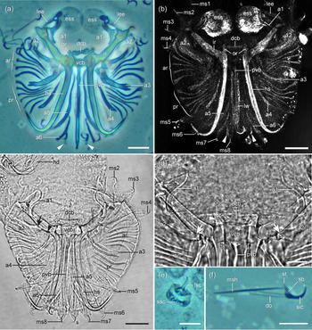
Armature of the haptor and male copulatory organ of Polyclithrum ponticum in phase contrast after being cleared in 5% aqueous solution of sodium dodecyl sulphate (a, e, f), in reflected light (b) and in brightfield (c, d): (a, b) haptor; (c) haptor and posterior body proper. Note that the inner hamular root is articulated with the A2 (arrow) and A3 (arrowhead); (d) central portion of the haptor. Note knob-shaped protuberances at the lateral ends of the dorsal bar (arrowhead) and the grooves at the medioanterior edges of the A2 that articulate with the inner roots of the hamuli (arrows); (e) male copulatory organ; and (f) marginal hook. Scale bars: (a–c) 20 μm; and (d–f) 10 μm. Abbreviations: a1–6, accessory bars 1–6; ar, anterior haptoral ribs; dcb, dorsal connecting bar; do, domus; ess, ear-shaped sclerite; hd, hamulus in daughter individual; hs, hamular shaft; ir, inner hamular root; lee, lateral extension of ear-shaped sclerite; lsc, large spine of male copulatory organ; lw, lateral wings of posterior process of ventral bar; ms1–8, sickles of marginal hooks 1–8; msh, shaft of marginal hook; or, outer hamular root; pr, posterior haptoral ribs; pvb, posterior process of ventral bar; sb, sickle base; sic, sickle of marginal hook; ssc, small spines of male copulatory organ; st, sickle toe of marginal hook; and vcb, ventral connecting bar.

The female gonophore is positioned at the ventral midline slightly posterior to the male copulatory organ. Much of the internal space of the body in most worms was occupied by a uterus (ut, fig. 2b) containing a daughter embryo in a U-folded position. The uterus appears to lack intrinsic musculature, but is enclosed by regularly arranged dorsoventral muscles (dv, fig. 2b). Both ventral and dorsal ends of these muscles are anchored on the body wall near the body midline and the muscles are applied closely to the uterine wall giving the impression of the uterus being enclosed by circular muscles.
Sclerites and musculature of the haptor
The armature of the haptor consists of a pair of large ventral hamuli, two connecting bars (one ventral and one dorsal), a pair of ESSs, six pairs of accessory bars (A1–6), two paired groups of lateral ribs and 16 marginal hooks.
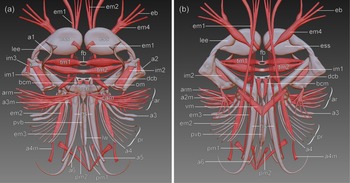
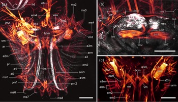
The hamuli have a long, straight shaft (hs, figs 4a, b, 5, 6a and 7b) with a recurved tip and two roots: a short outer root (or, fig. 4a, b, d) that forms the proximal extension of the shaft; and a long inner root (ir, figs 4a, b–d, 5b and 6a) that extends anterolaterally from the base of the shaft. The tips of the inner roots of the opposite hamuli are connected by two unpaired transverse muscles, one located slightly anterior (tm1, figs 2a, b, 5 and 6b) to the other (tm2, figs 5 and 6a, b). Each muscle consists of two lateral spindle-shaped bellies joined to each other medially by a fibrous bridge. This bridge is more prominent in the anterior muscle (fb, figs 5 and 6b), where it is oval in shape and is probably perforated by a hole. It appears to be highly stretchable differing in length in different preparations depending on the state of muscle contraction.
Schematic representation of haptoral sclerites and musculature: (a) ventral view; and (b) dorsal view. Abbreviations: a1–6, accessory bars 1–6; a2m, muscle connecting A2 to haptoral tegument; a3m, muscle connecting A3 to hamular base; a4m, muscle connecting A4 to haptoral tegument; ar, anterior haptoral ribs; arm, separate muscle fibres of anterior haptoral ribs; bcm, muscle connecting dorsal and ventral bars; cam, combined muscle bundle of anterior haptoral ribs; dcb, dorsal connecting bar; eb, branches of extrinsic muscles em4; em1–4, extrinsic haptoral muscles; ess, ear-shaped sclerite; fb, fibrous bridge of anterior transverse muscle; hs, hamular shaft; im1–3, inner hamular root muscles; ir, inner hamular root; lee, lateral extension of ear-shaped sclerite; lw, lateral wings of posterior process of ventral bar; om, muscle connecting outer hamular root and A3; pm1–2, muscles of posterior process of ventral bar; pr, posterior haptoral ribs; pvb, posterior process of ventral bar; tm1–2, transverse hamular muscles; vcb, ventral connecting bar; and vm, muscle connecting posterior process of ventral bar to A3.

Confocal images of haptoral sclerites in reflected light (white) and phalloidin-stained haptoral musculature (orange). Anterior is toward the top: (a) overall view of the haptor and posterior body proper; (b) anteromedial portion of the haptor showing the ear-shaped sclerites and transverse muscles; and (c) dorsal view of the anterior portion of the haptor (sclerites are not shown). Scale bars: 20 μm. Abbreviations: a2m, muscle connecting A2 to haptoral tegument; a3–5, accessory bars 3–5; a3m, muscle connecting A3 to hamular base; a4m, muscle connecting A4 to haptoral tegument; ar, anterior haptoral ribs; arm, separate muscle fibres of anterior haptoral ribs; bcm, muscle connecting dorsal and ventral bars; cam, combined muscle bundle of anterior haptoral ribs; eb, branches of extrinsic muscles em4; em1–4, extrinsic haptoral muscles; ess, ear-shaped sclerite; fb, fibrous bridge of anterior transverse muscle; hd, hamulus of daughter individual; hs, hamular shaft; im1–3, inner hamular root muscles; ir, inner hamular root; lee, lateral extension of ear-shaped sclerite; ms1–8, sickles of marginal hooks 1–8; om, muscle connecting outer hamular root and A3; pm1–2, muscles of posterior process of ventral bar; pr, posterior haptoral ribs; tm1–2, transverse hamular muscles; and vm, muscle connecting posterior process of ventral bar to A3.

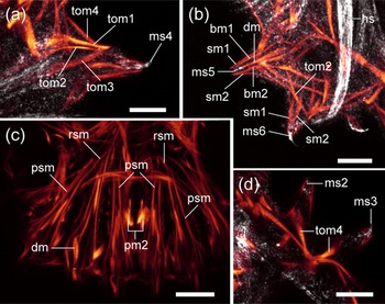
Confocal images of marginal hooks of Polyclithrum ponticum in reflected light (white) and associated phalloidin-stained musculature (orange). Anterior is toward the top: (a) marginal hook 4 in an anterior group; (b) marginal hooks 5 and 6 in the posterior group; (c) posterior portion of the haptor showing muscles of the marginal hooks in the posterior group; and (d) marginal hooks 2 and 3 in an anterior group showing tom4 muscles of the four hooks coming together to form a single bundle of muscle fibres. Scale bars: (a) 5 μm; and (b–d) 10 μm. Abbreviations: bm1–2, base muscles of marginal hooks; dm, domus muscle; hs, hamular shaft; ms2–6, sickles of marginal hooks 2–6; psm, shaft protractor muscles of marginal hook; pm2, muscle 2 of posterior process of ventral bar; rsm, shaft retractor muscle of marginal hook; sm1–2, sickle muscle of marginal hooks; and tom1–4, toe muscles of marginal hooks.

The ventral connecting bar (VCB; vcb, figs 4a, c, d, 5 and 8a) is roughly trapezoid in shape and is hinged between the hamuli, its lateral sides forming articulating surfaces with protuberances at the base of the inner root of each hamulus. The central part of the VCB is strongly arched dorsally and has a long, slender medial process (pvb, figs 4a–d and 5a) that extends to the posterodorsal margin of the haptor ending below the posterior group of marginal hooks. Dorsally, the posterior process expands to form lateral wings (lw, figs 4b and 5a); the process is slightly forked at the end and each prong serves as attachment for two muscles. One pair of these muscles (pm1, figs 5 and 6a) extends diagonally in the anterolateral direction and attaches to the dorsolateral wall of the haptor near the tip of the process of the A4, the other (pm2, figs 5, 6a and 7c) runs anteriorly and terminates on the dorsal wall in front of the posterior marginal hooks.
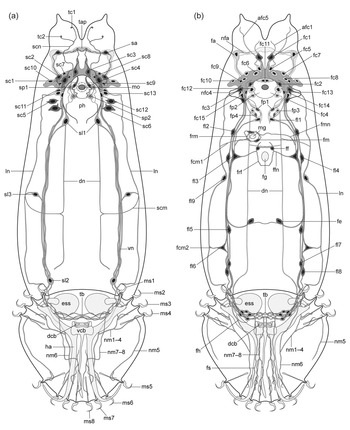
Schematic drawing of the nervous system of Polyclithrum ponticum: (a) 5HT-IR; and (b) FaRP-IR. Abbreviations: afc1, axon of neuron fc1; afc5, axon of neuron fc5; dcb, dorsal connecting bar; dn, neurite in dorsal nerve cord; ess, ear-shaped sclerite; fa, anterior FaRP-IR neurons; fc1–15, FaRP-IR neurons associated with cerebral ganglia; fcm, commissural FaRP-IR neurite; fe, medial neurite of presumable egg cell-forming region; ff, FaRP-IR neurons of female gonopore; fg, female gonopore; fh, haptoral FaRP-IR neurons; fl1–9, FaRP-IR neurons associated with nerve cords; fm, FaRP-IR neurons of male copulatory organ; fmn, medial FaRP-IR neurite to male gonopore; fp1–4, pharyngeal FaRP-IR neurons; frf, FaRP-IR nerve ring around female gonopore; frm, FaRP-IR nerve ring around male gonopore; fs, submedial haptoral FaRP-IR neurite; ha, hamulus; ln, neurite in lateral nerve cord; mg, male gonopore; mo, mouth; ms1–8, sickles of marginal hooks 1–8; nfa, neurite of neuron fa; nfc4, arcuate neurite of neuron fc4; nm1–8, neurites supplying marginal hooks 1–8; ph, pharynx; sa, anterior transverse 5HT-IR neurons; sc1–13, 5HT-IR neurons associated with cerebral ganglia; scm, commissural 5HT-IR neurite; scn, 5HT-IR neurites innervating cephalic lobes; sl1–3, 5HT-IR neurons associated with nerve cords; sp1–2, pharyngeal 5HT-IR neurons; tap, transverse anterior plexus; tb, transverse neurite bundle; tc1–2, 5HT-IR sensory terminals on cephalic lobes; vcb, ventral connecting bar; and vn, neurites in ventral nerve cord.

The dumbbell-shaped dorsal connecting bar (DCB; dcb, figs 4a–d, 5 and 8) is fit tightly between the outer roots of the hamuli, to which it appears to be flexibly connected (arrowhead, fig. 4d). A pair of short muscles (bcm, figs 5 and 6a, c) extends from the posterior margin of the DСB to the base of the posterior process of the VCB and a slender muscle (im1, figs 5 and 6a) runs from each lateral end of the DCB to the tip of the inner root of each hamulus.
The ESSs (ess, figs 4a, b, 5, 6a, b and 8) are large, paddle-shaped sclerites that reinforce the anterior extremity of the haptor; the oval main bodies of these sclerites lie medially anterior to the inner roots of the hamuli on the ventral side of the haptor, while their lateral handle-shaped extensions (lee, figs 4a, b, 5 and 6b) overlap with the tips of the inner roots. In all individuals of P. ponticum, the lateral tip of the handle is located over the anterolateral corner of the A2 and appears to be connected or articulated with it. Since a distinct suture is visible between the main bodies of the ESSs and their lateral handles, the latter may actually be separate sclerites partially fused to the ESSs. Each ESS is attached to the hamulus via the A1 (a1, figs 4a–d and 5a), S-shaped sclerites that form a bracket-like connection between the dorsal side of the ESS and the inner root of the hamulus. The attachment to the hamulus probably allows the ESSs a certain degree of mobility as the ESSs are oriented at somewhat various angles to the hamulus in different preparations.
The A2 (a2, figs 4a–d and 5) are L-shaped sclerites positioned near and roughly parallel to the inner roots of the hamuli, with the narrow end of the sclerite directed medially and the wider end overlapping the tips of the inner roots at the anterolateral margin of the haptor. The medial end of the A2 forms a groove (arrows, fig. 4d) into which fits the posterior edge of the inner root so that the two sclerites are articulated by a gliding joint. Two muscles connect the A2 to the inner root of the hamulus: a large sheet-like muscle (im2, figs 2b, c, 5 and 6a, c), which inserts on a shelf-like projection near the medial end of the A2 and originates from a similar projection near the tip of the root; and a smaller muscle (im3, figs 5a and 6a) connecting the terminal projection of the inner root to the lateral end of the A2. Another muscle (a2m, figs 5b and 6a, c) arises from the posterior corner of the A2 just lateral to the im2 muscle; it runs posteriorly and ramifies into several branches that anchor on the dorsal tegument of the haptor in front of the posterior group of marginal hooks.
The irregular-shaped A3 (a3, figs 4a–c, 5 and 6a) lie near the base of the hamuli; each sclerite gives rise to two parallel rib-like projections that extend to the lateral margins of the haptor. Like the A2, the A3 have a groove, which is articulated with the inner root of the hamulus to form a gliding joint (arrowhead, fig. 4c). Three muscles arise from the A3: the medioanterior edge of the sclerite is connected to the tip of the outer hamular root by a short spindle-shaped muscle (om, figs 5a and 6a); a wide muscle (a3m, figs 5a and 6a, c) extends from the A3 medially to attach to a ridge at the base of the hamular shaft; and a strap-shaped muscle (vm, figs 5b and 6c) runs diagonally toward the posterior process of the VCB to end near its base.
Posteriorly, the A3 are firmly joined to the A4 (a4, figs 5a–c, 6 and 7a, b, d), small sclerites with two slender processes that extend posteriorly at a slight angle to the shafts of the hamuli. The A4 are anchored to the haptoral wall by a slender muscle (a4m, figs 6 and 7d) that runs from the distal tip of the sclerite to the tegument near the opening of the hamulus.
The A5 (a5, figs 4a–c, 5a and 6a) are a pair of long rod-shaped sclerites lying medially between the hamuli parallel to their shafts. The anterior ends of the A5 are articulated with the bases of the hamular shafts immediately posterior to their articulation with the VCB.
The A6 (a6, figs 4a–c and 5) are short rods that lie medial to the posterior ends of the A5 and are not attached directly to any other skeletal elements. Reflected light and SDS-cleared preparations also reveal a pair of small triangular sclerite-like elements (arrowheads, fig. 4a, b) lying ventrally at the posterior margin of the haptor between the A6 and the posterior process of the VCB.
The rod-shaped lateral ribs are oriented radially in the lateral flaps of the haptor and arranged in two groups: the anterior group (ar, figs 4a–d, 5 and 6a) is located between the A2 and processes of the A3; and the posterior (pr, figs 4a–c, 5 and 6a) between the processes of the A3 and A4. The number of ribs in each group can vary from four to six, with five being the most common number. Two muscle fibres (arm, figs 2b, 5 and 6a, c) are inserted on each rib in the anterior groups near their lateral ends; from there, these fibres run medially merging into a single muscle (cam, figs 5a and 6c) and attach to the VCB near the base of its posterior process.
The extrinsic musculature of the haptor comprises several muscles derived from the longitudinal body-wall musculature. A pair of fibres (em1, figs 5 and 6b) in the ventral bundles of longitudinal body-wall muscles run along the ventral surfaces of the ESSs and then wrap around their posterior edges to attach to the dorsal sides of these sclerites. Another pair of extrinsic muscles (em2, figs 1e, 5 and 6a, c) have their insertions on the processes of the A4 near their tips and run anteriorly to transition into the ventrolateral bundles. A slender muscle (em3, figs 5 and 6a, c) arises from the distal tip of each A5 and proceeds forward and dorsally to continue in the body proper as dorsal bundles of longitudinal body-wall muscles. A pair of large spindle-shaped muscles (em4, figs 2b, 5 and 6a) are located in the posterior part of the body proper and converge diagonally toward the haptor midline. Anteriorly, these muscles split into several branches (eb, figs 5 and 6a) that anchor on the ventral longitudinal body-wall muscles in the posterior portion of the body. The position of the posterior attachment of these muscles is uncertain as their myofibres end in the interior of the body at the anterior boundary of the haptor, but it is possible that they may bear tendinous processes that proceed into the haptor and insert on one of the haptoral sclerites.
Each marginal hook is borne on a finger-like papilla (fig. 4c) and consists of a needle-shaped internal shaft (msh, fig. 4f) and a crescent-shaped sickle (sic, fig. 4f), which is movable relative to the shaft. The sickle bears a thin trough-shaped plate called the domus (=filament loop) (do, fig. 4f) and the proximal end of the sickle is enlarged into a triangular base (sb, fig. 4f), the anterior end of which is tapered into a knob called the sickle toe (st, fig. 4f). The hooks are arranged in three groups (fig. 4b, c): two groups of four hooks each lie at the anterolateral margins of the haptor; and the other eight form a medial cluster at the posterior extremity of the haptor.
Each marginal hook is operated by 12 muscles. Two protractor muscles (psm, fig. 7c) arise from the proximal end of the shaft and extend diagonally to the body surface to anchor on the haptor's wall on each side of the hook. A single retractor muscle (rsm, fig. 7c) is inserted at the proximal end of the shaft and proceeds anteriorly in the posterior group of hooks and medially in each lateral group to attach to the dorsal tegument of the haptor. The fourth muscle (dm, fig. 7b, c) arising from the proximal tip of the shaft connects it to the domus. Two muscles (sm1–2, fig. 7b) extend from the mid-portion of the sickle and run down each side of the hook papilla to attach to the tegument between the adjacent papillae. Another pair of muscles (bm1–2, fig. 7b) runs proximally from the posterior corner of the sickle base to anchor on the ventral tegument some distance from the hook. Four muscles originate from the toe of the sickle. Two of these (tom1–2, fig. 7a, b) extend horizontally toward the two adjacent hooks to attach to the tegument between the hook papillae. The third fibres (tom3, fig. 7a) of all marginal hooks in each anterolateral group run medially, merging together as a single muscle to attach to the lateral side of the A3. The presumably homologous fibres in the posterior cluster of marginal hooks do not end on a sclerite but proceed anteriorly and attach to the dorsal tegument of the haptor. The fourth fibres (tom4, fig. 7a, d) of all marginal hooks in one group converge to a single point where they attach to each other and possibly to the ventral wall of the haptor.
Major features of the nervous system
The brain consists of a pair of cerebral ganglia (cg, fig. 9a, i, j) positioned on each side of the pharynx and connected in front of the pharynx by a broad commissure (cec, fig. 9a, c, k). A pair of submedial finger-like extensions containing both nerve cells and neurites arise anteriorly from the cerebral commissure. A semicircular connective arises laterally from each cerebral ganglion and loops in front of the brain at the bases of the cephalic lobes. Three longitudinal nerve cords (ventral, lateral, and dorsal) originate separately from the cerebral ganglia and extend throughout the body proper, with the ventral cords being significantly thicker than the lateral and dorsal pairs. The ventral cords bear a pair of large prehaptoral ganglia just in front of the haptor. The three nerve cords unite in the haptor, where they form another pair of ganglia and the combined contralateral cords become connected by a transverse bundle of neurites in the space between the hamuli and the ESSs.
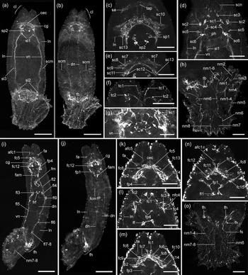
Confocal images of the 5HT-IR (a–h) and FaRP-IR (i–o) nervous systems of Polyclithrum ponticum. Anterior is toward the top: (a) overall view of the worm on the ventral side; (b) overall view of the worm on the dorsal side; (c) anterior end of the body showing the brain and the precerebral plexus (tap); (d) ventral view of the anterior half of the body; (e) brain region showing the neurons of the cerebral ganglia; (f) anterior end of the body on the ventral side showing sensory terminals of neuron sc1; (g) anterior end of the body on the ventral side showing innervation of the pharynx. Arrows point to two of the eight neurites of the pharyngeal neurons that ramify at the bases of the pharyngeal lobes; (h) ventral view of the haptor showing innervation of marginal hooks; (i) overall view of the worm on the ventral side; (j) overall view of the worm on the dorsal side; (k–n) anterior end of the body showing cerebral neurons and their neurites; and (o) ventral view of the haptor. Scale bars: (a, b, i, j) 50 μm; and (c–h, k–o) 20 μm. Abbreviations: afc1, axon of neuron fc1; afc5, axon of neuron fc5; cec, cerebral commissure; cg, cerebral ganglion; cl, cephalic lobe; dn, neurite in dorsal nerve cord; fa, anterior FaRP-IR neurons; fam, cluster of FaRP-IR neurons associated with male copulatory organ; fc1–15, FaRP-IR neurons associated with cerebral ganglia; fcm1, commissural FaRP-IR neurite 1; ff, FaRP-IR neurons of female gonopore; fh, haptoral FaRP-IR neurons; fl1–9, FaRP-IR neurons associated with nerve cords; fm, FaRP-IR neurons of male copulatory organ; fp1–4, pharyngeal FaRP-IR neurons; frf, FaRP-IR nerve ring around female gonopore; fs, submedial haptoral FaRP-IR neurite; ln, neurite in lateral nerve cord; nfc4, arcuate neurite of neuron fc4; nm1–8, neurites supplying marginal hooks 1–8; ph, pharynx; sa, anterior transverse 5HT-IR neurons; sc1–13, 5HT-IR neurons associated with cerebral ganglia; scm, commissural 5HT-IR neurite; scn, 5HT-IR neurites innervating cephalic lobes; sl1–3, 5HT-IR neurons associated with nerve cords; sp1–2, pharyngeal 5HT-IR neurons; tap, transverse anterior plexus; tb, transverse neurite bundle; tc1–2, 5HT-IR sensory terminals on cephalic lobes; and vn, neurites in ventral nerve cord.

Serotoninergic nervous system
The arrangement of 5HT-immunoreactive (IR) cells and their processes was essentially the same in all the individuals studied (n = 3). Serotonin immunoreactivity was present in the cerebral ganglia and all three longitudinal nerve cords (fig. 9a–h). The cerebral ganglia contain six ventral and seven dorsal pairs of 5HT-IR cell bodies, most of which are likely to be bipolar. One ventral pair (sc1, figs 8a and 9d, g) lies submedially in front of the pharynx. The axon of these neurons projects toward the anterior end of the body where it divides into two branches: one continuing forward to end in a sensory terminal at the medial side of the cephalic lobe (tc1, figs 8a and 9f); and the other proceeding laterally to a sensory ending at the lobe's ventral side (tc2, figs 8a and 9f). Both terminals form a distinctly stained bulb-shaped distension slightly projecting from the body surface. Another pair of ventral cells (sc2, figs 8a and 9d) located laterally just in front of the pharynx send one neurite anteriorly within the semicircular connective where these neurites come in contact with a pair of bipolar neurons (sa, figs 8a and 9c) positioned transversally at the bases of the cephalic lobes. These latter neurons send their other neurite medially toward one another to supply a small plexus (tap, figs 8a and 9c) located within the transverse anterior portion of the semicircular connective. This plexus is also innervated by the neurites located within the anterior extensions of the cerebral commissure; these neurites arise from the third pair of ventral cerebral neurons, whose somata (sc3, figs 8a and 9d) lie medially at the anterior extremity of the brain. The fourth pair of neurons (sc4, figs 8a and 9d) lies on the wall of the anterior region of the pharynx and its anterior neurite projects into the cerebral commissure. Two pairs of ventral cells (sc5 and sc6, figs 8a and 9d) flank the posterior pharyngeal region; their neurites were not visualized in any of the individuals studied, but these cells appear to be multipolar. Of the dorsal pairs of cerebral neurons, four cells (sс7–10, figs 8a and 9c, e) lie in the dorsolateral part of the cerebral ganglia and three (sc11–13, figs 8a and 9c, e) lateral to the posterior pharyngeal region; one of the latter cells (sc13, figs 8a and 9c, e) gives rise to the neurite of the lateral nerve cord (ln, fig. 9c), but the neurites of the other dorsal cells were difficult to trace.
The posterior region of the pharynx contains two pairs of bipolar neurons located between the inner and outer muscle layers, one slightly anterior (sp1, figs 8a and 9c) to the other (sp2, figs 8a and 9a, c). One neurite of each neuron (arrows, fig. 9g) proceeds into the anterior region of the pharynx where they divide into two branches to form eight sensory endings at the base of the pharyngeal lobes between each two adjacent lobes. The other neurite of both neurons on each side of the pharynx converges together and projects into the dorsal nerve cord.
Three pairs of bipolar somata are arranged along the length of the longitudinal nerve cords. Two are located within the ventral nerve cord: one just behind the cerebral ganglion (sl1, figs 8a and 9a, d); and the other at the posterior end of the body proper (sl2, figs 8a and 9a). The third pair of neurons (sl3, figs 8a and 9a) form a transverse commissure (scm, figs 8a and 9a, b) that connects the three nerve cords in the posterior third of the body. The dorsal (dn, figs 8a and 9b) and lateral (ln, figs 8a and 9a, c, d) nerve cords contain each only one 5HT-IR neurite, while the ventral cord (vn, figs 8a and 9a, d, g) includes at least three.
The neurites of the ventral and lateral nerve cords enter the haptor separately, but both converge on an unpaired transverse bundle of neurites (tb, figs 8a and 9a, b, h) that forms a loop cross-linking the contralateral cords. Immunoreactivity of the dorsal nerve cords decreases in intensity in the posterior body proper and they cannot be traced in the haptor. In front of the haptor, one of the neurites of the ventral nerve cord gives rise to a lateral neurite that turns posteriorly and also enters the haptor (nm1–6, fig. 9h), where, at the level of the hamular inner roots, it divides into two branches. One branch (nm6, figs 8a and 9h) continues to the posterior end of the haptor and innervates marginal hook 6 (ms6, fig. 8a), the other, lateral, branch (nm1–4, figs 8a and 9h) ramifies into four further branches each innervating a marginal hook (ms1–4, fig. 8a) in the anterolateral group of hooks. The branch innervating the posterior-most hook in this group (ms4, fig. 8a) gives off another neurite (nm5, figs 8a and 9h) that proceeds posteriorly following the lateral contour of the haptor to innervate the anterior-most marginal hook in the posterior group (ms5, fig. 8a). Marginal hooks 7 and 8 in the posterior group are supplied by a pair of neurites (nm7–8, fig. 8a) arising laterally from the transverse neurite bundle. Extending posteriorly, each neurite bends sharply medially and posteriorly in a zigzag fashion and then splits into two branches (nm7 and nm8, fig. 9h) that innervate the hooks.
FaRP-IR nervous system
The FaRP-IR staining differed somewhat in intensity among individuals (n = 6) and the neuronal elements innervating the reproductive organs exhibited some differences depending on the extent of development of the latter. Staining showed a disjunctive pattern and neuronal perikarya were indistinctly stained in some of the individuals. The following text describes neuronal elements common to all individuals, with the observed differences explicitly noted.
The FaRP immunoreactivity was present in the cerebral ganglia and all three longitudinal nerve cords (fig. 9i–o). At least 15 pairs of bipolar or unipolar cell bodies (fc1–14, figs 8b and 9i–n) are located within the cerebral ganglia and cerebral commissure or in close association with them. Four pairs lie in the ventral positions. One of these (fc1, figs 8b and 9l–n) is located in the paired medial extensions of the brain; the anterior sensory axon of these bipolar neurons (afc1, figs 8b and 9i, n) runs to the medial side of each cephalic lobe ending there in a sensory terminal and their posterior dendrite projects into the cerebral commissure. The second ventral pair (fc2, figs 8b and 9l) flanks the anterior region of the pharynx; their neurites were not stained. The third pair (fc3, figs 8b and 9l), which is placed lateral to the second pair, gives rise to the single neurite of the lateral nerve cords. The fourth pair (fc4, figs 8b and 9l) are unipolar cells positioned lateral to the posterior region of the pharynx; their neurites (nfc4, figs 8b and 9l) run anteriorly in a conspicuous arc to enter the lateral portions of the cerebral ganglia. The remaining cells are located in the dorsal portion of the brain. Two pairs (fc6–7, figs 8b and 9m) lie submedially within the cerebral commissure, three (fc8–10, figs 8b and 9k, m, n) are positioned laterally in the cerebral ganglia and three (fc13–15, figs 8b and 9k, m, n) lateral to the posterior region of the pharynx; the neurites of these cells are difficult to trace because they intermingle with other neurites in the brain and most of them were only weakly stained. A pair of especially large and intensely stained cells (fc12, figs 8b and 9j, n) lies dorsolaterally on the wall of the anterior region of the pharynx; their single anterior neurite extends into the cerebral commissure. Two unipolar cells (fc11, figs 8b and 9n) are located dorsal to the anterior region of the pharynx overlapping at the body midline, with their neurites projecting laterally into the cerebral ganglion, and yet another dorsal pair (fc5, figs 8b and 9i, k) is located medially at the anterior margin of the cerebral commissure; these bipolar cells send their dendrite into the commissure and their long axon (afc5, figs 8b and 9k) extends anteriorly within the anterior extensions of the brain to end in a sensory terminal at the tips of the cephalic lobes. A pair of bipolar cells (fa, figs 8b and 9i–k, m) is located inside the semicircular connective; the posterior neurite of these cells enters the brain laterally and the other neurite (nfa, fig. 8b) extends medially within the transverse portion of the connective, but cannot be traced to its terminal.
The pharynx contains four pairs of neurons, all within the posterior pharyngeal region. One pair of large bipolar cells (fp1, fig. 8b and 9j, n) lies diagonally in the dorsal part of the pharynx; their anterior neurite passes through the pharyngeal wall into the cerebral ganglion and the other neurite leaves the pharynx posteriorly to come in contact with the anterior neuron of the ventral nerve cord (fl1, figs 8b and 9n). These neurons form synaptic connections with the other three pairs of pharyngeal neurons (fp2–4, figs 8b and 9i, k–m), one of which (fp2, figs 8b and 9k, m) sends a sensory projection into the anterior pharyngeal region.
Each ventral nerve cord includes three FaRP-IR neurites and eight neuronal somata: one located at the base of the pharynx (fl1, figs 8b and 9n); one at the level of the male copulatory organ (fl2, figs 8b and 9i); two laterally to the female gonopore (fl3–4, figs 8b and 9i); and four in the posterior half of the body proper (fl5–8, figs 8b and 9i). In the individuals with a well-developed male copulatory organ, the neurite arising from the cell at the base of the pharynx (fl1) gives rise to a medial projection (fmn, fig. 8b) that forms a nerve ring (frm, fig. 8b) around the male gonopore. The male copulatory organ, when present, is invested with a network of neurites that arise from two large neurons (fm, figs 8b and 9i). In one nongravid individual, a group of four cells (fam, fig. 9i, j) was also present slightly anterior to the copulatory organ.
A pair of triangular cells (fl3) in the anterior third of the ventral nerve cord sends a neurite medially to form a nerve ring (frf, figs 8b and 9i) around the female gonopore. In some gravid individuals, another pair of large cells (ff, figs 8b and 9l) is visible in the anterolateral position within this nerve ring; these cells project their neurites dorsally around the anterior extremity of the uterus.
Both lateral (ln, figs 8b and 9i, j, l) and dorsal (dn, figs 8b and 9i, j) nerve cords contain a single FaRP-IR neurite. Only two neuronal somata are arranged along the lateral nerve cord: anterior cell body (fc3) that gives rise to the single neurite of the nerve cord as described above; and a cell body (fl9, figs 8b and 9i) located slightly posterior to the female gonopore. The dorsal nerve cords appear to lack any distinctly stained neuronal somata.
Two commissures containing FaRP-IR neurites were visualized in some of the individuals: one connecting lateral and dorsal nerve cords at the level of the male gonopore (fcm1, figs 8b and 9j) and the other (fcm2, fig. 8b) connecting the ventral and lateral cords in the posterior third of the body proper, the latter neurite arising from a triangular neuron (fl6) within the ventral nerve cord. In non-gravid individuals, a pair of neurites extends medially from the ventral nerve cords to terminate in cell bodies (fd, fig. 8b) flanking the muscle fibres of the presumable egg cell-forming region.
The neurites of all three nerve cords enter the haptor separately, but, like 5HT-IR neurites, unite in a single bundle that forms a loop in the space between the ESSs and the hamuli. This bundle forms a pair of ganglia each containing at least five pairs of neuronal somata (fh, figs 8b and 9i, j, o). Three pairs of neurites extend posteriorly from the bundle: the medial pair (nm7–8, figs 8b and 9i, o) divides into two branches that innervate marginal hooks 7 and 8; the submedial pair (fs, figs 8b and 9o) runs posteriorly along the shaft of the hamuli; and the lateral pair supplies marginal hook 6 (nm6, figs 8b and 9o) and gives off a neurite (nm1–4, figs 8b and 9o) that ramifies to innervate hooks 1–4. The branch supplying marginal hook 4 gives rise to an offshoot (nm5, fig. 8b) that extends posteriorly along the contour of the haptor, but it was not stained as intensely as its 5HT-IR counterpart and was not traced all the way to marginal hook 5.
Discussion
This study used phalloidin staining and immunochemistry in combination with confocal microscopy to provide detailed information on the musculature and nervous system of P. ponticum adding to a growing body of confocal-microscopy research that has been published on monogenean neuromusculature over the last 30 years (Maule et al., Reference Maule, Halton, Johnston, Shaw and Fairweather1990; Cable et al., Reference Cable, Marks, Halton, Shaw, Johnston, Tinsley and Gannicott1996; Zurawski et al., Reference Zurawski, Mousley, Mair, Brennan, Maule, Gelnar and Halton2001, Reference Zurawski, Mousley, Maule, Gelnar and Halton2003; El-Naggar et al., Reference El-Naggar, Arafa, El-Abbassy, Stewart and Halton2004, Reference El-Naggar, Arafa, El-Abbassy, Stewart and Halton2007; Arafa et al., Reference Arafa, El-Naggar, El-Abbassy, Stewart and Halton2007; Petrov et al., Reference Petrov, Popyuk, Dmitrieva and Gerasev2015, Reference Petrov, Gerasev, Popyuk and Dmitrieva2016, Reference Petrov, Dmitrieva, Popyuk, Gerasev and Petrov2017; Mochalova et al., Reference Mochalova, Terenina, Poddubnaya, Yashin, Kuchin and Kreshchenko2019; Petrov & Gerasev, Reference Petrov and Gerasev2019). In Gyrodactylidae, the neuromusculature has been examined by confocal microscopy in three gyrodactylid species (El-Naggar et al., Reference El-Naggar, Arafa, El-Abbassy, Stewart and Halton2004, Reference El-Naggar, Arafa, El-Abbassy, Stewart and Halton2007; Arafa et al., Reference Arafa, El-Naggar, El-Abbassy, Stewart and Halton2007) and a variety of other microscopy techniques including histology (Braun, Reference Braun1966), histochemistry (Lyons, Reference Lyons1969), silver nitrate impregnation (Shinn et al., Reference Shinn, Sommerville and Gibson1998; Bakke et al., Reference Bakke, Nilsen and Shinn2004), immunohistochemistry (Reuter, Reference Reuter1987), and electron microscopy (Lyons, Reference Lyons1969, Reference Lyons and Dawes1973; Watson & Rohde, Reference Watson and Rohde1994; Bakke et al., Reference Bakke, Nilsen and Shinn2004, Reference Bakke, Cable, Harris, Baker, Muller and Rollinson2007; Arafa, Reference Arafa2011; Grano-Maldonado, Reference Grano-Maldonado2014; Grano-Maldonado et al., Reference Grano-Maldonado, de Sousa and Rodríguez-Santiago2018) have also been employed to study different aspects of gyrodactylid muscle, sensory and nervous systems.
Haptoral sclerites and muscle system
Both nervous system and musculature provide a set of phylogenetically informative characters that can be useful at different taxonomic levels. The arrangement of body-wall muscles is especially promising in this respect as it is highly phylogenetically conserved and may characterize groups of high taxonomic rank. Body-wall musculature of several monogenean species (Halton et al., Reference Halton, Maule, Mair and Shaw1998; Zurawski et al., Reference Zurawski, Mousley, Maule, Gelnar and Halton2003; Halton & Maule, Reference Halton and Maule2004) including gyrodactyloideans (El-Naggar et al., Reference El-Naggar, Arafa, El-Abbassy, Stewart and Halton2004, Reference El-Naggar, Arafa, El-Abbassy, Stewart and Halton2007; Arafa et al., Reference Arafa, El-Naggar, El-Abbassy, Stewart and Halton2007) has been described by confocal microscopy as having outer circular, intermediate longitudinal and inner diagonal muscle layers. This muscle arrangement is the same as in Trematoda (Mair et al., Reference Mair, Maule, Shaw, Johnston and Halton1998, Reference Mair, Maule, Fried, Day and Halton2003; Krupenko & Dobrovolskij, Reference Krupenko and Dobrovolskij2015; Petrov & Podvyaznaya, Reference Petrov and Podvyaznaya2016; Nefedova et al., Reference Nefedova, Terenina, Mochalova, Poddubnaya, Movsesyan, Gordeev, Kuchin and Kreshchenko2021), but differs from that of free-living flatworms (‘turbellarians’) where diagonal muscle fibres occupy an intermediate position between outer circular and inner longitudinal layers (Ehlers, Reference Ehlers1985; Hooge, Reference Hooge2001; Tyler & Hooge, Reference Tyler and Hooge2004). In Cestoda, the body-wall musculature appears to lack diagonal muscles, which is likely a derived condition (Halton & Maule, Reference Halton and Maule2004; Rozario & Newmark, Reference Rozario and Newmark2015; Mair et al., Reference Mair, Halton and Maule2020). Our results have shown that the body-wall muscles in P. ponticum are arranged as in free-living flatworms, and the same order of body-wall muscles has been reported in Gyrodactylus wageneri Nordmann, 1832 by Braun (Reference Braun1966) based on histological observations. The sequence of body-wall muscles seen in ‘turbellarians’ is apparently a plesiomorphic condition for the phylum and the occurrence of the same arrangement in monogeneans may have potential phylogenetic implications. The parasitic flatworms (Neodermata) have evolved within the ‘turbellarian’ ancestral stock and most of the recent molecular phylogenetic studies suggest that Monogenea is the sister group to all other neodermatans (Park et al., Reference Park, Kim, Kang, Kim, Eom and Littlewood2007; Perkins et al., Reference Perkins, Donnellan, Bertozzi and Whittington2010; Fromm et al., Reference Fromm, Worren, Hahn, Hovig and Bachmann2013; Egger et al., Reference Egger, Lapraz and Tomiczek2015). The innermost diagonal muscles have been tentatively presumed to represent a synapomorphy of all neodermatans (Tyler & Hooge, Reference Tyler and Hooge2004), but, given the presumable plesiomorphic condition present in Monogenea, it can be argued that either they diverged from other Neodermata before the diagonal muscles became the innermost muscle layer or that this reversal of body-wall muscles has occurred in the most recent ancestor of Trematoda and represents an apomorphy of only this group.
Gyrodactylids have been demonstrated to exhibit a variety of locomotory behaviours including leech-like creeping, scanning exploratory movements, self-cleaning, pendulum-like oscillations, rotatory movements and swimming, some of which are common to all species and others are species specific (El-Naggar, Reference El-Naggar2001, Reference El-Naggar2004; Bakke et al., Reference Bakke, Cable, Harris, Baker, Muller and Rollinson2007). Locomotory behaviours of the Polyclithrum species are unknown, but the possibility of some of them can be inferred from the arrangement of their body-wall muscles. Body-wall musculature of P. ponticum is clearly dominated by longitudinal muscles and a separate layer of oblique muscles, both of which are especially well developed on the ventrolateral aspects of the body. The longitudinal body-wall muscles are separated into five distinct pairs of bundles. A similar, although less pronounced, tendency was observed in other gyrodactylids studied by confocal microscopy (El-Naggar et al., Reference El-Naggar, Arafa, El-Abbassy, Stewart and Halton2004, Reference El-Naggar, Arafa, El-Abbassy, Stewart and Halton2007; Arafa et al., Reference Arafa, El-Naggar, El-Abbassy, Stewart and Halton2007), where longitudinal muscles were characterized as laterally concentrated or arranged into bundles in the posterior portion of the body. The longitudinal body-wall muscles apparently play a supreme role in the locomotion of P. ponticum, as they participate in the movement of both cephalic lobes and haptoral sclerites, on which they are inserted either directly or via extrinsic haptoral muscles. The division into bundles may confer a certain degree of independence to longitudinal muscles operating different structures involved in locomotion or attachment. The contraction of ventrolateral longitudinal muscles causes ventral flexion of the body and the presence of especially strongly developed bundles in these positions suggests that P. ponticum is capable of vigorous leech-like movements. Powerful lateral oblique muscles can also contribute to this locomotion enabling the worm to turn during crawling as has been described in other gyrodactylids (El-Naggar, Reference El-Naggar2001). Polyclithrum is probably also capable of other modes of locomotion involving oblique muscles such as twists and pendulum-like movements, and it is possible that in the attached worm rapid flexure caused by longitudinal and oblique muscles may lower the body profile to prevent dislodgement by water currents. Swimming has so far been observed only in Gyrodactylus rysavyi Ergens, 1973, in which a flat rectangular haptor acts as a paddle (El-Naggar, Reference El-Naggar2004). Since Polyclithrum has a large disc-shaped haptor that can be used as a paddle, it should not be completely discounted that the same kind of wriggling swimming by coordinated action of body-wall muscles and some haptoral muscles is also possible in this genus.
The gross morphology of the pharynx and organization of the pharyngeal musculature in P. ponticum are generally consistent with those observed in other gyrodactylids (Arafa et al., Reference Arafa, El-Naggar, El-Abbassy, Stewart and Halton2007, Reference Arafa, El-Naggar and Kearn2013; Bakke et al., Reference Bakke, Cable, Harris, Baker, Muller and Rollinson2007; El-Naggar et al., Reference El-Naggar, Arafa, El-Abbassy, Stewart and Halton2007). The pharynx of gyrodactylids commonly consists of two regions separated by a superficial constriction. The posterior region is predominantly glandular, while the anterior region is muscular and bears a ring of finger-like papillae. Pharyngeal papillae vary in number between species; eight papillae have been described, for example, in P. ponticum (this study), Gyrodactylus salaris Malmberg, 1957 (Bakke et al., Reference Bakke, Cable, Harris, Baker, Muller and Rollinson2007), G. transvaalensis Prudhoe & Hussey, Reference Prudhoe and Hussey1977 (Prudhoe & Hussey, Reference Prudhoe and Hussey1977), and Citharodactylus gagei Přikrylová et al., Reference Přikrylová, Shinn and Paladini2017 (Přikrylová et al., Reference Přikrylová, Shinn and Paladini2017), but there are six papillae in G. rysavyi (Arafa et al., Reference Arafa, El-Naggar, El-Abbassy, Stewart and Halton2007) and two species of Macrogyrodactylus (Prudhoe, Reference Prudhoe1957; Arafa et al., Reference Arafa, El-Naggar, El-Abbassy, Stewart and Halton2007, Reference Arafa, El-Naggar and Kearn2013; El-Naggar et al., Reference El-Naggar, Arafa, El-Abbassy, Stewart and Halton2007). These differences are likely to be mirrored by the corresponding variation in number of gland cells in the posterior pharyngeal region, which may also be either eight (Bakke et al., Reference Bakke, Cable, Harris, Baker, Muller and Rollinson2007) or six (Prudhoe, Reference Prudhoe1957; Arafa et al., Reference Arafa, El-Naggar and Kearn2013). Unfortunately, since the number of pharyngeal papillae is usually not specified in species descriptions, the available information is very limited and the taxonomic significance of this variation remains uncertain.
The apparent lack of intestinal musculature in P. ponticum is curious, because in the other three gyrodactylids studied by confocal microscopy, Macrogyrodactylus clarii Gussev 1961, Macrogyrodactylus congolensis (Prudhoe, Reference Prudhoe1957) and G. rysavyi (Arafa et al., Reference Arafa, El-Naggar, El-Abbassy, Stewart and Halton2007; El-Naggar et al., Reference El-Naggar, Arafa, El-Abbassy, Stewart and Halton2007), the intestinal wall is invested with circular and longitudinal muscles and in M. congolensis feeding is accompanied by vigorous peristaltic contractions (Arafa et al., Reference Arafa, El-Naggar and Kearn2013). It is possible that the intestinal musculature in the examined individuals of P. ponticum has been partially resorbed to provide nutrition to the embryo (some individuals were non-gravid, but they may have only recently given birth). Alternatively, the absence of intestinal musculature may be associated with some, as yet unknown, feeding habits of P. ponticum and food passage in this species may be aided by contraction of body-wall musculature and dorsoventral muscles.
Reflection mode of the confocal microscope has proved efficient in revealing new morphological details of haptoral sclerites in monogeneans and in identifying sclerites that are otherwise almost undetectable in light-microscopy preparations (Petrov et al., Reference Petrov, Gerasev, Popyuk and Dmitrieva2016; Petrov & Gerasev, Reference Petrov and Gerasev2019). In P. ponticum, this technique has also been useful in providing new information on haptoral sclerites. The ESSs, which are notoriously difficult to discern using conventional light-microscopy methods, had been overlooked in the first accounts of Polyclithrum (Rogers, Reference Rogers1967; Ernst et al., Reference Ernst, Whittington and Jones2000), but have been described later in P. mugilini and P. ponticum (Gerasev et al., Reference Gerasev, Dmitrieva and Gaevskaia2002) and are probably present in all species of the genus. The present study confirmed and supplemented the results of Gerasev et al. (Reference Gerasev, Dmitrieva and Gaevskaia2002); it showed that in addition to the previously described oval main body the sclerite has a rod-shaped process directed laterally toward the inner root of the hamulus and sclerite A2. The ESSs are not unique to Polyclithrum: structures strikingly similar to the ESSs both in their overall shape and position have been described as suction discs in Mormyrogyrodactylus gemini (Luus-Powell et al., Reference Luus-Powell, Mashego and Khalil2003) and additional ear-shaped structures are present at the tips of the inner hamular roots in three species of Gyrodactylus, Gyrodactylus proterorhini Ergens, Reference Ergens1967, Gyrodactylus. mulli Gerasev & Dmitrieva, Reference Gerasev and Dmitrieva2005 and Gyrodactylus thysi Vanhove et al., Reference Vanhove, Snoeks, Volckaert and Huyse2011 (Ergens, Reference Ergens1967; Gerasev & Dmitrieva, Reference Gerasev and Dmitrieva2005; Vanhove et al., Reference Vanhove, Snoeks, Volckaert and Huyse2011). In M. gemini, these structures were assumed to act as suction pads assisting in parasite attachment to the host (Luus-Powell et al., Reference Luus-Powell, Mashego and Khalil2003). Another aspect in which the description of haptoral sclerites of P. ponticum in this study differs from other descriptions of Polyclithrum species is the presence of wide lateral wings on the posterior process of the VCB. These wings are clearly seen in reflection mode, but are completely invisible in transmitted light, except in preparations cleared in SDS. The present study has also described a new, previously unknown, sclerite at the posterior extremity of the haptor, but no muscles were found attached to this small sclerite and it probably plays only a minor role in reinforcing the haptor. The shapes of the remaining sclerites seen in reflected light are generally as described in the earlier studies.
As argued by Gerasev et al. (Reference Gerasev, Dmitrieva and Gaevskaia2002), the arrangement of sclerites in Polyclithrum suggests that the worm folds the haptor longitudinally and clamps the host's skin surface by gripping it between the lateral flaps of the haptor. The possibility of this unusual attachment mode is supported by the results of this study showing that the arrangement of haptoral muscles is consistent with the longitudinal folding of the haptor. Some of the individuals (n = 3) have been fixed with their haptor folded in this manner (see, for example, fig. 9i, j) allowing the sclerites and associated muscles to be seen in the position they may occupy during attachment.
The primary role in folding the haptor appears to be played by two powerful transverse muscles (tm1 and tm2) connecting the inner roots of the hamuli. Their contraction brings the opposite hamular roots toward each other and with them the other sclerites attached to or articulated with the hamuli (A1–4 and ESSs). Muscle fibres (arm) connecting the anterior ribs to the VCB strengthen the grip by drawing together the lateral sides of the haptor. As the transverse and rib muscles contract, the hamuli are pivoted around the ventral and dorsal bars and the haptor is folded longitudinally, with the posterior process of the VCB and its lateral wings serving as a supportive strut for the folding line. The ESSs are movable relative to the hamuli and it is likely that muscles em1 wrapping around the main body of these sclerites may rotate them so that their anterior margins are tightly pressed against the host's surface. Since the handle-shaped processes of the ESSs appear to be articulated with sclerites A2, the latter may provide additional leverage for pivoting the ESSs. The inner hamular root forms an articulation with the A2 and partially fused sclerites A3 and A4 that could be interpreted as a sliding joint allowing these sclerites to move along the root. The arrangement of muscles attached to the A2 adds further credence to this possibility: contraction of im2 and im3 pulls the A2 toward the tip of the root, while muscle a2m acts as an antagonist drawing the sclerite medially. Muscle im2 is one of the largest muscles in the haptor and has already been described previously by Ernst et al. (Reference Ernst, Whittington and Jones2000) in P. alberti, which points to its importance for attachment on the host. When muscles im2 and im3 contract, the A2 is probably drawn forward and laterally pulling on the handle-shaped process of the ESS and pivoting this sclerite around sclerite A1. It is possible that the ESSs adhere to the host's surface by suction as posited by Luus-Powell et al. (Reference Luus-Powell, Mashego and Khalil2003) for the suction discs of M. gemini, but it seems more likely that the holding force is purely frictional and the ESSs are simply pressed tight against the host's skin by muscles.
The part extrinsic muscles em4 play in operating haptoral sclerites is difficult to deduce, because there is uncertainty about the location of their attachment points within the haptor. These muscles probably bear long tendinous processes attached either to one of the sclerites or to the tegument, but they cannot be visualized by any of the methods used in this study. Ernst et al. (Reference Ernst, Whittington and Jones2000) have described in P. alberti a pair of longitudinal extrinsic muscles that pass through a medial tissue ring at the anterior extremity of the haptor and then attach to the opposite hamular roots. It is possible that these muscles are homologous to em4 and that the medial tissue ring of P. alberti corresponds to the oval-shaped bridge that was described in this study between the lateral bellies of the tm1 muscle. If so, it is conceivable that the presumed tendinous processes of em4 thread through this bridge and then continue laterally to end at the tips of the inner hamular roots. The possible functional significance of this arrangement is that the contraction of em4 may draw the tm1 muscles anteriorly, thereby increasing their pull on the inner hamular roots. It also cannot be excluded that the muscle described by Ernst et al. (Reference Ernst, Whittington and Jones2000) represents two separate muscles corresponding to em4 and tm1 of P. ponticum that overlapped to give an impression of a single muscle.
The gyrodactylids possess articulated marginal hooks, in which the sickle is freely movable relative to the shaft. This imparts a high degree of mobility allowing protraction and retraction of the shaft to be performed independently of the lowering and raising of the sickle. Consequently, the musculature of marginal hooks is generally more complex in gyrodactylids (Arafa et al., Reference Arafa, El-Naggar, El-Abbassy, Stewart and Halton2007; Arafa, Reference Arafa2011) than in monopisthocotyleans with non-articulated hooks (Petrov et al., Reference Petrov, Popyuk, Dmitrieva and Gerasev2015, Reference Petrov, Gerasev, Popyuk and Dmitrieva2016, Reference Petrov, Dmitrieva, Popyuk, Gerasev and Petrov2017). Marginal hooks of P. ponticum have a more elaborate musculature compared even to G. rysavyi and M. congolensis, two other gyrodactylids in which the muscle system of marginal hooks has been studied. The shaft of marginal hooks in P. ponticum, as in other gyrodactylids, is operated by one retractor and two protractor muscles, but the movement of the sickle is controlled by six muscle fibres rather than two as in G. rysavyi (Arafa et al., Reference Arafa, El-Naggar, El-Abbassy, Stewart and Halton2007) or four as in M. congolensis (Arafa, Reference Arafa2011). Four of these six muscles are connected to the toe of the sickle base: three of them (tom1–3) probably pull the tip of the sickle downward to pierce the host's skin, while the fourth muscles (tom4) in each group of hooks link all marginal hooks in this group together apparently drawing the sickles toward one another and enabling them to act as a single unit. Polyclithrum ponticum is similar to M. congolensis in having two muscles anchored on the blade of the sickle (sm1–2), but their point of attachment in P. ponticum is closer to the base of the sickle than to its tip as in M. congolensis. The function of these muscles in P. ponticum is likely to raise the sickle releasing it from the host's tissue rather than rotate the sickle sideways as in M. congolensis. The contraction of muscle fibres attached to the posterior corner of the sickle base also apparently assists in releasing the sickle; there are two such muscles (bm1–2) in P. ponticum and only one in G. rysavyi and M. congolensis. In all studied species of gyrodactylids, one muscle (called the domus muscle in this study and the muscle filament in the other species) connects the tip of the domus to the shaft. The domus of the marginal hooks is traditionally called the filament loop, but it is in fact a thin trough-shaped plate, whose thickened edges impart an impression of a loop (Arafa, Reference Arafa2011). The precise role of the domus in the movement of marginal hooks remains obscure, but it might serve as a guide for the protrusion and rotation of the sickle (Shinn et al., Reference Shinn, Bron, Sommerville and Gibson2003).
The complexity of the musculature of marginal hooks in P. ponticum suggests that in this species they play a significant role in attachment to the host. It can be presumed that attachment is initiated by the central disc of the haptor that may fold longitudinally to grab a gill lamella or the host's skin and then the attachment is further secured by three groups of marginal hooks that anchor to the surface around the haptor. This stands in sharp contrast with the mode of attachment observed in some other gyrodactylids with a disc-shaped haptor, where the haptor either curves around the gill lamella as in M. clarii (Arafa et al., Reference Arafa, El-Naggar and El-Abbassy2009) or remains flat probably acting as a suction cup as in Fundulotrema (Cone & Odense, Reference Cone and Odense1988).
Nervous system
In flatworms, both FaRPs and 5-HT are known to be myoactive probably exerting excitatory control over the musculature and are also implicated in sensory perception (Halton & Maule, Reference Halton and Maule2004; Ribeiro et al., Reference Ribeiro, El-Shehabi and Patocka2005), which is in good agreement with the results of the present study. Both 5HT-ergic and FaRP-ergic sensory neurons are present at the anterior end of the body innervating the cephalic lobes, and the pharynx contains 5HT- and FaRP-IR nerve cells receiving sensory input from the mouth and pharyngeal papillae. 5HT and FaRPs are both involved in innervation of muscles of the marginal hooks and FaRP-ergic neurons probably also provide motor innervation to the pharynx and reproductive organs.
There are, however, significant differences in distribution of FaRP and 5HT nerve elements supporting the view that in flatworms they represent different subsets of neurons (Cable et al., Reference Cable, Marks, Halton, Shaw, Johnston, Tinsley and Gannicott1996; Halton & Gustafsson, Reference Halton and Gustafsson1996; Arafa et al., Reference Arafa, El-Naggar, El-Abbassy, Stewart and Halton2007). The pattern of FaRP immunoreactivity is more discontinuous and shows greater variability than 5HT, apparently varying according to the physiological conditions and reproductive status of the worm. FaRP-IR staining reveals about twice as many neuronal somata as in the serotoninergic nervous system (36 pairs in some worms vs. 19 pairs of 5HT-IR cells) and shows abundant innervation to both male and female reproductive organs, while no 5HT-IR nerve elements were observed in association with the reproductive system.
The central nervous system of P. ponticum adheres to the overall structural plan seen in monogeneans, with paired subpharyngeal cerebral ganglia connected by a commissure and three pairs of longitudinal nerve cords, of which the ventral pair is the most prominent. The nervous system of P. ponticum also displays features that appear to be common with other studied gyrodactylids (El-Naggar et al., Reference El-Naggar, Arafa, El-Abbassy, Stewart and Halton2004, Reference El-Naggar, Arafa, El-Abbassy, Stewart and Halton2007; Arafa et al., Reference Arafa, El-Naggar, El-Abbassy, Stewart and Halton2007). One of these features are the submedial anterior extensions of the cerebral commissure that participate in innervation of the cephalic lobes. Another common gyrodactylid trait is the presence of a semicircular connective arising laterally from the cerebral ganglia from which sensory neurites extend to the cephalic lobes. In P. ponticum, this nerve contained both 5HT- and FaRP-IR nerve elements and in both cases neuronal somata (sa and ra) were located in the anterolateral part of the arc probably forming a pair of ganglia in this part of the nerve. The elaborate neural architecture of the anterior extensions and semicircular connective both supplying the cephalic lobes highlights the important role that the cephalic lobes play in gyrodactylid biology. Their involvement as adhesive organs in leech-like movement and other types of locomotion have been well documented, but since gyrodactylids lack a free-living transmission stage these organs may also be directly implicated in host switching sensing the environment for the presence of potential host. The cephalic lobes could also provide a sensory input for a range of other activities such as the search for a partner or a better place for feeding, detection of water currents and aiding in self-cleaning, which explains the diversity of receptors found on these organs.
Several different types of tegumental receptors have been identified in gyrodactylids (Watson & Rohde, Reference Watson and Rohde1994; Arafa et al., Reference Arafa, El-Naggar and Kearn2003; Bakke et al., Reference Bakke, Nilsen and Shinn2004, Reference Bakke, Cable, Harris, Baker, Muller and Rollinson2007), most of them located at the anterior end of the body on or around the cephalic lobes. One of these, which appears to be commonly present in a number of monopisthocotylean groups (Bakke et al., Reference Bakke, Cable, Harris, Baker, Muller and Rollinson2007), is a large cone-shaped spike organ, a compound ciliary receptor opening at the extreme tips of the cephalic lobes (Lyons, Reference Lyons1969, Reference Lyons and Dawes1973). Spike organs are clearly seen protruding from the cephalic lobes in most preparations of P. ponticum and it seems likely that their innervation is supplied by FaRP-IR neurons rc5, because the axons of these nerve cells (arc5) terminate at the approximate location where the spike organs open to the exterior. The spike organs of gyrodactylids have been observed to retract into the cephalic lobes when the latter attach to the substratum (for example, during leech-like movement) (Lyons, Reference Lyons1969). In P. ponticum, this retraction appears to be accomplished by two muscles (sr1–2) originating from the tegument around the spike organs and inserted around the opening of the spike organs. Another type of tegumental receptor that has been described from the head region of G. salaris, the club sensilla, is elevated above the body surface and bears a characteristic terminal bulb (Bakke et al., Reference Bakke, Nilsen and Shinn2004). Two pairs of 5HT-IR sensory endings (tc1 and tc2) that have been found to extend from the brain to the cephalic lobes in P. ponticum have a conspicuous distended terminal, which raises the possibility that they belong to the same type of tegumental receptor as the club sensilla of G. salaris.
The nervous system of P. ponticum is different in certain important aspects from that of other gyrodactylids. In three other gyrodactylids studied by confocal microscopy (El-Naggar et al., Reference El-Naggar, Arafa, El-Abbassy, Stewart and Halton2004, Reference El-Naggar, Arafa, El-Abbassy, Stewart and Halton2007; Arafa et al., Reference Arafa, El-Naggar, El-Abbassy, Stewart and Halton2007), there are two pairs of prehaptoral ganglia, one slightly posterior to the other, and the lateral and dorsal nerve cords join the ventral nerve cords at the anterior prehaptoral ganglia. In P. ponticum, there is only one pair of prehaptoral ganglia containing 5HT-IR cells sl2 and FaRP-IR cells rl7–8 and the neurites of all three nerve cords enter the haptor separately. This distinction, however, is not so clear-cut, because P. ponticum does have a second pair of ganglia, which is located in the haptor between the hamuli and ESSs. Each of these ganglia contains at least five FaRP-IR cells (rh), but apparently no cells reactive to 5HT. These intrahaptoral ganglia are probably homologous to the posterior pair of prehaptoral ganglia of other gyrodactylids, which moved into the haptor from their initial position in the posterior body proper. All three nerve cords in P. ponticum come together within the haptor at this posterior pair of ganglia, which is distinct from the situation in other gyrodactylids, in which the nerve cords join each other at the anterior, rather than posterior, prehaptoral ganglia. A more posterior position of the ganglia in Polyclithrum is probably explained by a more complex haptoral musculature in this genus requiring shorter neural circuits for its operation than in other gyrodactylids. The relocation of the ganglia into the haptor has also probably been made possible by the presence of large ESSs that provide additional mechanical protection for the nerve cells.
The innervation pattern of the haptor in P. ponticum is somewhat unusual, as both 5HT-immunostaining and FaRP-immunostaining revealed almost exclusively the neurites that innervate the musculature of marginal hooks, while no nerve elements were found associated with muscles operating the hamuli and sclerites, with a possible exception of a pair of FaRP-IR neurites (rs) running along the hamular shafts. Aside from a difference in the position of neurites innervating marginal hooks 7 and 8, both serotoninergic and FaRP-ergic neurites had essentially the same arrangement within the haptor. Serotonin immunostaining has shown that marginal hook 5 is innervated by a long branch arising from the neurite that supplies marginal hook 4. A similar side branch could also be seen by anti-FaRP staining and although immunoreactivity fades away as this branch runs posteriorly, it is highly likely that it also supplies marginal hook 5. One possible interpretation for this unusual innervation pattern is that marginal hook 5 had initially been part of the anterior group of hooks, but has shifted later in the ancestral lineage of Polyclithrum to the posterior group, while retaining its innervation within the anterior group. More likely, however, is the opposite explanation, according to which marginal hook 4 has moved from the posterior to the anterior group together with the nerve that provided its innervation. The arrangement of marginal hooks in the genera with a disc-shaped haptor that could be related to Polyclithrum (Fundulotrema, Swingleus and Accessorius), in which hooks are also arranged into three separate groups, may be taken as indirect evidence that supports this evolutionary scenario. In these gyrodactylids, the two anterior groups include hooks 1–3, while marginal hook 4 belongs to the single posterior group as posited for the ancestral state of Polyclithrum.
Conclusions
Species of Polyclithrum possess arguably the most morphologically complex haptor of all gyrodactylids. The armament of the haptor in P. ponticum has been shown to be even more elaborate than had been previously thought, with ESSs, their handles and sclerites A1 and A2 probably working as a single functional unit during parasite attachment to the host. This study has shown the haptor to be operated by at least 16 muscles associated with hamuli and haptoral sclerites and 12 muscles have been described for each of the 16 marginal hooks. Although antibody staining for serotonin and FaRPs in the haptor revealed primarily the nerve elements innervating the marginal hooks, it is fairly certain that innervation of other haptoral musculature is also complex, but is mediated by other chemical messengers. The attachment of P. ponticum to the host is probably initiated by longitudinal folding of the haptor and then secured by marginal hooks, but the haptor's complexity suggests that the worm can employ different attachment strategies depending on the relief of the substrate and other external factors. Other gyrodactylids such as Fundulotrema, Swingleus and Accessorius that possess haptors of comparable complexity remain little studied and further confocal studies of their haptoral neuromusculature may help understand how the attachment organ has evolved within this group of gyrodactylids.
Financial support
This work was supported by the research programme of the Zoological Institute of the Russian Academy of Sciences (project number 122031100281-5) and the A.O. Kovalevsky Institute of Biology of the Southern Seas (project number 121030100028-0).
Conflicts of interest
None.
Ethical standards
The authors assert that all procedures contributing to this work comply with the ethical standards of the relevant national and institutional guides on the care and use of laboratory animals.