The five major types of child maltreatment (physical, sexual, emotional abuse, neglect, and exposure to intimate partner violence) are examples of adverse early life events associated with a substantial increase in vulnerability for physical health problems (Danese, Pariante, Caspi, Taylor, & Poulton, Reference Danese, Pariante, Caspi, Taylor and Poulton2007; Felitti et al., Reference Felitti, Anda, Nordernberg, Williamson, Spitz and Edwards1998) and general psychiatric impairments (Kessler et al., Reference Kessler, McGonagle, Zhao, Nelson, Hughes and Eshleman1994; MacMillan et al., Reference MacMillan, Fleming, Streiner, Lin, Boyle and Jamieson2001; Mullen, Martin, Romans, & Herbison, Reference Mullen, Martin, Romans and Herbison1996; Neumann, Houskamp, Pollack, & Briere, Reference Neumann, Houskamp, Pollack and Briere1996). Functionally, the experience of child maltreatment has been linked to multiple negative outcomes, including threat hypervigilance, depression, impaired self-regulation, impulsivity, substance abuse, emotional instability, interpersonal violence, antisocial tendencies, and borderline personality disorder (Felitti et al., Reference Felitti, Anda, Nordernberg, Williamson, Spitz and Edwards1998; Johnson, Cohen, Brown, Smailes, & Bernstein, Reference Johnson, Cohen, Brown, Smailes and Bernstein1999; Lewis, Reference Lewis1992; Pollak, Cicchetti, Klorman, & Brumaghim, Reference Pollak, Cicchetti, Klorman and Brumaghim1997; Rogosch & Cicchetti, Reference Rogosch and Cicchetti2004, Reference Rogosch and Cicchetti2005; van der Kolk, Reference van der Kolk2003; van der Kolk & Fisler, Reference van der Kolk and Fisler1994; Widom, Reference Widom1989).
More recently, the field has moved toward embracing measures that can enhance our knowledge of how early adverse experiences affect brain structure and function (e.g., Curtis & Cicchetti, Reference Curtis and Cicchetti2007). This shift is important, because it is understood that experience alters complex behavioral outcomes via its impact on more fundamental brain systems (Knudsen, Reference Knudsen2004). One hope is that the addition of biological indices can help elucidate of the neural bases of risk and resilience, as well as identify some of the possible mechanisms that are involved in mediating or moderating the effects of maltreatment on psychological outcomes (Cicchetti & Valentino, Reference Cicchetti, Valentino, Cicchetti and Cohen2006; Pollak, Reference Pollak2005). An inclusion of multiple levels of analysis from psychological to biological in a theoretical framework that does not privilege one over the other promises to provide the most useful insights into developmental psychopathology (Cicchetti & Tucker, Reference Cicchetti and Tucker1994; Rutter & Sroufe, Reference Rutter and Sroufe2000).
A wealth of studies with nonhuman animals (Harlow, Dodsworth, & Harlow, Reference Harlow, Dodsworth and Harlow1965; Maestripieri et al., Reference Maestripieri, Higley, Lindell, Newman, McCormack and Sanchez2006; Sanchez, Ladd, & Plotsky, Reference Sanchez, Ladd and Plotsky2001) and humans (De Bellis et al., Reference De Bellis, Keshavan, Clark, Casey, Giedd and Boring1999; Ito, Teicher, Glod, & Ackerman, Reference Ito, Teicher, Glod and Ackerman1998; Pollak, Reference Pollak2005; Teicher, Glod, Surrey, & Swett, Reference Teicher, Andersen, Polcari, Anderson, Navalta and Kim2003; Wismer Fries, Ziegler, Kurian, Jacoris, & Pollak, Reference Wismer Fries, Ziegler, Kurian, Jacoris and Pollak2005) indicate that the stress of early abuse and neglect is associated with altered structure and function of central nervous system components involved in socioaffective regulation. The most consistent neurobiological correlates of early abuse in humans include limbic hyperexcitability (Teicher et al., Reference Teicher, Glod, Surrey and Swett1993), dysregulated hypothalamic–pituitary–adrenal responding (Heim et al., Reference Heim, Newport, Heit, Graham, Wilcox and Bonsall2000; MacMillan et al., Reference MacMillan, Georgiades, Duku, Shea, Steiner and Niec2009; Tarullo & Gunnar, Reference Tarullo and Gunnar2006), and reduced hippocampal and corpus callosum volumes (Bremner et al., Reference Bremner, Randall, Vermetten, Staib, Bronen and Mazure1997; De Bellis et al., Reference De Bellis, Keshavan, Clark, Casey, Giedd and Boring1999, Reference De Bellis, Keshavan, Shifflett, Iyengar, Beers and Hall2002; Stein, Reference Stein1997; Teicher et al., Reference Teicher, Dumont, Ito, Vaituzis, Giedd and Andersen2004). At the level of the neocortex, common neurological impairments associated with maltreatment include deficient hemispheric integration (Schiffer, Teicher, & Papanicolaou, Reference Schiffer, Teicher and Papanicolaou1995) and left hemisphere electrophysiological abnormalities (Ito et al., Reference Ito, Teicher, Glod, Harper, Magnus and Gelbard1993, Reference Ito, Teicher, Glod and Ackerman1998). The full constellation of structural and functional psychobiological changes initiated by maltreatment has been conceptualized as an environmentally induced complex developmental disorder (De Bellis, Reference De Bellis2001). The ability to noninvasively index some of the relevant psychobiological processes in developmental populations provides a viable strategy for bridging the gap between early experience, brain, and behavior.
EEG Coherence in the Study of Brain Development and Function
One way to assess neocortical aberrations in children and adolescents who have experienced maltreatment is by the use of quantitative EEG techniques such as EEG coherence. A major strength of these techniques is that they are relatively inexpensive and noninvasive to use, making them particularly suitable for pediatric populations and developmental research.
In computational terms, EEG coherence represents a cross-correlation in the frequency domain between spatially separate neuroelectric signals. Coherence has been suggested to reflect the number and strength of corticocortical synaptic connections (Nunez, Reference Nunez1981; Thatcher, Krause, & Hrybyk, Reference Thatcher, Krause and Hrybyk1986). The possible values of EEG coherence range from 0 to 1. Greater values (more phase synchrony of oscillations) indicate increased neuronal coupling between brain regions and decreased values (less phase synchrony) indicate more cortical differentiation. Changes in coherence probably reflect multiple neurophysiological mechanisms, including axonal sprouting, synaptogenesis, synaptic pruning, myelination, as well as changes in pre- and postsynaptic neurotransmitter dynamics (Thatcher, Reference Thatcher1992). Unfortunately, EEG coherence cannot discriminate between these distinct physical processes.
Coherence can provide information regarding corticocortical connections within (intrahemispheric EEG coherence) and between (interhemispheric EEG coherence) cerebral hemispheres. Intrahemispheric corticocortical connections are subserved by white matter fibers that exhibit marked variation in length, whereas interhemispheric connections are subserved mostly by corpus callosum fibers (Pogarell et al., Reference Pogarell, Teipel, Juckel, Gootjes, Moller and Burger2005; Thatcher et al., Reference Thatcher, Krause and Hrybyk1986).
Investigators have used measures of EEG coherence to track developmental changes in brain circuitry (Fox & Bell, Reference Fox and Bell1990; Thatcher et al., Reference Thatcher, Krause and Hrybyk1986; Thatcher, North, & Biver, Reference Thatcher, North and Biver2008). One of the main findings to emerge from this work is that EEG coherence waxes and wanes in cyclical patterns across different stages of ontogenetic development, and that these cycles seem to have functional correlates in the psychological domain (Thatcher, Reference Thatcher1992). It is important to note, however, that high EEG coherence values are not necessarily reflective of greater maturation or optimal cortical organization. Thatcher and colleagues (2008) have argued that decreased EEG coherence is associated with more complex cortical networks that support improvements in the speed and efficiency of information processing. Studies of EEG coherence, intelligence, and cognition, both with developmental and adult populations, provide evidence for this hypothesis (Gasser, Jennen-Steinmetz, & Verleger, Reference Gasser, Jennen-Steinmetz and Verleger1987; Silberstein, Song, Nunez, & Park, Reference Silberstein, Song, Nunez and Park2004; Thatcher, McAlaster, Lester, Horst, & Cantor, Reference Thatcher, McAlaster, Lester, Horst, Cantor and Perecuman1983; Thatcher, North, & Biver, Reference Thatcher, North and Biver2005). Of interest, decreases in EEG coherence are also associated with the mastery of locomotor milestones in infancy (Bell & Fox, Reference Bell and Fox1996), possibly reflecting age-dependent synaptic pruning. These findings are consistent with reports that the brain's average connection density decreases with a concomitant rise in brain size and complexity, reflecting an increasingly modular architecture (Ringo, Reference Ringo1991).
Given its ability to provide temporally resolved information regarding dynamic cortical connectivity, EEG coherence has been used in a range of clinical populations, including studies of autism (Coben, Clarke, Hudspeth, & Barry, Reference Coben, Clarke, Hudspeth and Barry2008), schizophrenia (Wada, Nanbu, Kikuchi, Koshino, & Hashimoto, Reference Wada, Nanbu, Kikuchi, Koshino and Hashimoto1998), attention-deficit/hyperactivity disorder (Murias, Swanson, & Srinivasan, Reference Murias, Swanson and Srinivasan2006), childhood neglect (Marshall, Reeb, Fox, Nelson, & Zeanah, Reference Marshall, Reeb, Fox, Nelson and Zeanah2008), and extremely low birth weight (Miskovic, Schmidt, Boyle, & Saigal, Reference Miskovic, Schmidt, Georgiades, Boyle and MacMillan2009). Some of these studies have found measures of EEG coherence to be more sensitive to between-group differences in brain structure and function than the traditional measures of EEG spectral power.
Adverse Early Life Events and EEG Coherence
Ito and colleagues (1998) used intrahemispheric α-band EEG coherence to measure cortical development in hospitalized children with severe physical or sexual abuse and their age- and gender-matched nonabused peers. Compared with their healthy, nonabused counterparts, children who experienced maltreatment showed greater overall left hemisphere EEG coherence and a reversed hemispheric asymmetry, with left hemisphere coherence exceeding that of the right hemisphere. By contrast, normal children evidenced greater right than left hemisphere coherence, consistent with other studies (Thatcher et al., Reference Thatcher, Krause and Hrybyk1986; Tucker, Roth, & Bair, Reference Tucker, Roth and Bair1986) and with a higher ratio of white/gray matter and more axonal association fibers in the right hemisphere (Gur et al., Reference Gur, Packer, Hungerbuhler, Reivich, Obrist and Amarnek1980). Increased left hemisphere coherence in abused children was interpreted as reflecting a lack of cerebral differentiation and deficient cortical maturation (Ito et al., Reference Ito, Teicher, Glod and Ackerman1998). Moreover, these results were corroborated by findings of a greater prevalence of left hemisphere EEG abnormalities (Ito et al., Reference Ito, Teicher, Glod, Harper, Magnus and Gelbard1993) and left hemisphere deficits on neuropsychological tasks in abused populations (Navalta, Polcari, Webster, Boghossian, & Teicher, Reference Navalta, Polcari, Webster, Boghossian and Teicher2006; Teicher et al., Reference Teicher, Ito, Glod, Andersen, Dumont and Ackerman1997). However, other than the one preliminary study (Ito et al., Reference Ito, Teicher, Glod and Ackerman1998), there have been no attempts at replicating the finding of excessive left hemispheric EEG coherence in maltreated children using a larger sample size.
There have also been no studies examining interhemispheric EEG coherence in children and adolescents who experienced maltreatment. Nonhuman animal studies have shown that the development of both the rodent and primate corpus callosum is negatively influenced by social adversity (e.g., neglect; Berrebi et al., Reference Berrebi, Fitch, Ralphe, Denenberg, Friedrich and Denenberg1988; Denenberg, Reference Denenberg1981; Juraska & Kopcik, Reference Juraska and Kopcik1988; Sanchez, Hearn, Dung, Rilling, & Herndon, Reference Sanchez, Hearn, Dung, Rilling and Herndon1998). Similarly, clinical data from human studies have pointed to a reduction in areas of the midsaggital corpus callosum in maltreated children with (De Bellis et al., Reference De Bellis, Keshavan, Clark, Casey, Giedd and Boring1999, Reference De Bellis, Keshavan, Shifflett, Iyengar, Beers and Hall2002) and without (Teicher et al., Reference Teicher, Dumont, Ito, Vaituzis, Giedd and Andersen2004) a diagnosis of posttraumatic stress disorder (PTSD). Because interhemispheric EEG coherence is positively correlated with regional corpus callosum area (Pogarell et al., Reference Pogarell, Teipel, Juckel, Gootjes, Moller and Burger2005), it should be a sensitive measure of interhemispheric connectivity deficits associated with child maltreatment.
The Present Study
The present study sought to extend earlier work in at least four ways. First, we attempted to replicate the Ito et al. (Reference Ito, Teicher, Glod and Ackerman1998) findings of excessive left hemisphere coherence in a larger sample of nonhospitalized adolescent females who were exposed to one or more types of child maltreatment, including physical, emotional, and sexual abuse and neglect. Second, we examined interhemispheric EEG coherence differences between female adolescents exposed to child maltreatment and their age- and gender-matched peers. In accordance with previous evidence of corpus callosum atrophy in maltreated children (De Bellis et al., Reference De Bellis, Keshavan, Clark, Casey, Giedd and Boring1999, Reference De Bellis, Keshavan, Shifflett, Iyengar, Beers and Hall2002; Teicher et al., Reference Teicher, Dumont, Ito, Vaituzis, Giedd and Andersen2004), we predicted that the maltreated group would evidence abnormalities in interhemispheric connectivity. Third, we performed exploratory analyses in which we examined associations between EEG coherence and the different subtypes of abuse and neglect. Although different subtypes of abuse and neglect have been suggested to impact development in unique ways (Sullivan, Fehon, Andres-Hyman, Lipschitz, & Grilo, Reference Sullivan, Fehon, Andres-Hyman, Lipschitz and Grilo2006; Teicher et al., Reference Teicher, Andersen, Polcari, Anderson, Navalta and Kim2003), this relation has not been examined in previous studies, primarily because of the considerable amount of overlap across the various subtypes (Manly, Kim, Rogosch, & Cicchetti, Reference Manly, Kim, Rogosch and Cicchetti2001). Fourth, we attempted to link individual differences in cortical connectivity to measures of psychiatric adjustment. Although suggested, research has not empirically demonstrated that electrophysiological disturbances of the left hemisphere associated with child maltreatment (Ito et al., Reference Ito, Teicher, Glod and Ackerman1998) are directly related to psychological outcomes.
This latter hypothesis is based on at least two lines of research. First, Davidson et al. (Davidson, Reference Davidson2002; Davidson, Jackson, & Kalin, Reference Davidson, Jackson and Kalin2000) has implicated the left frontal cortex as exerting the principal inhibitory influence over the amygdala such that reduced left hemisphere function would lead to a release of subcortical centers and increased emotional lability as well as a reduced ability to successfully regulate stress. Studies using positron emission tomography imaging have demonstrated an inverse relation between the rate of glucose metabolism in the left anterior cortex and the amygdala (Abercrombie et al., Reference Abercrombie, Schaefer, Larson, Ward, Holden and Turski1996). Evidence from functional magnetic resonance imaging (fMRI) likewise implicates left anterior structures in the regulation of affect and behavior (Johnstone, van Reekum Urry, Kalin, & Davidson, Reference Johnstone, van Reekum, Urry, Kalin and Davidson2007; Ochsner, Bunge, Gross, & Gabrieli, Reference Ochsner, Bunge, Gross and Gabrieli2002).
Second, on the basis of contralateral inhibition between the two hemispheres, deficient maturation and development of one hemisphere may be expected to lead to a dominance of the opposing cerebral hemisphere. Accordingly, underdevelopment of the left cerebral hemisphere would be expected to release the negative affect bias of the right hemisphere, because of decreased modulatory tone (Kinsbourne & Bemporad, Reference Kinsbourne, Bemporad, Fox and Davidson1984). This idea has been demonstrated most recently in studies using slow frequency repetitive transcranial magnetic stimulation (rTMS) to transiently inhibit neural networks. The application of rTMS technology allows researchers to examine dynamic interactions between the two cerebral hemispheres. For instance, inactivation of the right prefrontal cortex leads to an increase in left frontal brain activity (Schutter, van Honk, d'Alfonso, Postma, & de Haan, Reference Schutter, van Honk, d'Alfonso, Postma and de Haan2001) and a reduced tendency to attend to fearful faces (van Honk, Schutter, d'Alfonso, Kessels, & de Haan, Reference van Honk, Schutter, d'Alfonso, Kessels and de Haan2002). By contrast, inactivation of the left frontal cortex increases attention for fearful faces (d'Alfonso, van Honk, Hermans, Postma, & de Haan, Reference d'Alfonso, van Honk, Hermans, Postma and de Haan2000). There is also structural MRI evidence in psychiatric patients that volumetric asymmetry involving a smaller left than right orbitofrontal cortex is related to increased impulsivity and aggression (Antonucci et al., Reference Antonucci, Gansler, Tan, Bhadelia, Patz and Fulwiler2006). Although maturational deficits of one hemisphere are more subtle than rTMS-induced inactivation or volumetric differences, it is possible that reduced contralateral inhibition of the right hemisphere might be associated with an increased prevalence of difficulties regulating stress and emotion. We predicted that deficits in left hemisphere development as reflected in abnormally elevated intrahemispheric EEG coherence would lead to impoverished regulatory abilities expressed in higher levels of psychiatric impairment.
To summarize, we tested the following five hypotheses:
1. Adolescents exposed to child maltreatment will show excessive left hemisphere EEG coherence, replicating previously published work (Ito et al., Reference Ito, Teicher, Glod and Ackerman1998).
2. They will also exhibit reduced interhemispheric EEG coherence, presumably because of reduced corpus callosum volume.
3. Within the maltreated group, distinct types of abuse and/or neglect will be associated with differential patterns of intra- and interhemispheric EEG coherence.
4. Atypical brain activity in the adolescents exposed to child maltreatment, as reflected in EEG coherence, will predict some of the variability in measures of psychiatric impairment.
5. Left EEG coherence will mediate the relation between exposure to child maltreatment and psychiatric impairment.
Method
Participants
Female youths between the ages of 12 and 16 from three local child protection agencies (CPAs) qualified for eligibility review in the south central region of Ontario, Canada. Only females were enrolled because the present study was part of a larger exploratory study that examined the cortisol response to a psychosocial stress test among maltreated youths as a predictor of subsequent depression (see MacMillan et al., Reference MacMillan, Georgiades, Duku, Shea, Steiner and Niec2009); because depression is two to five times more common among adolescent females than males, only females were recruited in the larger study.
Two trained undergraduate research assistants, who signed confidentiality agreements with the agencies, reviewed the 243 qualifying cases and summarized the files of the 201 females with open cases who had a history of maltreatment. Two members of the research team and two clinical researchers independently adjudicated the summaries for eligibility and to determine abuse exposures and severity. About half (108/201) were ineligible, based on the following exclusion criteria: a history of depression (25%), current or past use of antidepressants or medication affecting the hypothalamic–pituitary–adrenal axis (31%), cognitive impairment (20%), living in an unstable environment (12%), and no confirmed abuse. An additional 6% could not be contacted. A letter describing the study was mailed to the 93 eligible youths and their parent or guardian; 76 (82%) were enrolled in the larger study on the psychiatric and physiological sequelae of maltreatment carried out over a 2-year period, with assessments every 6 months. Nine were subsequently excluded based on eligibility criteria. Of the eligible youths, 38 (41%; M age = 14.47, SD = 1.00) female youths participated in the present psychophysiological study.
We included 38 maltreated participants from the larger sample of 67 maltreated females (see MacMillan et al., Reference MacMillan, Georgiades, Duku, Shea, Steiner and Niec2009) for the following reasons: (a) of the 55/67 maltreated participants who came to our laboratory for the psychophysiological portion of the study, an additional 15 (the oldest females of 55 total) participants were excluded from analysis because they were too old and could not be aged matched with the control females; and (b) the EEG data from 2 maltreated participants were not usable because of excessive artifact and/or technical malfunction.
A control group of 25 females (M age = 14.00, SD = 1.50) was recruited from an existing database of children in the Department of Psychology, Neuroscience & Behaviour at McMaster University whose mothers had agreed at the child's birth to be contacted for future participation in research studies. These children were born full term with no history of pre- or postnatal problems. Controls were matched with the maltreated sample, on a group level, with respect to age, gender, handedness and zip code (a proxy for socioeconomic status [SES]). Controls had no history of involvement with a CPA, and no maltreatment based on two self-report measures: the Childhood Trauma Questionnaire (CTQ; Bernstein & Fink, Reference Bernstein and Fink1998) and the Childhood Experiences of Violence Questionnaire (Walsh, MacMillan, Trocme, Jamieson, & Boyle, Reference Walsh, MacMillan, Trocme, Jamieson and Boyle2008). EEG data from one control participant were excluded from analysis because of excessive artifact. The control participants were a convenience sample and tested in the laboratory on only one occasion. For a thorough overview of the full maltreatment and control samples used in the present study, see MacMillan et al. (Reference MacMillan, Georgiades, Duku, Shea, Steiner and Niec2009).
Consent to participate in the study for youths in the care of the province was provided by the CPA; parental consent was provided for those in the care of their parents. The present research was approved by the Hamilton Health Sciences/McMaster University Research Ethics Board. Participants received $15 for each home assessment and $25 for each clinic and laboratory visit.
Procedures
Upon arrival to the laboratory, the procedures were explained to the participants and written informed consent for the EEG portion of the study was obtained. Participants had their resting EEG collected and then completed a battery of self-report personality questionnaires. All laboratory procedures were conducted under the supervision of trained research staff.
EEG data collection
EEG recording
Regional EEG was collected for 2 min (1 min eyes open, 1 min eyes closed) using a Lycra stretch cap (Electro-Cap International, Inc., Eaton, OH). Studies have shown that EEG power estimates as short as 2 min have proven reliable (Allen, Urry, Hitt, & Coan, Reference Allen, Urry, Hitt and Coan2004; Schmidt, Reference Schmidt2008). The cap electrodes were positioned according to the 10/20 system of the International Federation (Jasper, Reference Jasper1958). The experimenter used the blunt end of a Q-tip in combination with an abrasive gel (Omni-prep) and gently abraded each electrode surface. Each electrode site was then filled with a small amount of electrolyte gel that served as a conductor. Electrode impedances below 10 kΩ per site and within 500 Ω between homologous sites were considered acceptable.
EEGs were recorded from the left and right anterior, midline, and posterior regions of the scalp (i.e., midfrontal: F3, F4; central: C3, C4; parietal: P3, P3; occipital: O1, O2). All active EEG sites were referenced to Cz during acquisition.
A calibration signal (10 Hz/0.47 V rms sine wave) was input through each amplifier prior to each data collection. The output of this signal was 50 µV, with a gain of 10,000 (A/D input range ± 2.5 V). The nine channels were amplified by individual bioamplifiers (32-channel Custom SA Instrumentation Bioamplifiers, San Diego, CA), with filter settings for all channels set at 1 Hz (high pass) and 100 Hz (low pass). The data from all nine channels were digitized on-line at a sampling rate of 512 Hz.
EEG data reduction and quantification
The EEG data were rereferenced to an average reference in software and visually scanned for artifact because of movement (e.g., eye blinks, body movements) and edited out using software developed by James Long Company (EEG Analysis Program, Caroga Lake, NY). The EEG Analysis Program allows the operator to scroll through analog data and add/remove artifact marks with mouse and keyboard. Epochs marked as artifact are marked for all channels and not used in subsequent analyses.
The electrode array used in the present study was extensive enough to justify the use of an average reference (Marshall, Bar-Haim, & Fox, Reference Marshall, Bar-Haim and Fox2002). Although average referencing may lead to inflated EEG coherence values (Nunez et al., Reference Nunez, Srinivasan, Westdorp, Wijesinghe, Tucker and Silberstein1997), it provides more accurate coherence estimates than the Cz reference that is highly confounded by volume conduction effects (Fein, Raz, Brown, & Merrin, Reference Fein, Raz, Brown and Merrin1988).
Coherence was computed for the α-frequency band (7.5–12.5 Hz) using the algorithm developed by Saltzberg, Burton, Burch, Fletcher, and Michaels (Reference Saltzberg, Burton, Burch, Fletcher and Michaels1986). Measures of EEG coherence were derived for six intrahemispheric, left (F3–C3, F3–P3, F3–O1, C3–P3, C3–O1, P3–O1) and right (F4–C4, F4–P4, F4–O2, C4–P4, C4–O2, P4–O2), and four interhemispheric (F3–F4, C3–C4, P3–P4, O1–O2) electrode pairs.
To ensure that there were no between-group differences in the amount of EEG data removed because of artifacts, we computed a ratio reflecting the number of discrete Fourier transform windows included in the analyses, divided by epoch duration length. There was no significant difference on this EEG artifact ratio between the maltreated (M = 1.38, SEM = 0.05) and control (M = 1.33, SEM = 0.10) groups (p = .59).
Composite psychiatric measure
As part of the Youth Mood Project (see MacMillan et al., Reference MacMillan, Georgiades, Duku, Shea, Steiner and Niec2009, for more information), a trained clinician administered the Schedule for Affective Disorders and Schizophrenia for School-Aged Children—Epidemiologic Version 5 (Orvaschel, Reference Orvaschel1994) off site from the psychophysiology laboratory. The schedule was administered at several time points. To create a composite measure of overall psychiatric impairment, we coded no symptoms present as 0, some symptoms present as 1, and definitely present as 2 and then summed across the following measures: current depression, dysthymic disorder, mania, hypomania, cyclothymic disorder, separation anxiety disorder, social phobia (including generalized subtype), specific phobia, panic disorder (with and without agoraphobia), generalized anxiety disorder, PTSD, obsessive–compulsive disorder, recurrent thoughts of death, suicidal ideation/plan/attempt, substance abuse and dependence, alcohol abuse and dependence, cigarette use, bulimia and anorexia nervosa, and conduct and oppositional disorders. Symptoms that were current at the time of the present study, as well as at two separate assessment points (6 months later and 1 year later) were pooled. The composite measure was standardized using a Z-score transformation (range = 4.48).
Childhood abuse and neglect measure
The CTQ (Bernstein & Fink, Reference Bernstein and Fink1998) is a 28-item retrospective self-report scale that assesses abuse and neglect occurring during childhood or adolescence. The CTQ measures emotional (“People in my family criticized me”), physical (“People in my family hit me so hard that it left bruises or marks”), and sexual abuse (“Someone tried to touch me in a sexual way or tried to make me touch them”) and emotional (“My family was a source of strength and support”) and physical (“I didn't have enough to eat”) neglect. Items are scored on a 5-point Likert scale, ranging from never true to very often true. The CTQ has strong psychometric properties (Bernstein & Fink, Reference Bernstein and Fink1998; Bernstein et al., Reference Bernstein, Fink, Handelsman, Foote, Lovejoy and Wenzel1994).
Maltreatment was also indexed by the Childhood Experiences of Violence Questionnaire (Walsh et al., Reference Walsh, MacMillan, Trocme, Jamieson and Boyle2008; Wekerle, Miller, Wolfe, & Spindel, Reference Wekerle, Miller, Wolfe and Spindel2006). However, only data from the CTQ are included in the present study, because we required a continuous measure of maltreatment to test relations with EEG coherence.
Statistical analyses
To examine group differences on the sociodemographic and participant characteristic variables, we performed independent t tests (for continuous data) and chi-square analyses (for categorical data).
Measures of EEG coherence between all intrahemispheric pairs were averaged across the eyes-open and eyes-closed resting conditions to form a composite index of left and right intrahemispheric coherence. These grand mean measures were normalized using the Fisher z transform (Fisher z = 0.5[ln(1 + r) − ln(1 − r)]). To examine group differences in left and right hemispheric coherence, we performed a repeated-measures analysis of variance (ANOVA) using group (controls, maltreated) as the between-subjects factor and hemisphere (left, right) as the within-subjects factor, on the transformed coherence values. Differences in laterality were examined by computing the coherence laterality index on the nontransformed composite coherence values using the formula (laterality = 100 × (left – right)/[average(L + R)]) as described by Ito et al. (Reference Ito, Teicher, Glod and Ackerman1998). A positive laterality score indicates that left hemisphere coherence exceeds right hemisphere coherence.
To determine the region(s) accounting for any possible differences in intrahemispheric connectivity, we created region-specific coherence laterality indices. Regional coherence measures were calculated using the Besthorn et al. (Reference Besthorn, Forstl, Geiger-Kabisch, Sattel, Gasser and Schreiter-Gasser1994) method for the spatial averaging of EEG coherence. We performed a series of one-way ANOVAs on the coherence laterality measures, using group (controls, maltreated) as the between-subjects factor, separately for each region.
To examine group differences in interhemispheric connectivity, we derived regional measures of interhemispheric EEG coherence, because they have previously been demonstrated to positively correlate with the area of corresponding subregions of the corpus callosum (Pogarell et al., Reference Pogarell, Teipel, Juckel, Gootjes, Moller and Burger2005). We focused on the frontal (F3–F4) and occipital (O1–O2) electrodes as the most salient poles of the anterior–posterior axis, and combined the central and parietal electrodes into a common centroparietal site (C3–C4, P3–P4). These indices were normalized using the Fisher z transform. We then computed a repeated-measures ANOVA, using group (controls, maltreated) as the between-subjects factor and region (anterior, posterior) as the within-subjects factor. Greenhouse–Geisser corrected p values were applied where appropriate.
To examine the relations between child maltreatment, atypical left hemisphere connectivity (left hemispheric EEG coherence) and psychiatric impairment, we conducted a series of linear regression analyses.
Results
Sociodemographic and clinical data
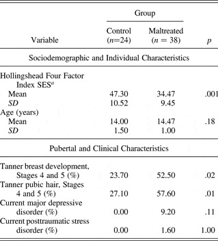
Table 1 presents the sociodemographic and clinical data for the control and maltreated groups. There were no significant group differences in age at assessment (p > .05). However, the maltreated group showed more pubertal development than the control group, on indices of pubic hair and breast development (ps < .05). As expected, females in the maltreated group come from lower SES families than females in the control group (p < .01). There were no between-group differences in the prevalence of current major depressive disorder (p > .05) or PTSD (p > .05).
Sociodemographic and clinical variables for the control and maltreated groups
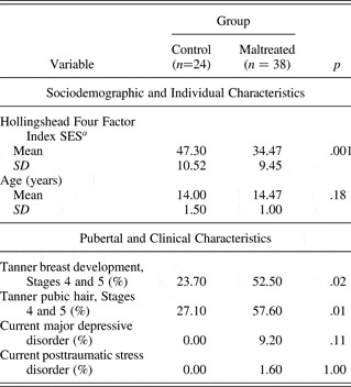
Note: SES, socioeconomic status.
aData were missing from five maltreated participants.
EEG coherence analyses
Intrahemispheric coherence
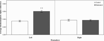
There was a significant Group × Hemisphere interaction on α-band intrahemispheric coherence, F (1, 60) = 35.77, p = .001. To decompose this interaction, we performed one-way ANOVAs with group as the between-subjects factor on α-band intrahemispheric coherence, separately for each hemisphere. As predicted, the maltreated group showed significantly higher left hemisphere coherence compared with the control group, F (1, 60) = 58.59, p = .001 (see Figure 1). There were no significant group differences for right hemisphere coherence (p > .05).
Mean differences between the maltreated (n = 38) and control (n = 24) groups on average resting intrahemispheric α-band EEG coherence shown separately for the left and right cerebral hemispheres. The composite coherence data were normalized using the Fisher z transform prior to analysis. Bars represent standard errors.

Across all scalp regions, the maltreated group (M = 22.56, SEM = 2.66) showed more left than right hemisphere EEG coherence than controls (M = −2.23, SEM = 2.42), as indicated by a greater coherence laterality index F (1, 60) = 33.55, p = .001. Positive coherence laterality index scores indicate more left than right hemisphere EEG coherence.
Regional coherence laterality
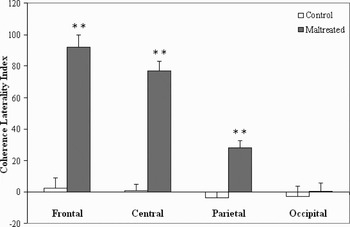
As predicted, the maltreated group showed significantly greater coherence laterality indices for the frontal, F (1, 60) = 63.72, p = .001, central, F (1, 60) = 91.08, p = .001, and parietal, F (1, 60) = 24.78, p = .001, regions than controls (see Figure 2). There was no significant group difference for occipital coherence laterality (p > .05).
Mean differences between the maltreated (n = 38) and control (n = 24) groups on regional EEG coherence laterality indices. Positive coherence laterality indices indicate left hemisphere > right hemisphere coherence, and negative scores indicate right hemisphere > left hemisphere coherence. Bars represent standard errors.

Interhemispheric coherence
There was a significant Group × Region interaction on α-band interhemispheric coherence, F (2, 59) = 22.39, p = .001. We performed one-way ANOVAs using group as the between-subjects factor, separately for the anterior, centroparietal, and posterior regions. As predicted, the maltreated group (M = 0.50, SE = 0.03) compared with the control group (M = 0.69, SE = 0.03) had lower anterior interhemispheric EEG coherence, F (1, 60) = 18.20, p = .001. By contrast, the maltreated group (M = 0.32, SE = 0.01) versus the control group (M = 0.20, SE = 0.02) had higher centroparietal interhemispheric EEG coherence, F (1, 60) = 31.64, p = .001. There was no significant difference in occipital coherence (p > .20).
EEG coherence analyses controlling for confounding variables
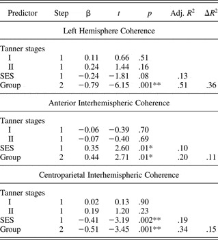
We also conducted hierarchical regression analyses to examine if group differences in EEG coherence revealed by the ANOVA analyses remained after controlling for the significant group differences in pubertal status and SES. For each equation, pubertal status and SES were entered in at Step 1 and group membership (1 = maltreated, 2 = control) was entered in at Step 2. Group continued to be a significant predictor of left hemisphere EEG coherence (group β = −0.79, p = .001), anterior interhemispheric EEG coherence (group β = 0.44, p = .01), and centroparietal interhemispheric EEG coherence (group β = −0.51, p = .001) and significantly improved all three regression models (ΔR 2s > .10, ΔFs > 6, ps < .05; see Table 2 for summaries).
Summary of hierarchical regression analyses controlling for pubertal status and SES
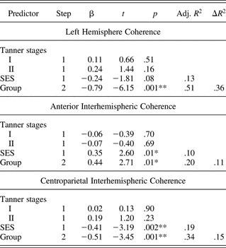
Note: SES, socioeconomic status.
*p < .05. **p < .01.
Association of types of abuse and neglect with brain activity
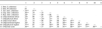
Associations between types of abuse and neglect and EEG coherence measures were determined using a series of Pearson zero-order correlation coefficients. Table 3 summarizes the results of these analyses. There was no association between right hemisphere coherence and childhood maltreatment (ps > .05). However, left hemisphere coherence and centroparietal interhemispheric coherence correlated positively with physical abuse and neglect.
Pearson zero-order correlations among measures of EEG coherence and different subtypes of abuse and neglect
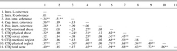
Note: Intra. L-coherence, intrahemispheric left hemisphere coherence; Intra. R-coherence, intrahemispheric right hemisphere coherence; Ant. inter. coherence, anterior interhemispheric coherence; Cnp. inter. coherence, centroparietal interhemispheric coherence; Post. inter. coherence, posterior interhemispheric coherence; CTQ, Childhood Trauma Questionnaire.
†p < .07. *p < .05. **p < .01.
Modeling early adversity, left hemisphere development, and psychiatric impairment
We next conducted a series of analyses to test whether left hemisphere EEG coherence significantly predicted the score of overall psychiatric impairment that was aggregated across three separate assessment points. Furthermore, if left hemisphere EEG coherence predicted psychiatric impairment, we were interested in testing whether it mediated the effects of maltreatment on this outcome. It is important to note that the following analyses included only the maltreated sample.
According to the standard mediation model (Baron & Kenny, Reference Baron and Kenny1986), mediation can be inferred when the following criteria are met: (a) the independent variable predicts both the putative mediator and the dependent variable, (b) the putative mediator predicts the dependent variable, and (c) the path from the independent variable to the dependent variable is made nonsignificant by the inclusion of the mediator into the equation. Because the CTQ total score provides a continuous measure of maltreatment, we used it as a predictor along with left hemisphere EEG coherence. We controlled for group differences in pubertal development and SES. All predictors were centered prior to regression analyses, as recommend by Aiken and West (Reference Aiken and West1991).
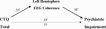
We first regressed the dependent variable (psychiatric impairment) and the putative mediator (left hemisphere EEG coherence) on the independent variable (child maltreatment, as indexed by the CTQ). High CTQ scores significantly predicted psychiatric impairment (β = 0.33, p = .03). High CTQ scores also significantly predicted left hemisphere EEG coherence (β = 0.35, p = .02). Next we regressed psychiatric impairment on left hemisphere EEG coherence. There was a direct association between left hemisphere coherence and psychiatric impairment (β = 0.42, p = .002). Increased left hemisphere coherence predicted increases in generalized psychiatric problems. Finally, we regressed psychiatric impairment on both left hemisphere EEG coherence and child maltreatment, resulting in a nonsignificant association between child maltreatment and psychiatric impairment (β = 0.19, p = .21), but a significant association with left hemisphere EEG coherence (β = 0.38, p = .02). Accordingly, despite the relatively small sample size, our results suggest that excessive left hemisphere EEG coherence mediated the effects of child maltreatment on the degree of psychiatric impairment (see Figure 3).
A model testing the relation among child maltreatment (as assessed by the Childhood Trauma Questionnaire), left hemisphere EEG coherence, and degree of psychiatric impairment (controlling for pubertal status and socioeconomic status). The weights represent standardized regression coefficients (β). The top numbers indicate direct associations, and the bottom number indicates an indirect association.

Discussion
This study demonstrated that child maltreatment was associated with atypical resting brain electrical activity in adolescent females. We found that maltreated females showed greater left hemisphere EEG coherence than their nonabused gender- and age-matched peers. The maltreated group also showed a reversal of the typical developmental pattern and exhibited more overall left than right hemisphere EEG coherence. Lateralized differences in EEG coherence were greatest in the most anterior scalp regions. These results support the preliminary report of aberrant cortical connectivity in abused children (Ito et al., Reference Ito, Teicher, Glod and Ackerman1998) and extend the findings to a larger sample of nonhospitalized adolescents.
We also found statistically significant differences in interhemispheric EEG coherence between the maltreated and control groups apparently for the first time. Females who were exposed to childhood maltreatment showed reduced frontal (anterior) interhemispheric coherence compared to controls, but increased centroparietal coherence. The severity of maltreatment (as indexed by the CTQ total score) correlated positively with left intrahemispheric EEG coherence and centroparietal interhemispheric EEG coherence. Of the different subtypes of abuse and neglect, the strongest relation emerged between physical neglect and EEG coherence. This finding is corroborated by recent evidence concerning the adverse effects of neglect on brain development and function in primates (Maestripieri et al., Reference Maestripieri, Higley, Lindell, Newman, McCormack and Sanchez2006) as well as human children (Marshall et al., Reference Marshall, Reeb, Fox, Nelson and Zeanah2008).
Of importance, we found that excessive left hemisphere EEG coherence mediated the effects of child maltreatment on the level of psychiatric impairment. These findings emerged even after controlling for significant group differences in pubertal development and SES.
What do differences in left hemisphere EEG coherence reflect?
Similar to Ito and colleagues (1998), we interpret greater left hemisphere coherence as reflecting decreased amounts of cerebral differentiation and development. There is some evidence to suggest that decreased coherence is related to optimal organization of cortical networks (Thatcher et al., Reference Thatcher, North and Biver2008). For instance, decreased EEG coherence predicts higher intelligence and efficiency of cognitive processing (Gasser, Verleger, Bacher, & Sroka, Reference Gasser, Jennen-Steinmetz and Verleger1987; Silberstein et al., Reference Silberstein, Song, Nunez and Park2004; Thatcher et al., Reference Thatcher, North and Biver2005) and average connection density decreases with a concomitant rise in brain size and complexity (Ringo, Reference Ringo1991). Although we did not administer any cognitive tasks, our results may explain some of the deficits on traditional “left hemisphere” neuropsychological tasks shown by maltreated individuals (Navalta et al., Reference Navalta, Polcari, Webster, Boghossian and Teicher2006; Teicher et al., Reference Teicher, Ito, Glod, Andersen, Dumont and Ackerman1997). Atypical circuitry of the left hemisphere may also account for some of the deficits and delays in language function among maltreated children (Culp et al., Reference Culp, Watkins, Lawrence, Letts, Kelly and Rice1991).
There is much room for ontogenetic sculpting of human brain structure and function, especially in the first two decades of postnatal life (e.g., Edelman, Reference Edelman1987). For example, anatomical, neurochemical, and electrocortical evidence suggests that the frontal lobes, in particular, continue to develop into adolescence and young adulthood (Benes, Reference Benes, Nelson and Luciana2001; Gasser et al., Reference Gasser, Verleger, Bacher and Sroka1988; Huttenlocher, Reference Huttenlocher1999; Segalowitz & Davies, Reference Segalowitz and Davies2004). Species-typical caregiving experiences are important for normal brain development. Child maltreatment represents an extreme deviation from an average expectable caregiving milieu and provides an inadequate environment for the establishment of normal brain structure and function.
It is not immediately obvious why maltreatment should be associated with atypical circuitry of the left cerebral hemisphere. Although EEG coherence potentially reflects processes such as axonal sprouting, synaptogenesis, synaptic pruning, myelination, as well as changes in pre- and postsynaptic neurotransmitter dynamics, electrophysiology alone cannot discriminate among these possibilities (Thatcher, Reference Thatcher1992).
One possibility is that adverse experiences occurring during periods of peak development are particularly likely to interfere with normal neurobiological maturation. The left and right cerebral hemispheres have unique EEG maturational profiles, with cyclical changes in the relative rates and peak periods of development across the first decade of postnatal life (Thatcher, Walker, & Giudice, Reference Thatcher, Walker and Giudice1987). Accordingly, some authors (e.g., Ito et al., Reference Ito, Teicher, Glod and Ackerman1998) have suggested that child maltreatment occurring between 6 months and 6 years may be especially likely to affect the left hemisphere. Unfortunately, the present study did not have adequate information regarding age of maltreatment onset to be able to formally test this hypothesis. However, it is interesting to note that a recent fMRI study found left basal ganglia dysfunction in young adults who were maltreated (Dillon et al., Reference Dillon, Holmes, Birk, Brooks, Lyons-Ruth and Pizzagalli2009), suggesting that lateralized effects are also present for subcortical structures, which have different developmental sensitive periods.
An alternative suggestion concerns the effects of species-atypical caregiving experience on the function of monoaminergic neurotransmitters (see also Dillon et al., Reference Dillon, Holmes, Birk, Brooks, Lyons-Ruth and Pizzagalli2009; Ito et al., Reference Ito, Teicher, Glod and Ackerman1998). For example, Rhesus monkeys who have experienced high levels of maternal rejection show reduced levels of serotonin (5-hydroxyindoleacetic acid) and dopamine (homovanillic acid) metabolites in their cerebrospinal fluid (Maestripieri et al., Reference Maestripieri, Higley, Lindell, Newman, McCormack and Sanchez2006). It is interesting that these monoaminergic systems are lateralized in the human brain (Arato et al., Reference Arato, Frecska, Maccrimmon, Guscott, Saxena and Tekes1991) and implicated in brain development (Sodhi & Sanders-Bush, Reference Sodhi and Sanders-Bush2004; Todd, Reference Todd1992). For example, serotonin metabolites are higher in the right than the left human cerebral hemisphere (Arato et al., Reference Arato, Frecska, Maccrimmon, Guscott, Saxena and Tekes1991). Depletion of serotonin, potentially induced by child maltreatment, may therefore lead to impaired maturation of the left hemisphere, which is not able to compensate for reduced serotonin levels. This is an interesting, although speculative proposition, which awaits future evidence from studies with maltreated human populations. It would be an obvious advantage to employ more direct indices of neural function (e.g., measurement of neurotransmitter metabolites) to better characterize the effects of child maltreatment on brain structure and function.
What do differences in interhemispheric EEG coherence reflect?
Given the positive correlation between EEG interhemispheric coherence and regional corpus callosum area (Pogarell et al., Reference Pogarell, Teipel, Juckel, Gootjes, Moller and Burger2005), as well as reports of reduced corpus callosum size in abused children (De Bellis et al., Reference De Bellis, Keshavan, Clark, Casey, Giedd and Boring1999, Reference De Bellis, Keshavan, Shifflett, Iyengar, Beers and Hall2002; Teicher et al., Reference Teicher, Dumont, Ito, Vaituzis, Giedd and Andersen2004), our results may provide electrophysiological evidence for anterior commissural fiber atrophy. However, our finding of increased centroparietal interhemispheric coherence in the maltreated group seems counterintuitive. There are a couple of factors that may help to explain this discrepancy. First, several studies have reported that child maltreatment is linked to smaller corpus callosum size particularly in males (De Bellis et al., Reference De Bellis, Keshavan, Clark, Casey, Giedd and Boring1999; Teicher et al., Reference Teicher, Ito, Glod, Andersen, Dumont and Ackerman1997), suggesting the need to consider potential sex effects when examining the influence of early adversity on callosal development. Similarly, a study examining the effect of prenatal stress on the development of the primate corpus callosum (Coe, Lubach, & Schneider, Reference Coe, Lubach and Schneider2002) suggests that there are both sex- and region-specific effects on the development of this brain structure. In particular, this study found that prenatal adversity led to increases in the posterior regions of the corpus callosum among females, but decreases in corpus callosum size among males. Thus, it is possible that there are complex interactions between sex and region that were not capable of being addressed by our study. Second, recent evidence (Davey, Victor, & Schiff, Reference Davey, Victor and Schiff2000) suggests that subcortical structures (thalamus, basal ganglia) contribute to sculpting patterns of EEG coherence, so that caution is urged in attributing any observed differences solely to corticocortical connections crossing the corpus callosum.
However, differences in interhemispheric EEG coherence between the maltreated and control females generally corroborate both the nonhuman animal (Berrebi et al., Reference Berrebi, Fitch, Ralphe, Denenberg, Friedrich and Denenberg1988; Denenberg, Reference Denenberg1981; Juraska & Kopcik, Reference Juraska and Kopcik1988; Sanchez et al., Reference Sanchez, Hearn, Dung, Rilling and Herndon1998) and human (De Bellis et al., Reference De Bellis, Keshavan, Clark, Casey, Giedd and Boring1999, Reference De Bellis, Keshavan, Shifflett, Iyengar, Beers and Hall2002; Teicher et al., Reference Teicher, Dumont, Ito, Vaituzis, Giedd and Andersen2004) evidence that development of the corpus callosum may be compromised by childhood rearing experiences. Enhanced glucocorticoid circulation, associated with the stress of maltreatment, may provide one potential mechanism linking early adversity to impaired callosal development (Kaur, Wu, Wen, & Ling, Reference Kaur, Wu, Wen and Ling1994). Such developmental alterations may eventually influence patterns of functional lateralization, including the lateralization of emotion and emotion regulatory systems (Miskovic, Schmidt, Georgiades, Boyle, & MacMillan, Reference Miskovic, Schmidt, Georgiades, Boyle and MacMillan2009).
Linking child maltreatment, the left hemisphere, and psychopathology
Our model has considered the links among early adversity, left brain function, and general psychopathology. We found preliminary evidence that atypical circuitry of the left hemisphere mediated the effects of child maltreatment on subsequent psychiatric impairment. Our study is correlational in nature and precludes us from drawing any causal conclusions. However, one potential interpretation of our findings is that connectivity deficits in the left hemisphere are associated with a reduced ability to regulate emotions, and in particular, negative emotions. The left frontal cortex has been suggested to exert a main inhibitory influence over subcortical regions such as the amygdala (Davidson, Reference Davidson2002; Davidson et al., Reference Davidson, Jackson and Kalin2000). In support of this suggestion, an inverse relation between left anterior cortex and amygdala metabolism has been reported in a previous positron emission tomography study (Abercrombie et al., Reference Abercrombie, Schaefer, Larson, Ward, Holden and Turski1996), whereas greater left frontal brain electrical activity predicts an attenuated startle response following affective challenge (Jackson et al., Reference Jackson, Mueller, Dolski, Dalton, Nitschke and Urry2003). Of importance, the startle reflex is modulated by the amygdala. Similarly, an fMRI study found evidence for the recruitment of a left-lateralized neural circuit during the regulation of negative affect (Oschner et al., 2002). Smaller left, relative to right, orbitofrontal cortex volume has also been related to increased impulsivity and aggression (Antonucci et al., Reference Antonucci, Gansler, Tan, Bhadelia, Patz and Fulwiler2006). Most recently, Johnstone and colleagues (2007) have reported left-lateralized brain activation during the downregulation of negative affect among healthy, nondepressed participants. It was interesting that nondepressed individuals showed an inverse relation between activity in the left ventrolateral prefrontal cortex and the amygdala, during an emotion regulation task. The results from that study also implicated a dysfunction of the left-lateralized neural circuit during emotion regulation in the pathophysiology of depression. Deviant cortical organization of the left hemisphere, associated with child maltreatment, may make these adolescents more vulnerable to subsequent stress and more likely to experience psychiatric symptoms.
One other parsimonious interpretation of our findings is that child maltreatment is associated with a depletion of the brain's differentially lateralized monoaminergic systems (e.g., serotonin). Subsequently, monoaminergic depletion may be a common factor that accounts for (a) a maturational lag of the left hemisphere (as a consequence, of the monoamines' neurotrophic effects) as well as (b) difficulties in successful emotion regulation, leading to increased susceptibility to psychiatric symptoms. It is important to note that serotonin function (especially serotonin depletion) has long been linked to behavioral impulsivity in nonhuman animal models (e.g., Brunner & Hen, Reference Brunner and Hen1997). However, until there is more evidence, we urge caution in interpreting this model and only suggest it as a tentative explanation for our findings.
At least another possible alternative model is that abusive parenting may be genetically cosegregated with atypical left hemisphere function and psychiatric problems (e.g., Green, Voeller, Gaines, & Kubie, Reference Green, Voeller, Gaines and Kubie1981). Similar issues have been raised in etiological theories of PTSD, where some controversy exists concerning the role of hippocampal volume, either as a risk factor for PTSD or a consequence of traumatic experience (Gilbertson et al., Reference Gilbertson, Shenton, Ciszewski, Kasai, Lasko and Orr2002).
A cautionary note should also be drawn regarding attempts to reduce the variability in complex developmental systems to one or more variables. Outcomes such as emotion regulatory problems and psychopathological risk are almost certainly determined by cumulative risk factors, originating at multiple, reciprocally interacting biopsychosocial levels (Sameroff, Reference Sameroff2000). Such nonlinear processes are among the core tenets of developmental psychopathology (Rutter & Sroufe, Reference Rutter and Sroufe2000; Sroufe & Rutter, Reference Sroufe and Rutter1984).
Limitations
The present study had several limitations. First, we were not able to tease apart potential sex-specific effects of maltreatment on brain function (Teicher et al., Reference Teicher, Andersen, Polcari, Anderson, Navalta and Kim2003), because we had an exclusively female sample. Second, we used measures of scalp EEG coherence to make inferences about functional connectivity. Scalp coherence can produce spurious coherence values as a result of volume conduction. Accordingly, future studies should use dense-array electrophysiology to allow for the computation of source coherence, thereby providing more accurate measures of corticocortical coupling (Hoechstetter et al., Reference Hoechstetter, Bornfleth, Weckesser, Ille, Berg and Scherg2004). Third, even though the adolescents were under CPA services, the reliance on retrospective self-report measures to define child maltreatment, rather than more objective methods such as court reports, was a limitation; however, these are also prone to bias. Fourth, we did not have reliable estimates of age of onset at which the maltreatment had begun. Because development is time dependent, early occurring insults are likely to lead to more dysfunctional outcomes. In the future, it will be increasingly important to characterize the timing and nature of sensitive periods in human socioemotional development, because this knowledge may eventually translate into better intervention efforts. Some of this important work is currently ongoing (e.g., the BEIP group studies) and will be vital to advancing the field. In a related vein, it will be important to assess the stability of the neurobiological correlates of child maltreatment to determine whether they reflect dynamic, short-term changes, or more long-lasting alterations. We have recently attempted to address some of these issues in our laboratory (e.g., Miskovic et al., Reference Miskovic, Schmidt, Boyle and Saigal2009).
A final limitation of our study concerns the relatively small sample size of maltreated adolescents. Issues of restricted sample size pose a statistical challenge, given the importance of large sample sizes for conducting reliable mediation analyses (MacKinnon, Fairchild, & Fritz, Reference MacKinnon, Fairchild and Fritz2007). However, such concerns are offset when considered in the context of the difficulty involved in conducting research with highly vulnerable populations and the potential theoretical and practical importance of such work.
Conclusions
Our results provide further evidence that the experience of child maltreatment can leave a lasting impact on resting brain function. Specifically, child maltreatment appears to be associated with deficient left hemispheric development and disturbed interhemispheric connectivity. Deficits in the circuitry of the left hemisphere may mediate the effects of child maltreatment on future psychiatric impairment. To be more precise, it is difficult to disambiguate whether the adverse effects on central nervous system development are due to the experience of maltreatment itself or the environmental stressors that are typically associated with maltreatment (e.g., SES differences, psychopathology). It should be noted that we attempted to control for some of these confounding factors. Studies of siblings discordant for childhood maltreatment and preclinical models can be useful in clarifying the mechanistic pathways linking early abuse with cerebral development and behavioral organization.
One of the implications of this study is that the use of noninvasive psychophysiological measures in developmental populations can index the middle level of analysis (Segalowitz & Schmidt, Reference Segalowitz, Schmidt, Schmidt and Segalowitz2008) that helps to link experience, brain, and psychopathology. Establishing such links is crucial for gaining insight into complex developmental processes, especially because experience does not program constellations of behaviors (normal or abnormal) but the brain systems that are involved in producing such behaviors (Knudsen, Reference Knudsen2004).
Finally, it is important to note that although we have proposed a putative pathway linking left hemispheric aberrations to psychiatric impairment, the pathways linking early adversity to atypical cerebral development are less clear. Although we have suggested several hypotheses (e.g., monoaminergic depletion), these should be considered only as suggestions for future research. Ultimately, such research will help us to identify the biological processes that mediate or moderate the relations between early adversity and future psychopathology.