Introduction
The new mineral species zincorinmanite-(Zn) described in this paper was discovered in a unique sulfide-free ore with chalcophile metals. All the chalcophile elements that occur in the ore are in oxygen-bearing minerals, including zincorinmanite-(Zn), whereas sulfides and sulfosalts are absent. The discovery of zincorinmanite-(Zn) provided an opportunity to better understand the patterns of cation ordering in isostructural minerals and compounds, and also served as an incentive for the development of the nomenclature and systematics of the nolanite supergroup, to which minerals of this structural type belong.
The nolanite supergroup approved by the Commission on New Minerals, Nomenclature and Classification of the International Mineralogical Association (IMA-CNMNC, Memorandum 127-SM24, Bosi et al., Reference Bosi, Hatert, Pasero and Mills2024) contains eight mineral species with the nolanite-type structure (hexagonal, space group P63mc and unit-cell parameters in the following ranges: a = 5.5–6.0 Å and c = 8.8–10.3 Å; Z = 2) and the general crystal-chemical formula M13M2M3O7X where M1, M2 and M3 are cation sites and X = OH or O. The crystal structure of nolanite-supergroup minerals is composed of alternating layers of two kinds: a layer of edge-sharing M1 octahedra (O layer) and a heteropolyhedral (H) layer of M2 octahedra and M3 tetrahedra connected via common vertices (Fig. 1). The X site is coordinated by M1 cations.
The general view of the crystal structure of a nolanite-group mineral drawn using the program Diamond, Version 3.2k. The unit cell is outlined.

The nolanite supergroup is subdivided into three groups (nolanite, kamiokite and rinmanite) in accordance with the largest charge of species-defining octahedral M cations (+3, +4 and +5, respectively). The general formulae of minerals belonging to the nolanite, kamiokite and rinmanite groups are M13+3M23+M33+O7(OH), M14+3M22+M32+O8 and (M13+2M12+)M25+M32+O7(OH), respectively.
The nolanite group contains three mineral species: nolanite, V3+3V3+Fe3+O7(OH), discovered in a hydrothermal uranium deposit on the shore of Fish Hook Bay, Lake Athabaska, Canada (Robinson et al., Reference Robinson, Evans, Schaller and Fahey1957); akdalaite, Al3AlAlO7(OH), discovered as imperfect elongate-tabular crystals in veinlets composed of muscovite and fluorite at the Solnechnoe fluorite deposit, Karaganda Region, Kazakhstan (Shpanov et al., Reference Shpanov, Sidorenko and Stolyarova1971); and ferrihydrite, Fe3+3Fe3+Fe3+O7(OH), which is widespread in soils and different weathered rocks (Chukhrov et al., Reference Chukhrov, Zvyagin, Gorshkov, Ermilova and Balashova1973).
Three members of the kamiokite group are known. Kamiokite, Mo4+3Fe2+Fe2+O7O, was described as a new mineral from the Kamioka mine, Japan by Sasaki et al. (Reference Sasaki, Yui and Yamaguchi1985). This mineral occurs in quartz–molybdenite stockwork veins associated with granite porphyry dykes. Iseite, Mo4+3Mn2+Mn2+O7O, was discovered in rhodochrosite veins cross-cutting Fe–Mn ore of the Shobu deposit, Shima Peninsula, Japan (Nishio-Hamane et al., Reference Nishio-Hamane, Tomita, Minakawa and Inaba2013). Majindeite, Mo4+3MgMgO7O, occurs as minute crystals (up to 1 μm across) in the Allende meteorite which is classified as a CV3 carbonaceous chondrite (Chi Ma and Beckett, Reference Chi and Beckett2016). However, despite the members of the nolanite and kamiokite groups all being isostructural, there are no similarities related to their genesis.
The rinmanite group contains two mineral species, namely, rinmanite, (Fe3+2Mg)SbZnO7(OH) (Holtstam et al., Reference Holtstam, Gatedal, Söderberg and Norrestam2001; IMA 127-SM24) and the new mineral zincorinmanite-(Zn), (Fe3+2Zn)SbZnO7(OH), described in this paper. Both minerals occur in sulfide-free metasomatic rocks with chalcophile metals.
A more detailed description of the members of the nolanite supergroup has now been published (Chukanov et al., Reference Chukanov, Gridchina, Rastsvetaeva, Zubkova and Pekov2025) describing the nomenclature and systematics of this group.
Zincorinmanite-(Zn) is named as the analogue of rinmanite with Zn as a charge-balancing component at the M1 site. The new mineral, its name and symbol (Zrnm-Zn) have been approved by the IMA CNMNC (IMA No. 2023-107 originally as ‘rinmanite-(Zn)’, Chukanov et al., Reference Chukanov, Gridchina, Rastsvetaeva, Varlamov, Kasatkin, Pekov, Vigasina, Virus, Jančev and Britvin2024, Bosi et al., Reference Bosi, Hatert, Pasero and Mills2024). The type specimen is deposited in the collections of the Fersman Mineralogical Museum of the Russian Academy of Sciences, Moscow, Russia with the registration number 6059/1.
Experimental methods and data processing
A Raman spectrum of zincorinmanite-(Zn) has been obtained from a polished section using an EnSpectr R532 spectrometer based on an OLYMPUS CX 41 microscope coupled with a diode laser (λ = 532 nm) at room temperature (Moscow State University, Faculty of Geology, Dept. of Mineralogy). Incident laser radiation was linearly polarised. The spectra were recorded in the range from 100 to 4000 cm–1 with a diffraction grating (1800 gr mm–1) and spectral resolution of ∼4–6 cm–1 (Raman Analyzer Specification). The Rayleigh line was corrected with a cutting-edge low pass filter. Scattered radiation was recorded with a Linear CCD Array detector type, with a number of pixels 3648 and pixel size 8 μm × 200 μm. The output power of the laser beam was ∼16 mW. The diameter of the focal spot on the sample was 15 μm. The back-scattered Raman signal was collected with a 40× objective; entrance aperture 20 μm wide slit; signal acquisition time for a single scan of the spectral range was 1 s, and the signal was accumulated over 50 scans. Crystalline silicon was used as a standard.
Chemical analyses (9 spots) were carried out for a polished section using an Oxford INCA Wave 700 electron microprobe (WDS mode, 20 kV and 1 μm beam diameter). The current of the absorbed electrons on the studied polished sample was 10 to 13 nA. The spectrum acquisition time was 40 s for each spot. The SEM is equipped with detectors of secondary and back-scattered electrons based on YAG. The following standards were used: diopside for Mg; albite for Al; CaF2 for F; and pure elements for the other components.
For the determination of the oxidation state of iron in zincorinmanite-(Zn), a method based on X-ray spectroscopy was used. Previously, this method, based on the dependences of ratios of intensities of different K-lines of X-ray spectra of an element (generally Fe) on its oxidation degree δ, has been described and employed in numerous works (Kupriyanova et al., Reference Kupriyanova, Filippov and Lyamina2003; Mukhanova et al., Reference Mukhanova, Kupriyanova and Morgunova2008; Chubarov et al., Reference Chubarov, Finkelshtein and Amirzhanov2009; Finkelshtein and Chubarov, Reference Finkelshtein and Chubarov2010; Murashko et al., Reference Murashko, Chukanov, Mukhanova, Vapnik, Britvin, Krivovichev, Polekhovsky and Ivakin2011; Chukanov et al., Reference Chukanov, Mukhanova, Rastsvetaeva, Belakovskiy, Möckel, Karimova, Britvin and Krivovichev2011a, Reference Chukanov, Rastsvetaeva, Britvin, Virus, Belakovskiy, Pekov, Aksenov and Ternes2011b). In this work, we used the ratio of the intensities I of the FeKβ5 and FeKβ1 lines. The X-ray spectra of the FeKβ5 and FeKβ1 lines were obtained at the D.S. Korzhinskii Institute of Experimental Mineralogy using a scanning electron microscope Tescan Vega XMU equipped with an INCA wave 700 X-ray spectrometer (Oxford INCA Wave 700). The spectra recording was carried out for 1 min in WDS mode at an accelerating voltage of 20 kV, an absorbed electron current of 100 nA and a beam diameter of 3.5 μm. A LiF crystal was used as an analyser.
The corresponding correlation based on the X-ray spectra of hematite, Fe2O3, magnetite, Fe3O4, and siderite, FeCO3, as well as Fe3+-bearing atencioite, ideally Ca2Fe2+3Mg2Be4(PO4)6(OH)4·6H2O (Chukanov et al., Reference Chukanov, Rastsvetaeva, Möckel, Zadov and Levitskaya2006), and Fe-bearing perraultite, (Ba,K)2CaNa(Mn,Fe2+,Fe3+)8Ti4(Si2O7)4O4(F,OH,O)6 (Rastsvetaeva et al., Reference Rastsvetaeva, Еskova, Dusmatov, Chukanov and Schneider2008), in which the Fe2+:Fe3+ ratio was determined by means of Mössbauer spectroscopy, as shown in Fig. 2. Its linear approximation corresponds to the equation δ = 128.2×[I(FeKβ5)/I(FeKβ5)] – 0.4359. The estimated error of the determination of δ from this correlation is ±0.08.
Correlation between oxidation degree of iron in minerals and the ratio of intensities of the FeKβ5 and FeKβ1 lines in X-ray spectra.

Reflectance values for zincorinmanite-(Zn) were measured in air using a MSF-R (LOMO, St. Petersburg, Russia) microspectrophotometer. Silicon was used as a standard. The photometric aperture diameter was 0.3 mm, monochromator exit slit size 0.1 mm and spectral interval 6 nm.
Powder X-ray diffraction data were collected using a Rigaku R-AXIS Rapid II diffractometer (image plate), CoKα, 40 kV, 15 mA, rotating anode with the microfocus optics, Debye-Scherrer geometry, r = 127.4 mm and exposure 15 min. The raw powder XRD data were collected using the program suite designed by Britvin et al. (Reference Britvin, Dolivo-Dobrovolsky and Krzhizhanovskaya2017). Calculated intensities were obtained by means of STOE WinXPOW v. 2.08 program suite based on the atomic coordinates and unit-cell parameters.
Single-crystal data, data collection information and structure refinement details are given in Table 1. The structure was refined in the space group P63mc by the full-matrix least squares method for F 2. In the refinement of the crystal structure of zincorinmanite-(Zn), the rinmanite structure model (Holtstam et al., Reference Holtstam, Gatedal, Söderberg and Norrestam2001) was used as starting one. Integration of diffraction peaks, correction for the Lorentz factor, and radiation polarisation were carried out using the CrysAlis Pro 1.171.42.80a (Rigaku Oxford Diffraction, 2022) and JANA2006 (Petřiček et al., Reference Petřiček, Dušek and Palatinus2006) software packages. The parameters of atomic displacements of atoms at the M1, M2 and M3 sites, as well as oxygen atoms, were refined in the harmonic approximation. The H atom was localised to a geometrically calculated position and refined using a ‘riding model’ (Busing and Levy, Reference Busing and Levy1964; Sheldrick and Schneider, Reference Sheldrick and Schneider1997).
Crystal data, data collection information and structure refinement details for zincorinmanite-(Zn) (upper and lower lines correspond to the routes with fixed and refined occupancies, respectively)

Results
Occurrence, general appearance and physical properties
The new mineral was found ∼4.5 km NW of the village of Nežilovo and ∼25 km WSW of the city of Veles, Republic of North Macedonia (41°41′N, 21°25′E). The associated minerals are quartz, baryte, gahnite, Zn-bearing phlogopite, together with accessory hematite, almeidaite, a Pb-analogue of hydroxycalcioroméite and an insufficiently studied högbomite-supergroup mineral (presumably, an analogue of zincohögbomite–2N2S with Sb > Ti). Zincorinmanite-(Zn) occurs in the orogenic zone related to the ‘Mixed Series’ metamorphic complex situated in the Pelagonian massif.
Zincorinmanite-(Zn) forms lamellar to tabular subhedral single-crystal grains up to 0.5 mm across and up to 40 μm thick (Figs 3 and 4), some of which form epitaxial intergrowths with a högbomite-supergroup mineral (Fig. 5) and hematite.
Grains of zincorinmanite-(Zn), mainly forming intimate intergrowths with hematite (black) in quartz–baryte rock. A fragment of the specimen 6059/1 deposited in the Fersman Mineralogical Museum of the Russian Academy of Sciences, Moscow, Russia. FOV width: 7 mm.

(a and b) Fragments of metasomatic rock containing zincorinmanite-(Zn) (1), quartz (2), baryte (3), a högbomite-supergroup mineral (4), hematite (5), gahnite (6) and a Pb-analogue of hydroxycalcioroméite (7). SEM (BSE) images of polished sections.

Epitaxial overgrowth of a högbomite-supergroup mineral (4) on zincorinmanite-(Zn) (1) in association with baryte (3), a högbomite-supergroup mineral (4) and hematite (5). SEM (BSE) image of a polished section.

The colour of zincorinmanite-(Zn) is black, the streak is black, and the lustre is submetallic. The new mineral is opaque. Zincorinmanite-(Zn) is brittle. Based on scratch tests, the Mohs hardness is ca. 6. No cleavage is observed. The fracture is uneven. Density calculated using the empirical formula and unit-cell volume refined from single-crystal XRD data is equal to 5.446 g·cm–3.
Raman spectroscopy
The Raman spectrum of zincorinmanite-(Zn) (Fig. 6) shows a strong luminescence which may be due to the presence of Fe3+. The luminescence curve has inflection points in the range 2800–3300 cm–1. Application of a standard procedure of baseline correction results in the appearance of the peaks at 2840 and 3132 cm–1 which may correspond to O–H stretching vibrations coordinated by (Fe3+Fe3+Fe3+) and (Fe3+Fe3+M 2+), respectively, where M 2+ = Zn and Mg.
Raman spectrum of zincorinmanite-(Zn). The inset shows a fragment of the spectrum in the range 2500–3500 cm–1 after baseline correction. Corresponding inflection points are indicated with vertical lines.

The strongest bands of Sb–O stretching vibrations of the SbO6 octahedron in Raman spectra of Sb-bearing oxides and hydroxides are observed in the range 500–670 cm–1 [509–518 cm–1 for roméite-group minerals and their synthetic analogues (Kendix et al., Reference Kendix, Moscardi, Mazzeo, Baraldi, Prati, Joseph and Capelli2008; Bahfenne and Frost, Reference Bahfenne and Frost2010; Atencio et al., Reference Atencio, Ciriotti and Andrade2013; Biagioni et al., Reference Biagioni, Orlandi, Nestola and Bianchin2013), 617–625 cm–1 for mopungite, Na[Sb(OH)6, brandholzite, Mg[Sb(OH)6]2·6H2O and bottinoite, Ni[Sb(OH)6]2·6H2O (Rintoul et al., Reference Rintoul, Bahfenne and Frost2011), 660 cm–1 for brizziite, NaSbO3 (Frost and Bahfenne, Reference Frost and Bahfenne2010) and 670 cm–1 for ordoñezite, ZnSb2O6 (Husson et al., Reference Husson, Repelin, Brusset and Cerez1979). Based on these data, it can be supposed that the strong band at 651 cm–1 and, possibly, the band at 622 cm–1 are related to stretching vibrations involving the Sb–O bond, whereas the bands in the range 360–470 cm–1 correspond to stretching vibrations involving mainly lower force-strength bonds, Fe3+–O and M2+–O. However, the band at 622 cm–1 can also correspond to the combination mode (∼162+463 cm–1). The strongest band at 162 cm–1 corresponds to a bending lattice mode.
Weak bands in the range 690–810 cm–1 cannot be ascribed to any overtones or combination modes, but this range corresponds to typical wavenumbers of (Fe3+,M2+)–O–H bending vibrations.
Optical properties
In reflected light, zincorinmanite-(Zn) is grey (substantially darker than associated hematite but brighter than associated högbomite-supergroup mineral). Bireflectance is weak, ∆R = 1.0% (589 nm). No pleochroism is observed. Anisotropy is weak. Internal reflections are not observed.
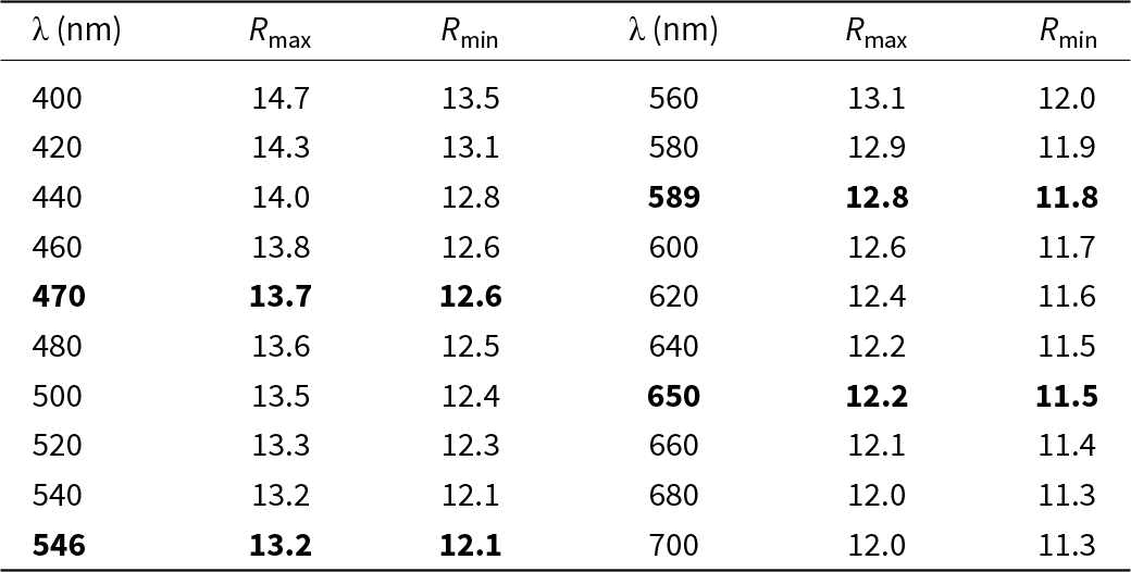
Reflectance values of zincorinmanite-(Zn) are given in Table 2 and plotted in Fig. 7 in comparison with the published data for rinmanite (Holtstam et al., Reference Holtstam, Gatedal, Söderberg and Norrestam2001).
Reflectance curves for (1) zincorinmanite-(Zn) and (2) rinmanite (Holtstam et al., Reference Holtstam, Gatedal, Söderberg and Norrestam2001).

Reflectance values for zincorinmanite-(Zn) and rinmanite (COM standard wavelengths are given in bold).

Chemical data
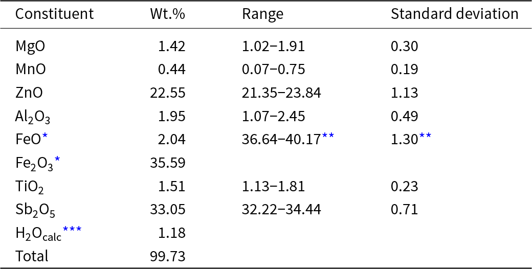
The analytical data are given in Table 3. The contents of other elements with atomic numbers higher than that of carbon, including F, are below detection limits.
Chemical data for zincorinmanite-(Zn).

* Iron was divided into Fe2+ and Fe3+ based on charge balance
** For total iron determined as Fe2O3
*** Calculated based on BVS for the O1 site
For zincorinmanite-(Zn), the I(FeKβ5)/I(FeKβ5) ratio is equal to 0.02682 which corresponds to the oxidation degree of iron δ = 3.00 ± 0.08. Thus, a major part of iron in zincorinmanite-(Zn) is trivalent. This result is not unexpected, taking into account extremely oxidising conditions under which sulfide-free ores of the Nežilovo area formed. Note that, according to Mössbauer spectroscopy data, even in rinmanite that crystallised under lower oxidising conditions, all iron is trivalent (Holtstam et al., Reference Holtstam, Gatedal, Söderberg and Norrestam2001).
The charge-balanced empirical formula of the studied zincorinmanite-(Zn) is [(Fe3+2.12Al0.18)(Zn0.32Mg0.16Fe2+0.13Mn0.03)Ti0.06]Σ3.00(Sb0.97Ti0.03)Σ1.00Zn1.00O7[(OH)0.61O0.39]Σ1.00.
The H2O content was not measured because of the scarcity of available pure material (most individuals of zincorinmanite-(Zn) form epitaxial or random intergrowths with hematite or a högbomite-supergroup mineral). Therefore, the content of OH-groups was calculated taking into account the bond valence sum (BVS) for the O1 site partly occupied by OH groups as well as direct measurement of the oxygen content in the mineral by electron microprobe. The BVS for the O1 site is equal to 1.38 vu (see below), which corresponds to the dominance of OH over O at this site and is consistent with the following empirical formula derived on the basis of electron microprobe analyses and structural data (see below). According to the empirical formula, the mean oxidation degree of Fe is 2.94, which agrees with the value of 3.00±0.08 determined by X-ray spectroscopy. Direct measurement of oxygen by means of electron microprobe resulted in 27.26 wt.% of O which is close to the value of 27.05 wt.% calculated from the empirical formula.
Thus, the simplified formula of zincorinmanite-(Zn) is (Fe3+,Zn,Al,Mg,Mn,Ti)3(Sb,Ti)ZnO7(OH,O) and the ideal, end-member, formula is (Fe3+2Zn)SbZnO7(OH).
X-ray diffraction and crystal structure
Powder X-ray diffraction data of zincorinmanite-(Zn) are given in Table 4. The hexagonal unit-cell parameters refined from the powder data are: a = 5.979(1), c = 9.372(2) Å and V = 290.2(1) Å3.
Powder X-ray diffraction data (d in Å) of zincorinmanite-(Zn)

Note: The strongest lines are given in bold
* For the calculated pattern, only reflections with intensities ≥0.5 are given.
** For the unit-cell parameters calculated from single-crystal data.
Two routes of refinement have been carried out. In the first route (R = 0.0194), the site occupancies have been fixed in accordance with the empirical formula. The corresponding crystal chemical formula is M 1[(Fe3+4.23Al0.36)(Zn0.63Mg0.33Fe2+0.27Mn0.06)Ti0.12] M 2(Sb1.94Ti0.06) M 3Zn2 O2–O4O14 O1[(OH)1.23O0.77].
In the second route (R = 0.0191), the occupancies of the M1, M2 and M3 sites have been modelled by the pairs Fe + Al, Sb + vacancy, and Zn + vacancy, respectively, which resulted in M 1(Fe5.384Al0.624) (24.68 electrons per site), M 2Sb1.902 (48.50 electrons per site) and M 3Zn1.944 (29.16 electrons per site). Correspondingly the charge-balanced crystal chemical formula is M 1[(Fe3+4.50Al0.36)(Zn0.72Mg0.36Mn0.06)] M 2(Sb1.82Ti0.18) M 3(Zn1.91Mg0.09) O2–O4O14 O1[(OH)1.32O0.68] with 24.85, 48.50 and 29.16 electrons per the M1, M2 and M3 sites, respectively. In this case, all iron is trivalent, in accordance with X-ray spectroscopy data. The two obtained crystal-chemical formulae are in good agreement: the differences between them are within limits determined by chemical heterogeneity and analytical errors. The position of H in zincorinmanite-(Zn) is close to that in rinmanite (Holtstam et al., Reference Holtstam, Gatedal, Söderberg and Norrestam2001).
The coordination polyhedra in the structure of zincorinmanite-(Zn) and their mutual arrangement in the layer of octahedra and heteropolyhedral layer are shown in Figs 8 and 9. Site coordinates, site occupancies, site multiplicities and equivalent atomic displacement parameters in Table 5, anisotropic atomic displacement parameters in Table 6, selected interatomic distances in Table 7 and bond valence calculations in Table 8. The crystallographic information file has been deposited with the Principal Editor of Mineralogical Magazine and is available as Supplementary material (see below).
(a) Layer of octahedra and (b) heteropolyhedral layer in the structure of zincorinmanite-(Zn). The unit cell is outlined.

Coordination polyhedra in the structure of zincorinmanite-(Zn).

Atom coordinates, site multiplicities (Q), site occupancy factors (s.o.f.) and equivalent-isotropic atomic displacement parameters (U eq) for zincorinmanite-(Zn)

Anisotropic atomic displacement parameters (Å) in the structure of zincorinmanite-(Zn) with site occupancies fixed in accordance with the empirical formula

Selected interatomic distances (Å) in the structure of zincorinmanite-(Zn) with site occupancies fixed in accordance with the empirical formula

Bond-valence (in valence units) analysis for zincorinmanite-(Zn)

Note: Bond-valence parameters are taken from Brese and O’Keefe (Reference Brese and O’Keeffe1991) with Fe3+ at the M1 site, Sb5+ at the M2 site and Zn at the M3 site.
Discussion
The ‘Mixed Series’ is a metamorphosed volcano-sedimentary formation composed mainly of albite augen gneisses and meta-rhyolites around which there are lenses of dolomitic marbles metasomatically replaced by very unusual and complex mineral assemblages including various members of the spinel, högbomite, pyrochlore and epidote supergroups, magnetoplumbite, crichtonite and hollandite groups, Cu, Zn and Pb arsenates, Zn-bearing pyroxenes, amphiboles, micas and talc, as well as numerous potentially new minerals (Bermanec et al., Reference Bermanec, Chukanov, Boev, Šturman, Zebec and Bermanec2021; Chukanov et al., Reference Chukanov, Jančev and Pekov2015, Reference Chukanov, Vorobei, Ermolaeva, Varlamov, Plechov, Jančev and Bovkun2019, Reference Chukanov, Varlamov, Ermolaeva and Jančev2020a, Reference Chukanov, Zubkova, Jančev, Pekov, Ermolaeva, Varlamov, Belakovskiy and Britvin2020b; Ermolaeva et al., Reference Ermolaeva, Varlamov, Jančev and Chukanov2019a, Reference Ermolaeva, Varlamov, Chukanov and Jančev2019b; Jančev, Reference Jančev1997; Jančev et al., Reference Jančev, Chukanov, Varlamov and Ermolaeva2021; Varlamov et al., Reference Varlamov, Ermolaeva, Jančev and Chukanov2018, Reference Varlamov, Ermolaeva, Chukanov and Jančev2022 and references therein). Post-magmatic fluids related to meta-rhyolites are considered as a possible source of a number of specific, ore and rare elements (Pb, Zn, Sb, As, Cu, Ba, REE, etc.) in contact metasomatic rocks widespread in the Nežilovo area (Jančev, Reference Jančev1997; Chukanov et al., Reference Chukanov, Jančev and Pekov2015).
Ore occurrences in the Nežilovo area formed under the occurence of two factors: extremely high oxygen fugacity and immobilisation of sulfur in baryte due to a high activity and mobility of barium at all stages of ore formation. As a result, chalcophile elements (S, As, Sb, Zn, Cu and Pb) are concentrated mainly in the form of oxides and oxysalts, whereas sulfides and sulfosalts are present only locally and in trace amounts.
The sulfide-free metasomatic rocks with chalcophile metals from the Nežilovo ore field belong to a rare kind of ore. The mineralogy and petrology of these rocks were studied in detail and now they can be considered as a standard for ores of this type. A characteristic feature of Nežilovo-type ores is a wide variety of accessory oxide minerals containing chalcophile elements (Zn, Pb, Sb, Cu and As). Similar ores are also known in Långban, Nordmark (including Jakobsberg) and Pajsberg (including Harstigen) Fe-Mn deposits in the Bergslagen ore province, Värmland, Sweden (Palache, Reference Palache1929a; Holtstam and Langhof, Reference Holtstam and Langhof1999) as well as in the Garpenberg Norra mine, Dalarna, Sweden and the Franklin and Sterling Hill Fe-Zn deposits in New Jersey, USA (Tarr, Reference Tarr1929; Palache, Reference Palache1929a, Reference Palache1929b; Wilkerson, Reference Wilkerson1962).
The empirical formula of the holotype rinmanite sample from the Garpenberg Norra mine in Dalarna, Sweden, written taking into account both structure data and the Mössbauer spectrum showing Fe3+ as the only valence state of iron (Holtstam et al., Reference Holtstam, Gatedal, Söderberg and Norrestam2001), is [(Fe3+3.88Al0.15)Mg1.95)]Σ5.98Sb2.03(Zn1.58Mn0.31Mg0.06)Σ1.95O14.01(OH)1.99 (Z = 1). The H2O content in this sample was not determined. Recalculation on the sum of 10 cations results in the charge-balanced formula [(Fe3+3.91Al0.15)Mg1.95Sb0.04)]Σ6.00Sb2(Zn1.59Mn0.31Mg0.10)Σ2.00O14[(OH)1.75O0.25]Σ2.00 (according to crystal-chemical criteria, the excess of Sb was placed at the octahedrally coordinated Fe3+-dominant M1 site). Both variants of the empirical formula lead to the end-member formula (Fe3+2Mg)SbZnO7(OH) (Z = 2), in accordance with the accepted rules of chemical identification and classification of minerals (Bosi et al., Reference Bosi, Hatert, Hålenius, Pasero, Miyawaki and Mills2019a, Reference Bosi, Biagioni and Oberti2019b). Thus, zincorinmanite-(Zn) is an analogue of rinmanite with Zn as a charge-balancing component at the M1 site.
The crystal-chemical formula of the holotype zincorinmanite-(Zn) sample corresponds to the end-member formula (Fe3+2Zn)SbZnO7(OH). Comparative data for zincorinmanite-(Zn) and rinmanite are given in Table 9.
Comparative data for zincorinmanite-(Zn) and rinmanite.

Supplementary material
The supplementary material for this article can be found at at https://doi.org/10.1180/mgm.2024.99
Acknowledgements
The study has been carried-out in accordance with the state task of Russian Federation, registration number 124013100858-3. The single-crystal X-ray diffraction data were obtained using the equipment of the Shared Research Center Kurchatov Complex of Crystallography and Photonics NRC “Kurchatov Institute”. The crystal structure investigation was carried out within the state assignment of NRC “Kurchatov Institute”. The powder XRD pattern of zincorinmanite-(Zn) was obtained at the Research Centre for X-ray Diffraction Studies of St Petersburg University in accordance with the State Task of the Russian Federation No. AAAA-A19–119091190094. Electron microprobe and X-ray spectroscopy data were obtained under Research program FMUF-2022-0002 of the D.S. Korzhinskii Institute of Experimental Mineralogy
Competing interests
The authors declare none.