Introduction
Epistaxis is a common presentation in accident and emergency departments in the UK. The estimated incidence is 108/100 000 population per year.Reference O'Donnell, Robertson and McGarry1 Whilst conservative management is effective in the majority of cases, intractable epistaxis remains a significant cause of morbidity and mortality.Reference Leong, Roe and Karkanevatos2 It has been estimated that 5 to 15 per cent of patients requiring hospital admission for this condition will need some form of surgical treatment if the bleeding continues despite repeated packing.Reference Kotecha, Fowler, Harkness, Walsmsey, Brown and Topham3 Posterior nasal packing, including balloon tamponade, has a high failure rate, ranging from 26 to 52 per cent.Reference Schaitkin, Strauss and Hounck4,Reference Shaw, Wax and Wetmore5
Historically, internal maxillary artery ligation via a transantral approach and ligation of the ethmoidal vessels and the external carotid artery has been the treatment of choice when conservative management failed. Over the past decade, with widespread popularization of endoscopic sinus surgery and a greater understanding of local regional anatomy, endoscopic control of the sphenopalatine artery has been advocated as an effective alternative for the control of posterior epistaxis.Reference Sharp, Rowe-Jones, Biring and Mackay6–Reference Winstead8 The sphenopalatine artery is the terminal branch of the internal maxillary artery and the dominant source of posterior nasal blood supply. Endoscopic ligation of the sphenopalatine artery causes interruption of the nasal vasculature at a point distal enough to prevent direct, retrograde and anastomotic blood flow from ipsilateral and contralateral carotid systems.Reference Shah7
Endoscopic sphenopalatine artery ligation is technically straightforward to perform, allows direct, secure ligation of the major vessel supplying the posterior nasal cavity, and avoids the complications associated with pterygopalatine fossa surgery.Reference Winstead8,Reference Simpson, Janfaza and Becker9 This technique reduces the potential for intra-orbital and intracranial complications and does not require any additional endoscopic equipment other than a standard endoscopic sinus surgery set and a clip applicator.Reference Pothier, MacKeith and Youngs10 In addition, although difficult to substantiate, the surgical and anaesthetic time may be less than that required for more traditional methods of arterial ligation.Reference Feusi, Holzmann and Steuer11 These factors are especially important in a patient group that is often elderly, frail and with multiple concomitant medical problems.
The aim of this prospective study was to assess the immediate and late post-operative cessation of bleeding in patients treated with ligation of the sphenopalatine artery.
Methods
A prospective audit was conducted of patients who had undergone endoscopic treatment for persistent posterior epistaxis in three east Scotland hospitals over the three year period between April 2003 and April 2005. All patients had suffered intractable posterior epistaxis despite conservative treatment with nasal packing. Patients with any bleeding diatheses, including those receiving anticoagulant therapy, were excluded. Late outcome assessment was performed by clinic visit and/or telephone interview.
Surgical technique
The surgical technique has been described previously.Reference Ram, White, Saleh, Odutoye and Cain12 The operation was performed under general anaesthesia. Nasal packing was removed and topical vasoconstriction achieved using pledgets soaked in 1/1000 adrenaline or similar. In most cases, an endoscopic middle meatal antrostomy was performed and enlarged posteriorly. If the middle meatus was wide, an antrostomy was avoided and access gained via a vertical incision in the posterior middle meatus. A mucoperiosteal flap was developed with a Freer elevator and dissected posteriorly, creating a mucosal tunnel under the sphenopalatine neurovascular bundle. The sphenopalatine artery trunk or branches were then identified just posterior to the ethmoidal crista.
The sphenopalatine artery was either clipped (using Ethicon 2 mm Liga-clips; ETHICON INC, New Jersey, USA) and/or diathermized with bipolar diathermy, either as a single emerging trunk or as its posterior lateral and posterior septal branches, depending upon the individual anatomy. The mucoperiosteal flap was repositioned, and the nasal cavity was usually left unpacked.
Results
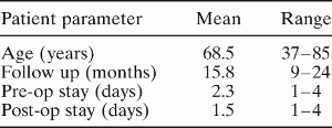
Forty-three patients (30 men and 13 women) had 45 arteries operated upon, as two patients required bilateral ligation. The cohort had an average age of 68.5 years, ranging from 37 to 85 years (Table I). Patients were admitted for an average of 2.3 days before going to theatre and were discharged on average 1.5 days after surgery. Twenty-seven procedures (60 per cent) involved clipping alone, 10 (22 per cent) involved both clipping and diathermy, and eight (18 per cent) involved diathermy alone. All patients were followed up for 15.8 months on average (range nine to 24 months).
Average Hospital Stay and Follow-up Period
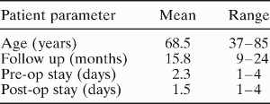
Pre-op=pre-operative; post-op=post-operative
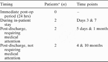
In the present study, no patients suffered recurrent epistaxis within the first 24 hours of surgery (Table II). Four patients subsequently suffered recurrent epistaxis requiring further intervention. Two of these patients suffered recurrence whilst still in hospital, on day three and day seven. Each required a return to theatre and responded to endoscopic diathermy of the bleeding sites and septoplasty. Two different patients had recurrent epistaxis within one month of discharge: one bled from a long-standing septal perforation and the other from Little's area, and each responded to conservative measures comprising local cautery and anterior nasal packing. An additional two patients experienced minor epistaxis at four and 10 months post-discharge; medical intervention was not required in either case.
Epistaxis Recurrence

* n=45. Hrs=hours; post-op=post-operative
Two mortalities occurred during the follow-up period. One patient succumbed to congestive heart failure due to long-standing ventricular aneurysm, and the other to acute respiratory distress syndrome. Neither of these patients had suffered recurrence of epistaxis after treatment.
Discussion
In recent years, in line with advances in surgical technology, techniques to access the sphenopalatine artery have been continually refined. Early descriptions involved endoscopic ligation of the sphenopalatine artery by means of canine fossa antroscopy.Reference White13 Sharp et al. simplified access to the sphenopalatine artery via a middle meatal antrostomy, whilst El-Guindy described a trans-septal approach to the sphenopalatine artery.Reference Sharp, Rowe-Jones, Biring and Mackay6,Reference El-Guindy14
We report the first long-term study which shows that good long-term control of intractable posterior epistaxis can be achieved by sphenopalatine artery ligation. While the local complication rate is extremely low, the not insignificant post-operative mortality rate seen in this series is indicative of the poor cardio-respiratory status of many of these patients. We support the view that endoscopic ligation of the sphenopalatine artery is superior to other procedures, such as ligation of the external carotid artery and the internal maxillary artery via a trans-antral approach.Reference Kumar, Shetty, Rockey and Nilssen15
Like all forms of surgery, a safe and effective intervention benefits from a sound knowledge of local anatomy. The failure rate for sphenopalatine artery ligation varies between zero and 30 per cent, although most large studies like our own show rebleeding rates in the order of 10 per cent.Reference Feusi, Holzmann and Steuer11 While rebleeds can be caused by a multitude of factors, including clip dislodgement and the presence of rich collateral vessels, the presence of more than one branch at the point of emergence of the sphenopalatine artery from its foramen may in part explain this fairly consistent failure rate. Lee et al. found that the sphenopalatine artery divided into two major branches in 76 per cent of cases, into three branches in 22 per cent and into four branches in 2 per cent.Reference Lee, Kim, Kim, Son, Kim and Cho16 Techniques that involve clipping the artery without further posterior dissection can easily result in failure to ligate the posterior septal branch. We recommend techniques that involve a formal dissection beyond the first trunk (usually the posterior lateral nasal artery) seen emerging from the foramen. In this way, ligation or diathermy of any posterior branches between the first trunk encountered and the anterior face of the sphenoid is achieved. Should the sphenopalatine artery be unidentified within the sphenopalatine foramen, further dissection posteriorly is also recommended, as 42 per cent of sphenopalatine arteries in one study were found to exit from a distinct foramen directly posterior to the sphenopalatine foramen.Reference Schwartzbauer, Shete and Tami17
In the United Kingdom to date, there has been no large cohort study on the surgical treatment of posterior epistaxis. In the present study, both the post-operative and the long-term success rates for ligation of the sphenopalatine artery were comparable with those in previously published reports (Table III). Three of these studies achieved 100 per cent success. However, the numbers of patients in these studies were small and the follow-up periods short. Our results show that recurrence of epistaxis requiring active intervention occurred within one month of surgery.
Complications and Success Rates: Comparison Between Studies

SPA=sphenopalatine artery
Apart from recurrent epistaxis, ligation of the sphenopalatine artery is associated with few complications. Snyderman et al. reported that one-third of patients had increased nasal crusting following treatment, and palatal numbness was reported in 13 per cent of patients. No patients in the present series reported any of these complications.
Sphenopalatine artery ligation has been shown to be associated with a shorter hospital stay compared with other surgical modalities.Reference Srinivasan, Sherman and O'Sullivan21 As yet, there are no known UK studies comparing the cost effectiveness of sphenopalatine artery ligation with that of other surgical procedures. However, North American studies comparing the average cost per patient showed that early surgical intervention was more economical than medical treatment or arterial embolization.Reference Klotz, Winkle, Richmon and Hengerer22,Reference Miller, Stevens and Orlandi23 These authors suggested that early surgical intervention be included in the treatment algorithm for intractable epistaxis.
Sphenopalatine control is an effective and time efficient method of controlling epistaxis and has a low complication rate. Assuming there are no contraindications to surgery, we recommend that control of posterior epistaxis via ligation of the sphenopalatine artery be considered as immediate second line treatment when the first attempt at conservative treatment fails.
• Posterior nasal packing, including balloon tamponade, has a high failure rate, ranging from 26 to 52 per cent
• Posterior epistaxis usually occurs in a patient group that is often elderly, frail and with multiple concomitant medical problems
• Endoscopic sphenopalatine artery ligation is technically straightforward to perform and allows direct, secure ligation of the major vessel supplying the posterior nasal cavity
• Control of posterior epistaxis via sphenopalatine artery ligation should be considered as immediate second line treatment when the first attempt at conservative treatment fails
• Sphenopalatine artery control is an effective and time efficient method of controlling epistaxis and has a low complication rate