Introduction
Wax is one of the most important products of honey bees. It is used in the pharmaceutical industry, dentistry and cosmetics. Wax contains many nutrients, pollen and honey, and therefore attacks the combs by various pests which destroy them (Ebadi, Reference Ebadi1975). The wax moth, Galleria mellonella L., is one of the most destructive and economically important pests of wax in the world, the primary concern for beekeepers is how to adequately store combs without them being destroyed or damaged by the larval stages of the moths. Destruction or damage to combs can also occur within weak hives, which have died or have low populations. The most successful control measure has been the use of insecticides at the larval stage. But the extensive use of insecticides has led to the development of resistance in many insect pests to them and has resulted in destructive effects on the environment. Also, the insecticides residue in the wax combs is a result of the frequent use of them (Colter, Reference Colter1995). Substitutional, eco-friendly methods to control insect pests include the sterile insect technique (SIT), or the partial male sterility technique (also called inherited sterility – IS or F 1 sterility). In SIT and Fl sterility, mass production and release of large numbers of genetically altered, large numbers of sterile males are released into the treated area in quantities that surpass the estimated population size of wild males (also called the critical flooding ratio). The sterile males outcompete the local wild males, and their mating with the females results in non-viable embryos (using SIT) or sterile, highly male-biased offspring (using IS) (Carpenter et al., Reference Carpenter, Bloem, Marec, Dyck, Hendrichs and Robinson2005). The advantages of using IS were previously studied by North (Reference North1975) and LaChance (Reference LaChance1985), they described several attributes that are common to IS in Lepidoptera: it is species-specific; has no off-target effects on the environment or on human health, F 1 male and female offspring are more sterile than the irradiated parental (P 1) generation, and more F 1 male progeny than female progeny are produced. Other attributes may include longer developmental time and reduced sperm quality in the F 1 generation. Radiation-induced deleterious effects that can be inherited for several generations; however, the majority of the inherited deleterious effects are expressed in the F 1 generation. Tothová & Marec (Reference Tothová and Marec2001) declared that transmission of various complexes of chromosome translocations is responsible for the production of genetically unbalanced gametes in F 1 progeny of treated males and, as such, represent the main chromosomal mechanism of IS. The ability of using SIT for controlling wax moth was laboratory investigated by Sayed (Reference Sayed2008). She stated that 250 Gy caused complete sterility in male when irradiated as full grown pupae. Furthermore, Jafari et al. (Reference Jafari, Goldasteh and Afrogheh2010) reported that 350 Gy was the most effective dose capable of sterilizing the male pupae of the wax moth for field application, and the best release ratio was at four sterile males, one normal male for each normal female (4:1:1).
The male tract consists of paired testes, each containing a series of testicular tubes or follicles (in which spermatozoa are produced), which open separately into the mesodermally derived sperm duct or Vas deferens, which expands posteriorly to form a sperm storage organ or seminal vesicle. Tubular paired accessory glands are formed as diverticula of the vasa deferentia. Sometimes the vasa deferentia themselves are glandular and fulfill the functions of accessory glands. The paired vasa deferentia unite where they lead into the ectodermally derived ejaculatory duct (the tube that transports the semen or the sperm to the gonopore). Accessory glands are 1–3 pair; either mesodermal of ectodermal in origin that is associated with vasa deferentia or ejaculatory duct. Its function is to produce seminal fluid and spermatophores (sperms).
In ecotoxicological studies, the random amplified polymorphic DNA (RAPD) assay based on polymerase chain reaction (PCR) has been used as a powerful tool for detecting DNA damage and mutations (Atienzar et al., Reference Atienzar, Cheung, Jha and Depledge2001; Ercan, Reference Ercan2015), species classification, genetic mapping and phylogeny studies (Garner & Slavicek, Reference Garner and Slavicek1996; Singh et al., Reference Singh, Bajpai and Tewari2011; Kumar et al., Reference Kumar, Kumar, Thakur, Rajneesh, Mattu and Seth2015). RAPD-PCR was first developed by Williams et al. (Reference Williams, Kubelik, Livak, Rafalski and Tingey1990) and based on amplifies random DNA fragments with short primers of arbitrary nucleotide sequence.
Therefore, the present study aimed to determine the effect of sub sterilizing doses of γ radiation at the pupal stage on some biometrically measured changes of the male testes. Also, the intent of this study was to find a genetic marker made by comparing the RAPD-DNA patterns of normal and irradiated parents and the F 1 generation, which would serve as a sterility fingerprint that enable to study the inheritance of sterility.
Materials and methods
Insect
The greater wax moth, G. mellonella L. strain used was obtained from the National Research Center (NRC), Giza, Egypt. The larval stage was reared on artificial media according to Wiesner (Reference Wiesner1993) at 28 ± 2°C and a relative humidity of 65 ± 5%.
Irradiation technique
Irradiation of G. mellonella full grown pupae was carried out using γ Cell Irradiation Unit (Co60 source) located at the National Center for Radiation Research and Technology (NCRRT). The dose rate was 2.08 kGy h−1. Full-grown pupae were irradiated with two sub sterilizing doses 80 and 160 Gy (Sayed, Reference Sayed2008). Unirradiated pupae were kept as control for comparison. The emerged adult males’ moths were caged till they were used. Five replicates were conducted for each treatment and the control.
Morphological studies of the gonads
Males emerged from irradiated pupae and F 1 males emerged from crossing irradiated males and normal females were dissected in Ringer physiological solution Humason (Reference Humason1972). After the gonads were freed from any fat body, the average length and width of the testes was estimated. Measurements were made by graduated binocular eye lens using magnification 160×. The radius and volume were calculated using Excel program for Windows 7.
DNA Extraction
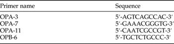
Total body tissue samples were ground with liquid nitrogen using a mortar and pestle. Extraction and purification of samples were carried out with use of DNeasy mini spin columns as described by the manufacturer (Qiagene, Hilden, Germany) stored at −80°C. RAPD-PCR reactions were performed according to the protocol of Williams et al. (Reference Williams, Kubelik, Livak, Rafalski and Tingey1990). Reactions were performed in a total volume 50 µl reaction buffer (100 mMKcL, Tris HCL ph 8.3), 3 mM MgCl2, 200 mM dNTPs (Promega Biotech. Inc.), 50 p mole−1 primers and 1 µ Taq polymerase (obtained from Operon Technologies). This reaction was added to 0.1 µl genomic DNA. Tubes containing mixes were placed in a thermo cycler (Perkin Elmer 2400) and the DNA was amplified using the following temperature profile (modified from Black et al., Reference Black, Nancy, Gary, James and Jennifer1992). Ten arbitrary primers were used, four of them only gave polymorphism with the samples (table 1). Amplification was carried out for 30 cycles. The temperature profiles were 94°C for 5 min, 94°C for 40 s, 72°C for 2 min, 72°C for 7 min, 36°C for 1 min and 4°C for hold. After the reaction, mixture was mixed with DNA loading buffer and electrophoresed on 1% Agarose gel. DNA ladder mix was used as standard DNA with molecular sizes of 2500, 2000, 1500, 1000, 900, 800, 700, 600, 500, 400, 300, 200, and 100 bp.
The nucleotide sequences of the primers used for RAPD-PCR analysis.

The electrophoresis gel images were recorded using a gel Documentation system (UVP, UK) and analyzed using CLIQS 1D Pro (Totallab) to reveal the molecular sizes (bp) of the bands. Bands in each treatment were compared using the similarity index formula of Nei & Li (Reference Nei and Li1979).
Statistical analysis
Measurements of the testes are presented as mean ± SEM and analyzed using one way analysis of variance (ANOVA) followed by Duncan's T test. Differences were considered significant at probability levels P > 0.05 using Statistical Package for Social Science (SPSS) version 15.0 for Windows.
Results
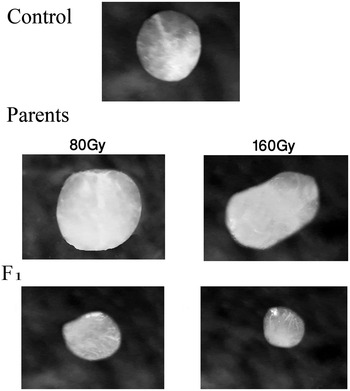
Table 2 and fig. 1 represented a significant increase in length, width, radius and volume of the parents’ testes of G. mellonella irradiated as pupae with 80 and 160 Gy as compared with control. On the other hand, some shrinkage and deformation were noticed in shapes of testes of both irradiated parents and F 1 males with 80 and 160 Gy. Whereas length, width, radius and volume of the tesets of F 1 irradiated with 160 Gy were significantly decreased than control. The tesets of F 1 irradiated with 80 Gy presented a non significant decrease in length, width, radius and volume in comparison with control.
Testes of untreated, treated parents and F 1 males with 80 and 160 Gy.

Effect of γ irradiation on the volume of tests of parents and F 1 Galleria mellonella males.

Data are represented as mean ± SE (SEM) of five replicates.
Radius = (Length + width)/4.
Volume = 4/3π r 3.
* Statistically significant at P < 0.05 with reference to normal control (Duncan's T test).
RAPD finger print profiles were generated for screening the genomic DNA extracted from unirradiated male, parent males were treated with 80 and 160 Gy and first generation. Banding patterns were scored as present (+) or absent (−) according to their molecular sizes. The number of RAPD amplicons was 35 distributed as 9, 11, 7, and 8 with primer OPA-3, OPA-7, OPA-11, and OPB-6, respectively.
In the present investigation irradiated males parents and F 1 males were compared with untreated ones to analyze any change in the DNA sequence, which might be induced by irradiation treatment.
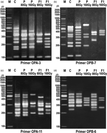
Only 8, 8, 7, and 6 bands were observed in control G. mellonella in case of OPA-3, OPA-7, OPA-11, and OPB-6 primers, respectively (fig. 2). From (table 3) the percentage of polymorphism was 77.14%, whereas primer OPA-3, OPA-7, OPA-11, and OPB-6 showed 6, 10, 4 and 7 polymorphic bands respectively.
RAPD-PCR pattern resulting from amplification of genomic DNA of G. mellonella males.

A list of the numbers of polymorphic and monomorphic bands of the different treatments with the listed five random primers.

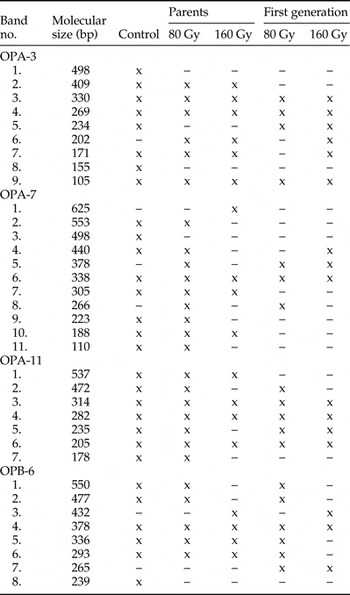
The results in (table 4) showed that RAPD markers using primer OPA-3 indicate the presence of one new band in parents irradiated as full grown pupae with 80 and 160 Gy and F 1 of irradiated parents with 160 Gy in comparing with control. In addition, two bands were present in control only and disappeared in all irradiated samples. RAPD analysis using primer OPA-7 represented appearance of three new bands in irradiated samples when compared with unirradiated control. One band was present only in parent irradiated with 160 Gy, the second one was in parent irradiated with 80 and 160 Gy and in F 1 of irradiated parents with 160 Gy and the third band was in parent and F 1 irradiated with 80 Gy. By using primer OPA-11; three bands were common in control and irradiated samples. The fragments produced by RAPD analysis using primer OPB-6 revealed an appearance of two new bands; the first was in parent and F 1 of irradiated pupae with 160 Gy; the another band was in F 1 generation of irradiated parent males pupae with 80 and 160 Gy in comparison with control. On the other hand, one band disappeared after irradiation. Regarding the four used primers, it was observed that the number of disappeared bands was four and the appeared extra new bands were six (table 4).
DNA polymorphism using randomly amplifying DNA (RAPD) with four primers for untreated control, parents treated with 80 and 160 Gy and F 1.

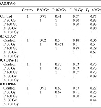
DNA profile generated by using RAPD-PCR was analyzed by using primer OPA-3. The similarity index analysis of value 1 revealed no changes in DNA structure (table 5). The obtained data in table (5A) showed that the highest similarity among different samples when using primer OPA-3 was found between the parents irradiated with 80 Gy and parents irradiated with 160 Gy (1), followed by the similarity obtained from parents irradiated with 80 or 160 Gy and F 1 males produced from parent males irradiated with 160 Gy (0.83), while the lowest value was between the control and parents irradiated with 160 Gy. Table 5 revealed the similarity among different samples when using OPA-7. The parents irradiated with 80 Gy and the control represented highest value (0.82), while the lowest value was between the control and F 1 males produced from parent males irradiated with 80 Gy. The similarity indexes analyzed using primer OPA-11 in table 5 were ranged from 0.67 to 1; where parents irradiated with 80 Gy was similar to the control. On the other hand, the lowest value was between parents irradiated with 160 Gy and F 1 80 Gy. On comparing the analyzed similarity matrix by using primer OPB-6, the highest value was found between parents irradiated with 80 Gy and the control with 0.91. In contrast, the lowest value was between control and F 1 males produced from parent males irradiated with 160 Gy (table 5).
Similarity index among G. mellonella male DNA pattern.

Discussion
As implied from the obtained results, there was a significant malformation in the testes of parent and F 1 Galleria mellonella irradiated as full grown pupae with 80 and 160 Gy, which was correlated with increasing the dose applied to the male parents. Our results agree with that obtained by Sallam et al. (Reference Sallam, El-Dossouki, El-Naggar and Shibel2000) on Spodoptera littoralis. Also, Seth et al. (Reference Seth, Rao and Kaur2000) stated that irradiation declined the testicular volume in F 1 progeny of S. litura and the effect was significant at a dose of 130 Gy. But, the obtained results differ with Abdel Baki & Al Khalaf (Reference Abdel Baki and Al Khalaf2011) who found that irradiated full grown pupae Plodia interpunctella (Hubn) with 50, 150, 250 and 350 Gy and showed a decrease in the overall size of the testes in some of the emerged moths (parents). Whereas, Sallam et al. (Reference Sallam, AbdelL-Gawad, Assar, Ibrahim and EL-Naggar1996) who found a significant change in testes volume of F 1, F 2 and F 3 generations of S. littoralis when parents male less than 24 h irradiated with 100, 150 and 200 Gy.
These effects of radiation were correlated with reproductive competence of F 1 moths. Thus, process of optimization is necessary to balance sterility level and competitiveness, taking into consideration that substerilizing doses are the most favorable condition for IS programme (North, Reference North1975).
The study of the RAPD – PCR profiles of normal and irradiated parent males and F 1 males of G. mellonella showed variation between them. The appearance of some extra bands and the disappearance of others, as a result of irradiation, were recorded throughout this study causing variations in the PCR patterns among different samples. Also, the number of new bands was found to be higher than that of lost bands. These induced variations differ according to the dose of irradiation given and differ from parent males and F 1 males. Similar findings were recorded by Alexandrov et al. (Reference Alexandrov, Lapidus, Alexandrov, Korablinova and Levkovich1998) who stated that 62.5% of γ rays induced mutants’ revealed deficiency of one or more fragments produced. Similar results were obtained by Abulyazid et al. (Reference Abulyazid, Ibrahim, El-Shikh and Abdlhamid2007) on S. littoralis and Abd-El Aziz & Haiba (Reference Abd-El Aziz and Haiba2008) on Phithoremaea operculella. Also, El-said (Reference El-said2013) on her study on S. littoralis males (emerged from irradiated pupae) at various generations (parents, F 1 and F 2) showed variation between them.
These results were explained by Atienzar & Jha, Reference Atienzar and Jha2006 who reported that the DNA damage, point mutations or complex chromosomal rearrangements may be induced by genotoxic factors. Also, supposing that radiation induced DNA damages by reactive oxygen species and (HO−) generated by the radiolysis of water or due to interaction of DNA with radiation (von Sonntag, Reference von Sonntag1987). The variations in the PCR patterns depend on the primer used, the dose of γ irradiation given to the insect, the kind of the irradiation source and differ from parent, F 1 and F 2. This variation may related to the hypothesis of Lea (Reference Lea1956), who stated that ionizing radiation deposits energy in irradiated biological specimens at discrete loci randomly distributed within the radiation field and that ionization, which occurs a cellular changes depends on the dose of radiation and on the size of irradiated specimens.
Alterations of DNA patterns of the sterile males provide an explanation for the importance of the DNA in the transfer of genetic materials from cell to cell. Therefore, it could be concluded that sterility can be inherited from parents to their offspring and increased in first generation than in their parents. This can be noticed from dissection of male reproductive system and from the size and malformations of the testes in first generation more than in their parents.
Acknowledgement
All Authors are thankful to Egyptian Atomic Energy Authority.