Introduction
In vitro embryo production (IVEP) in water buffaloes has some limitations, such as blastocyst rates between 9.5 and 30%, worse than the average normally observed for cattle, which ranges from 30 to 45% (Gasparrini, Reference Gasparrini2002; Ferraz et al., Reference Ferraz, Watanabe, Joaquim, Watanabe, Accorsi, Meirelles, Sá Filho and Baruselli2005; Sá Filho et al., Reference Sá Filho, Carvalho, Gimenes, Torres-Júnior, Nasser, Tonhati, Garcia, Gasparrini, Zicarelli and Baruselli2009). Even though the efficiency of IVEP has improved in recent years in buffaloes, the cleavage rate is still low compared with other domestic species (Gasparrini et al., Reference Gasparrini, De Rosa, Attanasio, Boccia, Di Palo, Campanile and Zicarelli2008). This result can be associated with the high concentration of lipids present in the cytoplasm of the oocytes and embryos of this species (Gasparrini, Reference Gasparrini2002).
Fetal bovine serum (FBS), a protein supplement normally used in IVEP, is a compound containing a variety of substances, the majority undefined, including fatty acids, growth factors, energy substrates, amino acids and vitamins. It is widely used in culture medium to promote higher in vitro production rates of bovine embryos (Ali and Sirard, Reference Ali and Sirard2002). However, this compound can also cause alterations in the ultrastructure, along with compaction and blastulation of embryos and changes in the expression of mRNA, increasing the incidence of stillbirths and calf mortality rates (Abe and Hoshi, Reference Abe and Hoshi2003). Other negative aspects related to FBS are lipid accumulation, which increases the concentration of fatty acids and cytoplasmic lipid droplets in the blastocysts and decreases embryo cryotolerance (Rizos et al., Reference Rizos, Gutierrez-Adan, Perez-Garnelo, De La Fuente, Boland and Lonergan2003; Leivas et al., Reference Leivas, Brum, Fialho, Saliba, Alvim, Bernardic, Rubin and Silva2011).
Therefore, in recent years several substances have been tested to replace FBS, such as polyvinylpyrrolidone (PVP), polyvinyl alcohol (PVA) and synthetic serum substitutes, both for in vitro maturation (IVM) of oocytes and in vitro culture (IVC) of bovine embryos (Ali and Sirard, Reference Ali and Sirard2002; Lim et al., Reference Lim, Jang, Ko, Lee, Park, Kim, Kang and Lee2008; Sayirkava et al., Reference Sayirkava, Misirlioglu, Kaya, First, Parrish and Memili2007). However, in buffaloes, the effects of reducing the FBS used in IVEP are still unknown.
In furtherance of the effort to reduce lipid accumulation, recent studies with bovine embryos have assessed the use of lipid metabolism regulators during IVEP. The results have shown that addition of these substances to culture medium can influence the expression of genes that regulate lipid metabolism and improve the development and quality of embryos (Ghanem et al., Reference Ghanem, Ha, Fakruzzaman, Bang, Lee and Kong2014; Spricigo et al., Reference Spricigo, Morato, Arcarons, Yeste, Dode, Lopez-Bejar and Mogas2017).
In this context, l-carnitine (3-hydroxy-4-N-trimethyl amino butyrate), a quaternary amine with a fundamental role in cell energy generation, has been extensively studied for its activity in the reactions that transfer long-chain free fatty acids from the cytosol to mitochondria, facilitating their oxidation and generation of adenosine triphosphate (ATP) (Dunning and Robker, Reference Dunning and Robker2012; Spricigo et al., Reference Spricigo, Morato, Arcarons, Yeste, Dode, Lopez-Bejar and Mogas2017). Although the positive effects of l-carnitine on oocytes and embryos have been described in cattle (Yamada et al., Reference Yamada, Imai and Yamada2006; Chankitisakul et al., Reference Chankitisakul, Somfai, Inaba, Techakumphu and Nagai2013; Phongnimitr et al., Reference Phongnimitr, Liang, Srirattana, Panyawai, Sripunya, Treetampinich and Parnpai2013a; Held-Hoelker et al., Reference Held-Hoelker, Klein, Rings, Salilew-Wondim, Saeed-Zidane, Neuhoff, Tesfaye, Schellander and Hoelker2017; Knitlova et al., Reference Knitlova, Hulinska, Jeseta, Hanzalova, Kempisty and Machatkova2017; Spricigo et al., Reference Spricigo, Morato, Arcarons, Yeste, Dode, Lopez-Bejar and Mogas2017), sheep (Reader, et al., Reference Reader, Cox, Stanton and Juengel2015; Mishra et al., Reference Mishra, Reddy, Gupta and Mondal2016), swine (Somfai et al., Reference Somfai, Kaneda, Akagi, Watanabe, Haraguchi, Mizutani, Dang-Nguyen, Geshi, Kikuchi and Nagai2011), mice (Moawad et al., Reference Moawad, Xu, Tan and Taketo2014; Zare et al., Reference Zare, Farahani, Salehi, Piryaei, Novin, Fathabadi, Mohammadi and Dehghani-Mohammadabadi2015; Khanmohammadi et al., Reference Khanmohammadi, Movahedin, Safari, Sameni, Behpour Yousefi, Jafari, Zarbakhsh and Zarbakhsh2016), camels (Fathi and El-Shahat, Reference Fathi and El-Shahat2017), and buffaloes (Phongnimitr et al., Reference Phongnimitr, Liang, Srirattana, Panyawai, Sripunya, Treetampinich and Parnpai2013b), to the best of our knowledge no studies are available describing the action of this lipid metabolism regulator associated with reduced use of FBS for in vitro maturation (IVM) on the IVP of buffalo embryos.
Considering the potentially positive impacts of strategies to substitute/reduce the employment of FBS during IVEP, associated with the use of metabolic regulators that increase the lipid metabolism of oocytes and embryos, and that the IVM step is critical in this process, we evaluated the effects of reducing the FBS concentration together with the use of l-carnitine in IVM on the development and lipid accumulation of bubaline embryos.
Materials and methods
Experiments and reagents
The experiments described were performed in the Laboratory for Analysis of Sustainable Systems (LASS) of Embrapa Eastern Amazon, located in Belém, Pará, Brazil.
Reagents and culture medium were purchased from Sigma Chemical Co. (St. Louis, MO, USA), unless indicated otherwise.
Experimental design
Experiment I. Effects of reducing FBS during IVM on the embryos rates
In the first experiment, we aimed to determine the lowest concentration of FBS (Cripion; Andradina, Brazil; cat. no. FB 0010S) in the IVM medium able to maintain the embryo development rate obtained by the control group (10% FBS). Bubaline oocytes were submitted to IVM medium supplemented with 0%, 2.5%, 5% or 10% FBS. We evaluated the cleavage rates and blastocyst production levels in five replicates.
Experiment II. Effects of adding L-carnitine during IVM on the embryo development and lipid accumulation
After defining the lowest effective concentration, we performed a second experiment in which the 0%, 5% and 10% FBS groups were also evaluated regarding the addition of 5 mM of l-carnitine (Sigma, Cat no: C0158) in the IVM medium. The blastocysts produced were submitted to lipid quantification tests, involving staining followed by observation by optical microscopy (OilRed O staining) and confocal microscopy (BODIPY 493/503 staining). In each lipid analysis, embryos produced from three replicates were evaluated.
Obtaining oocytes and embryos produced in vitro
Selection and in vitro maturation of oocytes
Bubaline oocytes from the ovaries of slaughtered animals were used in the experiments. The ovaries were collected and taken to the laboratory in under 3 h in an isothermal receptacle containing sterile saline solution, and kept at a temperature of 30–35ºC. Ovarian follicles measuring between 3 and 8 mm in diameter were aspirated using an 18-G needle attached to a 20 ml disposable syringe, and the aspirated fluid was transferred to a 50 ml polypropylene tube. After decanting for 15 min, the sediment was transferred to a polystyrene dish and visualized using a stereoscopic microscope to find and select the oocytes. Cumulus–oocyte complexes (COCs) presenting at least three layers of cumulus cells and homogenous cytoplasm were selected under a stereomicroscope. The oocytes were washed in TCM-199 medium buffered with 5 mM of sodium bicarbonate and 20 mM HEPES, supplemented with 5 mg/ml bovine serum albumin (BSA), 0.20 mM sodium pyruvate and 83.4 μg/ml amikacin (washing solution).
The base medium utilized for in vitro maturation (IVM medium) was composed of TCM-199 buffered with 25 mM sodium bicarbonate and supplemented with 1.0 μg/ml FSH, 50 μg/ml hCG, 1.0 μg/ml estradiol, 0.20 mM sodium pyruvate, 83.4 μg/ml amikacin and antioxidants (Sigma cat. no. A1345). The groups were supplemented with 0%, 2.5%, 5.0% or 10% FBS in experiment I and 0%, 5.0% or 10% FBS in experiment II. Oocytes were cultured in 100 µl droplets, in groups of 15–20, covered in mineral oil and kept in an incubator at 38.5ºC and in an air atmosphere with 5% CO2 for 22 h. In the second experiment, the same conditions were employed, with the addition of 5 mM of l-carnitine to the IVM medium, based on reports in the published literature.
In vitro production of embryos
After IVM for 22 h, the oocytes were fertilized in vitro with semen from a single bull (previously tested) in IVF-TALP (Tyrode’s albumin lactate pyruvate) medium supplemented with 0.6% BSA, 10 µg/ml heparin, 18 µM penicillamine, 10 µM hipotaurine and 1.8 µM epinephrine for up to 24 h. Then, the probable zygotes were washed and in vitro cultured in modified synthetic oviductal fluid (Del Collado et al., Reference Del Collado, Saraiva, Lopes, Gaspar, Padilha, Costa, Rossi, Vantini and Garcia2016) supplemented with 1.5% FBS and 6 mg/ml of BSA fatty acid free. All structures were maintained in an incubator in an air atmosphere with 5% CO2 for 7 days, until cells reached the blastocyst stage (day 0 = day of fertilization).
Between 10 and 20 zygotes were cultured per microdroplet (100 µl), in 35 mm polystyrene dishes covered with mineral oil. Cleavage and blastocyst rates were evaluated on the day 2 and day 7 of in vitro culture (IVC), respectively. Both the cleavage rate and blastocyst rates were calculated from the total number of viable oocytes (utilized in the IVM). The development medium was replaced by half on the fourth day of embryo development.
Evaluation of the lipid accumulation in the embryos produced in vitro
Lipid accumulation test with optical microscopy
Blastocysts evaluated on day 7 of in vitro culture (18 embryos from five groups: 0% FBS = 3, 5% FBS = 6, 10% FBS = 3, 5% FBS–l-carnitine (FBS–LC) = 3, 10% FBS–LC = 3) were fixed in 4% paraformaldehyde and stained with Oil Red O (Sigma cat. no. O0625), a liposoluble dye that reveals neutral lipids, enabling visualization of the cytoplasmic lipid droplets under an optical microscope. The structures were fixed in 4% paraformaldehyde for 20 min and then stored in PBS at 4ºC until staining. To analyze the lipid droplets, the fixed embryos were washed in 60% isopropanol and then incubated in a working solution containing Oil Red O, according to the manufacturer’s instructions. After staining, the embryos were washed four times with distilled water and mounted on slides.
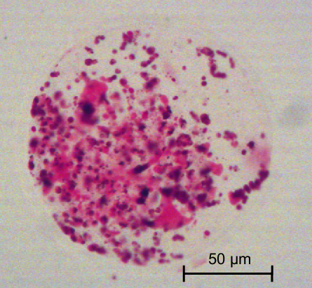
Slides were observed using an inverted optical microscope (Leica DMLS) and the images (Figure 1) were captured with a Moticam 5.0 MP camera and stored using the Motic Image Plus 2.0 program. The images were then analyzed with ImageJ software, to estimate the lipid droplets in buffalo embryos.
Image of the embryonic lipid droplets (neutral lipids) after staining with Oil Red O and visualization by optical microscopy.

Lipid accumulation evaluation with confocal microscopy
Eighty-seven D7 blastocysts from the five groups evaluated (0% FBS = 18; 5% FBS = 14; 10% FBS = 20; 5% FBS–LC = 22; 10% FBS–LC = 13) were fixed in 4% paraformaldehyde for 30 min, permeabilized with 0.1% saponin in PBS for 1 h and stained with 20 μg/ml of BODIPY 493/503 (Molecular Probes, Eugene, OR, USA), a specific probe for neutral lipids, for 30 min to analyse the lipid quantification by confocal microscopy. To visualize the structures, excitation of 330–385 nm and emission of 420–490 nm were used. To count the number of cells, the embryos were stained with 10 μg/ml of Hoechst 33342 for 20 min and then mounted on slides using Fluoromount (Sigma cat. no. F4680).
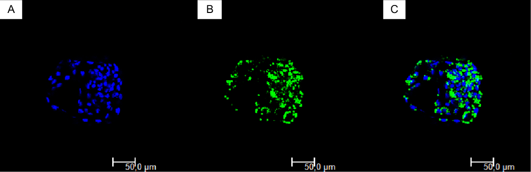
The embryos were analyzed using an LSM 710 confocal microscope (Carl Zeiss, Oberkochen, Germany) with argon laser for visualization of the lipid droplets, at a resolution of 1024 × 1024 pixels with 12 bits, at magnification of ×63 in oil, with excitation and emission of 488 and 516 nm, respectively (Figure 2). An image was captured of each embryo using the section with the greatest diameter, and the images were subsequently analyzed with the ImageJ program. The ratio between the area of the lipid droplets and total area of the embryo was calculated as described previously (Del Collado et al., Reference Del Collado, Saraiva, Lopes, Gaspar, Padilha, Costa, Rossi, Vantini and Garcia2016). To obtain the area of the lipid droplets, the nucleus counter tool was used, which allows the total area occupied by the droplets to be calculated. Then the area of the embryo was measured to compute the ratio between the areas of the lipid droplets and embryo, and the number of blastomeres was counted.
Processing of the original image. (A) Nucleus stained with Hoechst 33342. (B) Staining of the lipids with BODIPY 493/503. (C) Superimposed images for lipid quantification by confocal microscopy.

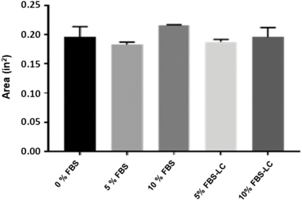
Total area of neutral lipids accumulated in bubaline day 7 blastocysts from different treatments during IVM, after staining with Oil Red O. There was no difference between groups (Kruskal–Wallis test, P > 0.05). LC, l-carnitine. Evaluated embryos were from three replicates.

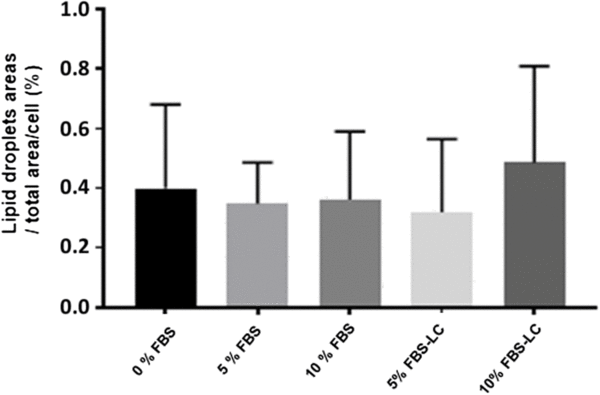
Effects of neutral lipids accumulation in bubaline day 7 blastocysts from different treatments during IVM. There was no difference between groups (Kruskal–Wallis test, P > 0.05). Evaluated embryos were from three replicates.

Statistical analysis
Embryo production data were submitted to analysis of variance (ANOVA) together with the post-hoc Tukey test. Lipid quantification data, as they were not normally distributed, were evaluated using the non-parametric Kruskal–Wallis test. All statistical analyses were performed at a 5% level of significance, using the SPSS version 22.0.0.0 software, except for the lipid data, which were evaluated using GraphPad Prism 7 software version 7.03.
Results
In total, 1394 oocytes were used to evaluate the effect of different FBS concentrations and the use of LC during IVM on the initial development of bubaline embryos.
Experiment I. Effects of reducing FBS during IVM on the embryo production rates
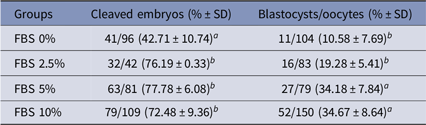
Table 1 shows the effects of reducing the FBS concentration during IVM on embryo development until blastocyst stage, comparing the FBS variable in four groups simultaneously. Cleavage rates were higher in all groups supplemented with FBS than in the serum-free group (P < 0.05; Table 1). In relation to blastocyst rates, the 10% FBS group did not present any difference in comparison with the 5% FBS group. However, both were superior (P < 0.05) to the 0% FBS and 2.5% FBS groups, which were similar to each other.
Cleaved embryos and blastocysts rates after IVM of bubaline oocytes in medium containing different concentrations of FBS

a,bMeans followed by different letters in the column indicate significant difference (P < 0.05) by analysis of variance (ANOVA) and Tukey test.
Cleaved embryos and blastocysts at day 7 were evaluated in five replicates.
FBS, fetal bovine serum; IVM, in vitro maturation; SD, standard deviation.
Experiment II. Effects of LC supplemented in IVM on the embryo development and lipid accumulation
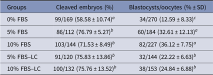
There was a decline in the cleavage rate only in the 0% FBS group, which differed significantly from the others (Table 2).
Embryo development (cleavage and blastocysts rates) under different concentrations of FBS, with or without L-carnitine (LC) during IVM of bubaline oocytes

a,b,cMeans followed by different letters in the column indicate significant difference (P < 0.05) by analysis of variance (ANOVA) and Tukey test.
Cleaved embryos and blastocysts in day 7 were evaluated in five replicates.
FBS, fetal bovine serum; IVM, in vitro maturation; LC, l-carnitine; SD, standard deviation.
Blastocysts development rates decreased in the 0% FBS group. The effects of treatments with 5% FBS and 10% FBS were similar to each other and superior to the groups supplemented with LC (Table 2).
The results obtained by optical microscopy allowed the quantity of lipids present in the structures to be calculated, expressed by averages of the total area (inch2). The result of the lipid quantification by confocal microscopy analysis was expressed as the ratio between the area occupied by the lipid droplets and the area of the embryo and by the number of blastomeres. There was no difference between groups in both techniques (Figs. 3 and 4).
Discussion
To date, this study is the first to evaluate the effects of reducing the concentration of FBS in association with the use of LC during IVM on the development as strategies to reduce the lipid accumulation in buffalo embryos.
Regarding the effect of the presence of FBS on the embryo development rate, several researchers have demonstrated its role in different phases of IVEP (Rizos et al., Reference Rizos, Gutierrez-Adan, Perez-Garnelo, De La Fuente, Boland and Lonergan2003; Leivas et al.; Reference Leivas, Brum, Fialho, Saliba, Alvim, Bernardic, Rubin and Silva2011; Del Collado et al., Reference Del Collado, Saraiva, Lopes, Gaspar, Padilha, Costa, Rossi, Vantini and Garcia2016). Our results show that the FBS concentration in the IVM medium could be reduced by half (from 10% to 5%) without affecting the rates of embryo development into blastocysts. Although the 2.5% FBS concentration provided almost double the blastocyst rate compared with the serum-free group, there was no significant difference between the lowest two concentrations (0% and 2.5% FBS) and both reduced the embryo development rates compared with the other groups. There was a dose–response effect on SFB concentration in the IVM medium up to 5%.
Various studies have been performed to investigate the effects of removing FBS in culture systems by replacing it, or lowering its concentration (Feugang et al., Reference Feugang, Camargo-Rodríguez and Memili2009), with the use of various supplements (Lim et al., Reference Lim, Jang, Ko, Lee, Park, Kim, Lee, Hwang, Lee and Kang2007; Mingoti et al., Reference Mingoti, Castro, Méo, Barreto and Garcia2011). Del Collado et al. (Reference Del Collado, Saraiva, Lopes, Cruz, Gaspar, Oliveira, Perecin and Garcia2014) used bovine serum albumin free of fatty acids (BSA–FAF), and a commercial product called embryonic fluid, alone or in different combinations and concentrations, and concluded that it is possible to diminish the concentration of FBS in the IVM medium for cattle to 3.5% without significantly impairing the nuclear and cytoplasmic maturation rates.
One of the mechanisms explaining the importance of lipids in events during nuclear maturation is the relationship between β-oxidation and meiotic resumption (Downs et al., Reference Downs, Mosey and Klinger2009; Dunning et al., Reference Dunning, Cashman, Russell, Thompson, Norman and Robker2010). The mechanism occurs by activation of mitogen-activated protein kinases (MAPKs) and promotes the breakdown of the germinal vesicle and resumption of meiosis (Chen et al., Reference Chen, Hudson, Chi, Chang, Moley, Hardie and Downs2006). In this event, acetyl-CoA carboxylase (ACC) is mainly responsible for MAPK activation (Downs et al., Reference Downs, Mosey and Klinger2009). Even though fatty acid oxidation is one of the main consequences of MAPK activation, by reducing the levels of malonyl-CoA, studies have demonstrated that inhibiting lipid oxidation impedes reactivation of meiosis (Downs et al., Reference Downs, Mosey and Klinger2009; Dunning et al., Reference Dunning, Cashman, Russell, Thompson, Norman and Robker2010).
Del Collado et al. (Reference Del Collado, Saraiva, Lopes, Gaspar, Padilha, Costa, Rossi, Vantini and Garcia2016) reported that culture medium used for IVM and containing a high concentration of FBS (10%) increased the total lipids content 18-fold, resulting in higher accumulation of lipids in bovine oocytes compared with medium containing only BSA. As FBS contains components that lead to lipid accumulation, the use of medium supplemented with this compound can result in embryos that are sensitive to cryopreservation. Furthermore, FBS can cause damage to the mitochondrial membrane and alter the expression of genes that are important for embryo development (Rizos et al., Reference Rizos, Gutierrez-Adan, Perez-Garnelo, De La Fuente, Boland and Lonergan2003; Wrenzycki et al., Reference Wrenzycki, Herrmann and Niemann2007).
We did not observe reduction in lipid accumulation in bubaline embryos produced in vitro irrespective of FBS concentration in the IVM medium. The behaviour of lipids during IVM in different species is controversial. For example, a reduced accumulation of c. 21% was observed during in vivo maturation of porcine oocytes (Romek et al., Reference Romek, Gajda, Krzysztofowicz, Kepczynski and Smorag2010), while an increase of nearly 25% was verified in mouse mature oocytes (Yang et al., Reference Yang, Dunning, Wu, Hickey, Norman, Russel, Liang and Robker2010). Recently, Del Collado et al. (Reference Del Collado, Saraiva, Lopes, Gaspar, Padilha, Costa, Rossi, Vantini and Garcia2016) found that supplementation of in vitro maturation medium with FBS caused an increase in oocyte lipids compared with medium supplemented with BSA alone. However, protein supplementation during IVM had an effect only on oocyte lipid accumulation and not on embryos produced from these treatments, indicating that IVC conditions are also crucial in the lipid accumulation process normally observed in in vitro produced embryos.
The harmful action of high FBS concentrations observed in some studies might be because serum is a compound rich in fatty acids that are easily transported to the oocyte cytoplasm, resulting in an increase in lipids in the oocytes (Del Collado et al., Reference Del Collado, Saraiva, Lopes, Cruz, Gaspar, Oliveira, Perecin and Garcia2014). That situation can be harmful to bubaline embryos, as the presence of abundant cytoplasmic granules, characterized as having a high lipid content, in oocytes of this species has been demonstrated in ultrastructural studies (Boni et al., Reference Boni, Sangella, Dale, Rovello, Di Palo and Barbieri1992; Gasparrini, Reference Gasparrini2002). It is likely that buffalo oocytes and embryos are more sensitive to oxidative stress due to their high lipid content (Gasparrini et al., Reference Gasparrini, Boccia, Marchandise, Di Palo, George, Donnay and Zicarelli2006), so decreasing the concentration of FBS during IVM can help to reduce this oxidative process in IVEP of the species.
Although some researchers have investigated the replacement of fetal serum in the medium used in IVEP, with the aim of reducing the lipid accumulation, it is important to maintain a proper balance between lipid quantity in oocytes and embryos, as their excess can impair the cryopreservation process and embryo quality, but their shortage can lead to a lower input of energy to the cells. In this respect, strategies have been proposed recently involving optimization of the β-oxidation process, such as the use of LC, for example, in the IVM and IVC steps. Therefore, we assessed in a second experiment the use of LC in association with reduction of FBS in the IVM medium, with the goal of improving the development and quality of bubaline embryos.
In our studies, we did not find a beneficial effect of LC during IVM of bubaline oocytes on embryonic development and quantity of lipids in embryos produced. In contrast, we observed decreased production of blastocysts that may be due to the dose used. Phongnimitr et al. (Reference Phongnimitr, Liang, Srirattana, Panyawai, Sripunya, Treetampinich and Parnpai2013b) reported an increase in the maturation rate (metaphase II) of bubaline oocytes given LC during IVM at 0.3 mg/ml but not at 0.6 mg/ml and 1.2 mg/ml. In sheep, Mishra et al. (Reference Mishra, Reddy, Gupta and Mondal2016) tested concentrations of 0, 2.5, 5, 7.5 and 10 mM LC and observed improved rates of cleavage and production of morulae and blastocysts only in the group treated with 10 mM LC. Chankitisakul et al. (Reference Chankitisakul, Somfai, Inaba, Techakumphu and Nagai2013), found that the use of 0.6 mg/ml (3.03 mM) LC for 21 h did not influence maturation rate (metaphase II) in bovine. More recently, Longobardi et al. (Reference Longobardi, Salzano, Campanile, Marrone, Palumbo, Vitiello, Zullo and Gasparrini2017) demonstrated that supplementation of freezing extender with carnitine significantly improved post thawing sperm motility and decreased capacitation-like damage in buffalo sperm. However, the study also showed a dose-dependent effect of carnitine on sperm capacitation status, in which the effect was greater at the highest concentration (7.5 mM LC). Therefore, it is clear that responses may vary depending on the concentration of LC used.
In our experimental conditions, the concentration of 5 mM LC in the IVM medium did not improve embryo production. This concentration was established based on other studies with cattle, as the number of dose–response studies with buffaloes is very limited. Therefore, we believe that more research is necessary to clarify the real molecular mechanisms of LC that affect the development of bubaline embryos, in addition to investigate other concentrations of this agent during different steps of IVEP. It is possible that the effects of LC would be better evidenced during embryo culture. In cattle, the addition of 2.5 mM LC starting on the fourth day of development resulted in greater embryo survival to vitrification (Lima, Reference Lima2015), and also better blastocyst development rates in the culture of 2-cell mouse embryos (Khanmohammadi et al., Reference Khanmohammadi, Movahedin, Safari, Sameni, Behpour Yousefi, Jafari, Zarbakhsh and Zarbakhsh2016). It is believed that the positive effect observed in the culture phase probably is due to the improved metabolism of mitochondrial lipids, mainly by the action of LC as an antioxidant, reducing oxidative stress (Abdelrazik et al., Reference Abdelrazik, Sharma, Mahfouz and Agarwal2009; Dunning et al., Reference Dunning, Akison, Russell, Norman and Robker2011).
In addition, more recent studies have pointed to the more intense effects of acetyl-l-carnitine (ALC), an ester of LC that transports long-chain fatty acids into mitochondria for use in energy metabolism (Ferreira and McKenna, Reference Ferreira and McKenna2017). ALC has been used more often than LC in clinical studies of metabolic effects on the brain, liver and other organs (Liu et al., Reference Liu, Head, Kuratsune, Cotman and Ames2004; Zhang et al., Reference Zhang, Zhang, Zhang, Wang, Niu, Cui and Xu2012; Malaguarnera, Reference Malaguarnera2013), and studies have also shown that ALC may have greater bioactivity than LC (Liu et al., Reference Liu, Head, Kuratsune, Cotman and Ames2004; Zhang et al., Reference Zhang, Zhang, Zhang, Wang, Niu, Cui and Xu2012). Recently, Xu et al. (Reference Xu, Yang, Lu, Liang, Lu, Zhang and Lu2018, Reference Xu, Geng, Li, Fu, Lu, Liang, Lu, Zhang, Yang and Lu2019), showed that the treatment ALC during in vitro maturation of buffalo oocytes improves oocyte quality and subsequent embryonic development and cryotolerance.
Our standard protocol was very satisfactory, as the control group (10% FBS) presented a high blastocyst production rate (36.12%) in comparison with previous reports in the literature such as 18.5% (Manjunatha et al., Reference Manjunatha, Ravindra, Gupta, Devaraj and Nandi2009), 30.1% (Gasparrini et al., Reference Gasparrini, Sayoud, Neglia, Matos, Donnay and Zicarelli2003) and 26.9% (Gasparrini et al., Reference Gasparrini, Boccia, Marchandise, Di Palo, George, Donnay and Zicarelli2006), in addition to observation of good results with half the FBS concentration for IVM than normally used. As we maintained FBS during IVC (at 1.5%), the possibility also exists that the effects during IVM were in some way annulled during development, as the two lipid testing techniques (staining with Oil Red O and analysis by confocal microscopy) did not show differences regarding lipid composition of structures obtained after different treatments, even between 0% FBS and 10% FBS.
We concluded that the FBS concentration could be reduced to 5% in IVM medium to produce bubaline embryos, and supplementation of the maturation medium with 5 mM LC did not cause an increase in embryo production of this species. Furthermore, greater alterations in lipid accumulation during IVEP were not found, with or without FBS and with addition of LC during the IVM, indicating the need for further research, mainly involving the in vitro culture step of bubaline embryos and the use of other esters of LC, such as ALC.
Financial support
This study was financed by the Empresa Brasileira de Pesquisa Agropécuária – Embrapa (SEG: 02.13.06.020.00.02.001), an agency connected to the Brazilian Ministry of Agriculture, Animal Husbandry and Supply.
Conflict of interest
None.
Ethical standards
Not applicable, as ovaries were obtained from slaughterhouses.