Introduction
Lipoid proteinosis, also known as Urbach–Wiethe disease, is a rare autosomal recessive disorder, first described in 1929 by dermatologist Erich Urbach and otolaryngologist Camillo Wiethe.Reference Di Giandomenico, Masi, Cassandrini, El-Hachem, DeVito and Bruno1, Reference Urbach and Wiethe2 To date, there are more than 300 documented cases in the literature;Reference Kumar, Sampath and Manoharan3 however, disease affecting the larynx is rarely discussed. It is caused by loss-of-function mutations in the extracellular matrix protein 1 (ECM1) gene, which has been implicated in epidermal differentiation, cell adhesion and wound healing.Reference Hamada, McLean, Ramsay, Ashton, Nanda and Jenkins4
The disease is characterised by deposition of hyaline material in the skin and mucous membranes, with a predilection for the upper aerodigestive tract.Reference Di Giandomenico, Masi, Cassandrini, El-Hachem, DeVito and Bruno1, Reference Acar, Eryilmaz, Gocer, Akmansu and Korkmaz5 Symptoms vary markedly amongst affected individuals.Reference Hofer6 Classically, the onset of disease occurs in early infancy, signalled by a weak cry or hoarseness of voice, reflective of laryngeal involvement.Reference Savage, Crockett and McCabe7 Dermatological manifestations include yellowish waxy papules and acneiform scarring.Reference Gutte, Sanghvi, Tamhankar and Khopkar8 However, the most typical finding is that of moniliform blepharosis: a beaded papular appearance of the upper eyelid.Reference Thappa and Gupta9 Neurological findings include bilateral symmetrical calcifications in the medial temporal lobes, which is associated with impaired memory, neuropsychiatric abnormalities and epilepsy.Reference Friedman, Mathews and Swanepoel10, Reference Kleinert, Cervos-Navarro, Kleinert, Walter and Steiner11
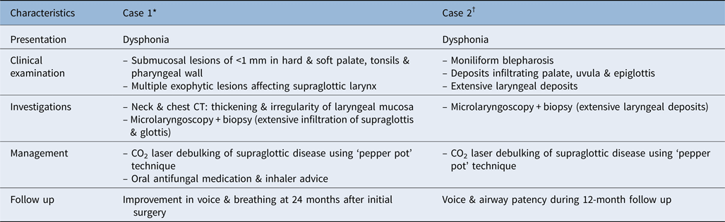
In this paper, we describe the clinical journey of two patients diagnosed with lipoid proteinosis affecting the larynx. A summary of this case series of two patients is provided in Table 1. Current literature is summarised, with an emphasis on the surgical management of laryngeal lipoid proteinosis to minimise morbidity and preserve function.
Summary of case series of two patients with lipoid proteinosis

*Fifty-one-year-old female; †56-year-old male. CT = computed tomography; CO2 = carbon dioxide
Case one
A 51-year-old Caucasian female presented with dysphonia. She was an ex-smoker and former intravenous drug user, with a background of asthma only. There was a family history of the condition, with her brother having lipoid proteinosis.
Examination of the oral cavity revealed submucosal lesions less than 1 mm in size, on the hard palate, soft palate, tonsils and pharyngeal wall. Flexible nasoendoscopy revealed multiple exophytic lesions mainly affecting the supraglottic larynx.
Investigations
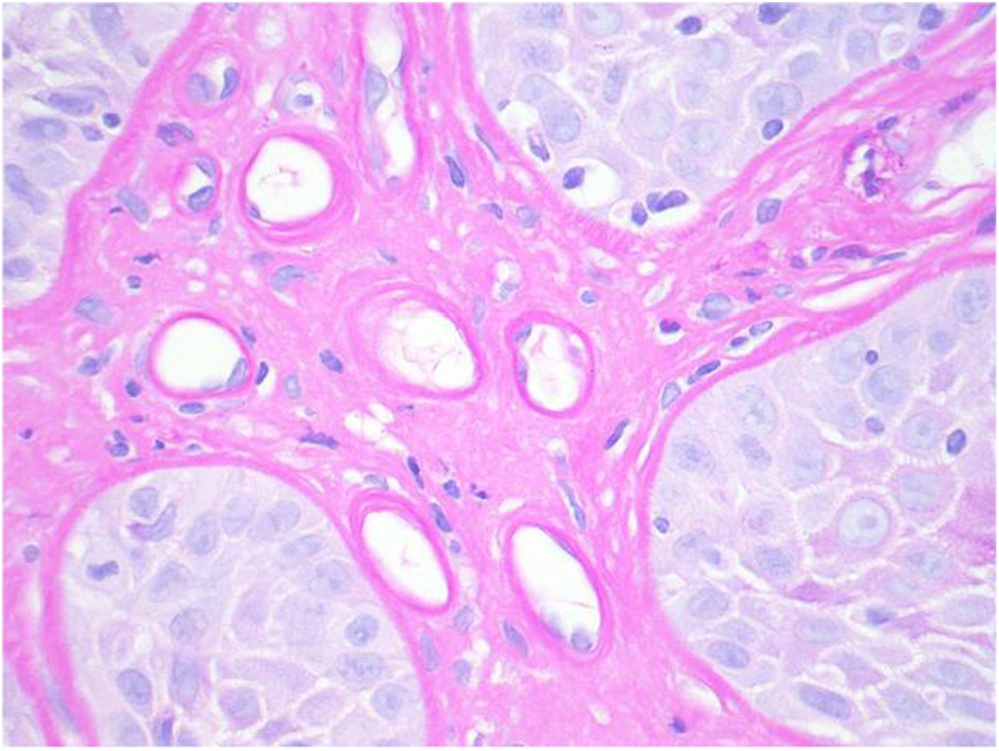
Computed tomography revealed a thickening and irregularity of the laryngeal mucosa. Microlaryngoscopy and biopsy performed under general anaesthesia confirmed severe infiltration of the supraglottis, extending from the petiole of the epiglottis, involving the aryepiglottic folds and false vocal folds. The subglottis and trachea were clear of any disease. Biopsies were taken from the supraglottis. Histopathological analysis included periodic acid-Schiff special stain. This revealed mucosa with characteristic stromal and perivascular hyalinisation (Figure 1), associated with acutely inflamed hyperplastic squamous epithelium and candida type organisms. There was no evidence of dysplasia or malignancy.
Histology in case one showed the characteristic bright pink stromal and perivascular hyalinisation associated with lipoid proteinosis. (Periodic acid-Schiff; ×100)

Diagnosis
Diagnosis is based on biopsy. The histology findings were reported as consistent with lipoid proteinosis, with superimposed acute inflammation and candida infection.
Management
The patient was initially treated with oral antifungal medication and inhaler advice (using a spacer and mouthwash after using a steroid inhaler for her asthma) to manage the co-existing fungal infection. In light of the ongoing dysphonia and some airway restriction, she consented to undergo debulking of the supraglottic disease under general anaesthesia.
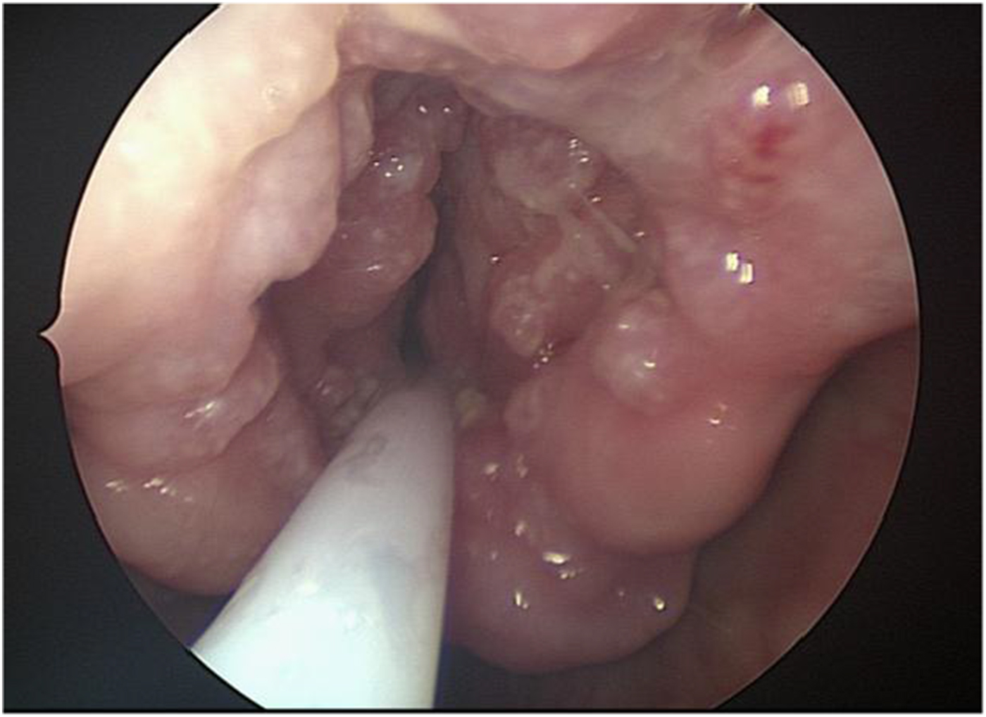
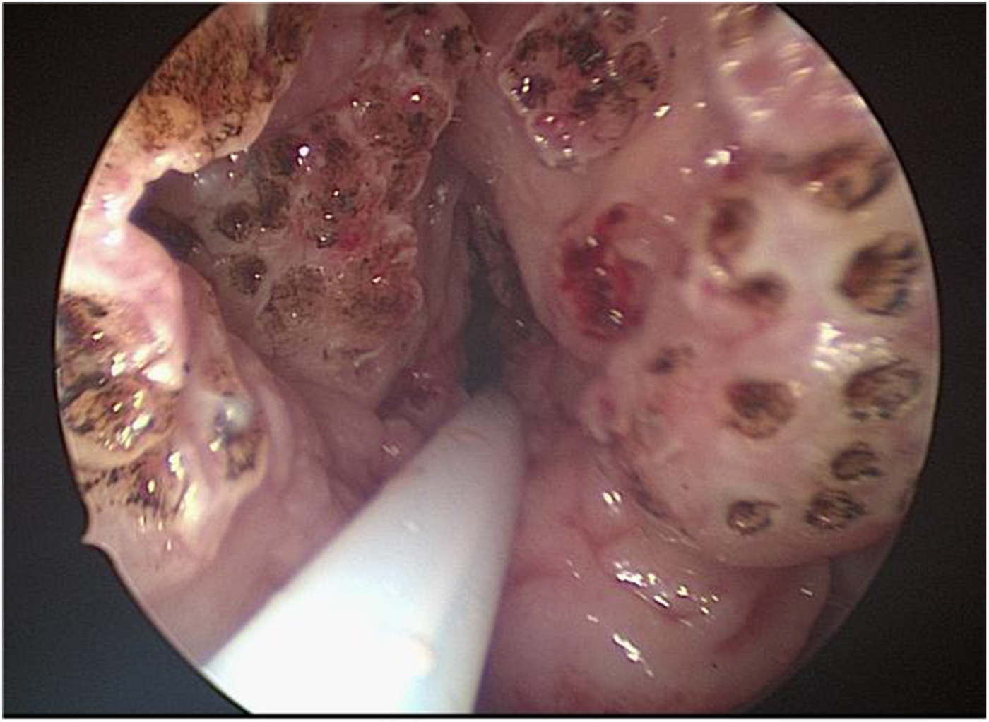
She underwent suspension microlaryngoscopy and ventilation via a subglottic jet catheter to enable unobstructed access for surgery. The supraglottic lesions were identified (Figure 2) and a biopsy was taken. Laser debulking to the supraglottis was performed using a carbon dioxide (CO2) laser at settings between 4 and 8 W (super-pulse, continuous wave laser setting). Large exophytic disease components were excised flush and the remainder of the disease mass was reduced using the ‘pepper pot’ technique. This involved creating multiple laser spots throughout, to a depth of 4–5 mm, at intervals of approximately 2 mm, thereby giving the ‘pepper pot’ appearance (Figure 3). This technique (preferred by author GSS) prevents airway stenosis by leaving bridges of mucosa.Reference Butler, Nouraei, Mace, Khalil, Sandhu and Sandhu12 The procedure was performed as day-case surgery. Repeat biopsies confirmed the diagnosis of lipoid proteinosis. The same surgical procedure has been performed on three separate occasions to control her symptoms.
Intra-operative endoscopic view of the larynx in case one.

Endoscopic view of the larynx in case one after laser debulking using a ‘pepper pot’ technique.

The patient has been followed up for 24 months since the initial surgery. She has reported an improvement in her voice and breathing at follow up. On nasoendoscopy in clinic, there remains some mucosal disease, but the airway is improved, with no evidence of stenosis.
Case two
A 56-year-old gentleman was referred with dysphonia. He had suffered voice problems since birth, which initially manifested as a hoarse cry. The patient had previously undergone two laryngeal procedures at another hospital, but felt no improvement in symptoms. This had been performed 6 years ago following a 12-month history of rhinorrhoea, increased phlegm, worsening hoarseness and weakness of voice.
On examination, the patient had moniliform blepharosis (beaded eyelid papules), and deposits infiltrating the palate, uvula and epiglottis. On laryngoscopy in clinic, there was evidence of extensively irregular and infiltrated vocal folds.
Investigations
A microlaryngoscopy and biopsy were performed under general anaesthesia. Biopsy samples were taken from the uvula, epiglottis, posterior oropharyngeal wall, left vocal fold, nasal sinus and left ear.
Diagnosis
The histopathological diagnosis based on samples from the larynx, oropharynx and skin was consistent with lipoid proteinosis.
Management
The patient underwent multiple microlaryngoscopies and laser debulking over an 18-month period. The ‘pepper pot’ technique was performed as described in the previous case.
When clinically stable, he experienced improvements in voice and airway patency. He was followed up regularly in the out-patient clinic, with no evidence of supraglottic stenosis for 12 months.
Discussion
The current literature on lipoid proteinosis relates predominantly to the dermatological manifestations of the disease. However, it has been reported that up to two-thirds of patients have dysphonia, a direct consequence of hyaline deposition of the vocal fold mucous membranes.Reference Savage, Crockett and McCabe7 At birth, this may present as a feeble cry; otherwise, dysphonia develops within the first few years of life. Thus, lipoid proteinosis is of particular relevance to the otolaryngologist, and should be considered a differential diagnosis, especially in infants and children with voice changes and dysphonia. Indeed, both our patients presented with dysphonia, with our male patient reporting a history dating to infancy, as outlined in a previous case report.Reference Virk, Tripathi, Sandison and Sandhu13
Although rarely a life-threatening condition, in very severe cases airway obstruction may culminate.Reference Hamada14 Indeed, diffuse deposition of the larynx and pharynx could result in respiratory distress, necessitating a tracheostomy. Infiltrates can form on the epiglottis, uvula and tonsils.Reference Caro15 Oral mucosal involvement is signified by the presence of yellowish pearly papular deposits. Thickening of the frenulum of the tongue limits movements, causing speech and deglutition difficulties.Reference Di Giandomenico, Masi, Cassandrini, El-Hachem, DeVito and Bruno1 Patients may have thickened lips and gingival hypertrophy. The salivary glands, particularly the submandibular and parotid gland, are frequently involved. This can lead to xerostomia and poor oral hygiene, as well as parotitis and parotid fistula.Reference Gutte, Sanghvi, Tamhankar and Khopkar8, Reference Kabre, Rani, Pai and Kamra16
In both cases presented here, confirmation of the diagnosis was achieved through histopathological analysis of laryngeal biopsy samples. Periodic acid-Schiff staining best demonstrated the characteristic deposition of perivascular eosinophilic material on biopsy.
Lipoid proteinosis is a multisystemic disorder with no known cure.Reference Kabre, Rani, Pai and Kamra16 Despite this, the prognosis is generally favourable and most patients have a normal life expectancy. Indeed, the disease follows a benign course. However, it is chronic and often progressive, and thus has the potential to significantly impact psychosocial wellbeing and quality of life.Reference Kabre, Rani, Pai and Kamra16
There is no ‘gold standard’ method of treatment for laryngeal lipoid proteinosis. Harper et al. demonstrated an improvement in phonation with microlaryngoscopy and dissection in 1983.Reference Harper, Duguid, Staughton and Moffat17 Since then, Xu et al. reported varying degrees of voice improvement in eight severely dysphonic patients who underwent microlaryngoscopic excision of deposits in the vocal folds.Reference Xu, Wang, Zhang, Han and Zhang18 It has been proposed that voice therapy alone is insufficient, as infiltration results in a hampered mucosal wave and vibratory amplitude, in addition to incomplete glottic closure.Reference Chen, Calawerts, Zhang, Jiang and Xu19
• Lipoid proteinosis is a rare autosomal recessive disorder
• It is characterised by deposition of hyaline in the skin and mucous membranes, resulting in a variety of symptoms
• Although there is no known cure, the overall prognosis is good
• This paper demonstrates how laryngeal symptoms can effectively be controlled with laser debulking using the ‘pepper pot’ technique
We have demonstrated that laryngeal symptoms can be effectively controlled with the judicious use of laser debulking. Whilst there are numerous open techniques for surgical debulking including laryngofissure procedures, as well as laryngectomy, they are associated with significant disability and morbidity.Reference Butler, Nouraei, Mace, Khalil, Sandhu and Sandhu12, Reference Chen, Calawerts, Zhang, Jiang and Xu19, Reference Neel and McDonald20, Reference Haar, Chaudhry, Kaplan and Milley21 Endolaryngeal surgery with microlaryngoscopy and CO2 laser ablation is less invasive, and has a reduced impact on voice and swallowing function when compared to open procedures.Reference Butler, Nouraei, Mace, Khalil, Sandhu and Sandhu12 There is already evidence to support the role of endolaryngeal surgery in establishing an adequate airway, and removing disease for both malignant and benign conditions.Reference Ruff and Bellens22, Reference Davis, Shapshay, Strong and Hyams23 However, there are risks of scarring and stenosis of the airway when using the CO2 laser in the supraglottis.Reference Ossoff24 In chronic conditions, such as sarcoidosis, debulking of supraglottis using the ‘pepper pot’ technique has been shown to help keep the airway patent, without causing long-term scarring or stenosis.Reference Butler, Nouraei, Mace, Khalil, Sandhu and Sandhu12 This technique spares mucosa and prevents stenosis, whilst controlling the disease, thereby preventing the need for open surgery or tracheostomy.
Competing interests
None declared