Introduction
Studies of adults (Sachs et al. Reference Sachs, Baldassano, Truman and Guille2000) and children (Biederman et al. Reference Biederman, Faraone, Wozniak and Monuteaux2000a, Reference Biederman, Mick, Faraone, Spencer, Wilens and Wozniakb, Reference Biederman, Mick, Wozniak, Monuteaux, Galdo and Faraone2003) with attention deficit hyperactivity disorder (ADHD) and bipolar disorder (BPD) have documented a substantial and bidirectional overlap between these disorders. Adults with BPD and co-morbid ADHD have an early age of BPD onset in childhood and adolescence (Sachs et al. Reference Sachs, Baldassano, Truman and Guille2000). Recent family studies documented co-segregation between BPD and ADHD, suggesting that BPD co-morbid with ADHD might be an early-onset, familially distinct subtype of BPD (Faraone et al. Reference Faraone, Biederman, Mennin, Wozniak and Spencer1997, Reference Faraone, Biederman, Mennin and Russell1998, Reference Faraone, Biederman and Monuteaux2001, Reference Faraone, Glatt and Tsuang2003; Biederman et al. Reference Biederman, Mick, Faraone, Spencer, Wilens and Wozniak2000b). Although these findings suggest that the co-morbidity between ADHD and BPD can help to identify more homogeneous subtypes with distinct neurobiological substrates, previous neuroimaging morphometric studies have not examined the impact of this co-morbidity.
Because BPD is primarily characterized by affective dysregulation whereas ADHD is primarily characterized by attention and executive function deficits, it is reasonable to hypothesize that these disorders contribute different neuroanatomical substrates to the co-morbid subgroup. Morphometric research on BPD has produced inconsistent results (McDonald et al. Reference McDonald, Zanelli, Rabe-Hesketh, Ellison-Wright, Sham, Kalidindi, Murray and Kennedy2004; Brambilla et al. Reference Brambilla, Glahn, Balestrieri and Soares2005; Strakowski et al. Reference Strakowski, Delbello and Adler2005) but there are strong theoretical reasons to hypothesize that dysfunctions in BPD occur in the prefrontal-limbic circuitry underlying affective regulation, such as the amygdala, orbital prefrontal cortex and thalamus (Soares et al. Reference Soares and Mann1997). Lesions in these areas can produce a disinhibition syndrome, with right-sided lesions producing classic manic symptoms (Soares et al. Reference Soares and Mann1997). Although a meta-analysis showed inconsistencies in the morphometric magnetic resonance imaging (MRI) literature on BPD (McDonald et al. Reference McDonald, Zanelli, Rabe-Hesketh, Ellison-Wright, Sham, Kalidindi, Murray and Kennedy2004), individual MRI studies of adults with BPD have shown a range of significant brain anatomic abnormalities consistent with this hypothesis, such as subcortical regions of interest (ROIs) in the amygdala, subgenual prefrontal cortex, thalamus, striatum, white matter and periventricular region, as well as in the ventricles, and cerebellum (Brambilla et al. Reference Brambilla, Glahn, Balestrieri and Soares2005; Strakowski et al. Reference Strakowski, Delbello and Adler2005; Adler et al. Reference Adler, Delbello, Jarvis, Levine, Adams and Strakowski2007; Rosso et al. Reference Rosso, Killgore, Cintron, Gruber, Tohen and Yurgelun-Todd2007). Functional imaging studies have also identified altered brain activation in these regions (Blumberg et al. Reference Blumberg, Stern, Ricketts, Martinez, de Asis, White, Epstein, Isenberg, McBride, Kemperman, Emmerich, Dhawan, Eidelberg, Kocsis and Silbersweig1999; Elliott et al. Reference Elliott, Ogilvie, Rubinsztein, Calderon, Dolan and Sahakian2004; Altshuler et al. Reference Altshuler, Bookheimer, Proenza, Townsend, Sabb, Firestine, Bartzokis, Mintz, Mazziotta and Cohen2005a, Reference Altshuler, Bookheimer, Townsend, Proenza, Eisenberger, Sabb, Mintz and Cohenb).
By contrast, morphometric studies of ADHD have produced a different and more coherent picture (Castellanos et al. Reference Castellanos, Lee, Sharp, Jeffries, Greenstein, Clasen, Blumenthal, James, Ebens, Walter, Zijdenbos, Evans, Giedd and Rapoport2002). In children with ADHD, structural differences have tended to be associated with the cortical attentional network, especially in the frontal lobe as a whole, the dorsolateral prefrontal cortex (DLPFC), and the splenium of the corpus callosum, caudate, and the cerebellum (Sowell et al. Reference Sowell, Thompson, Welcome, Henkenius, Toga and Peterson2003; Seidman et al. Reference Seidman, Valera and Makris2005; Valera et al. Reference Valera, Faraone, Murray and Seidman2007). Additionally, in the largest MRI study of adults with ADHD to date, we found significantly smaller overall cortical gray matter, DLPFC and anterior cingulate cortex (ACC) volumes associated with ADHD (Seidman et al. Reference Seidman, Valera, Makris, Monuteaux, Boriel, Kelkar, Kennedy, Caviness, Bush, Aleardi, Faraone and Biederman2006). In subsequent analyses of the same sample, the neocortex as a whole and also specific components of the cortical attentional network, including the ACC, were significantly thinner than in controls (Makris et al. Reference Makris, Biederman, Valera, Bush, Kaiser, Kennedy, Caviness, Faraone and Seidman2007). Thus, the available data suggest that BPD and ADHD are associated with relatively distinct neuroanatomical substrates primarily reflecting subcortical (‘limbic’) regions in BPD and cortical (‘cognitive’) regions in ADHD.
The main aim of the present study was to evaluate the morphometric MRI underpinnings of the co-morbidity between ADHD and BPD in adults. Based on the available literature and theoretical considerations, we hypothesized that the co-morbid condition would exhibit a combination of abnormalities affecting the amygdala, thalamus and orbital prefrontal cortex associated with BPD (Adler et al. Reference Adler, Delbello, Jarvis, Levine, Adams and Strakowski2007; Rosso et al. Reference Rosso, Killgore, Cintron, Gruber, Tohen and Yurgelun-Todd2007), and abnormalities affecting the DLPFC and ACC, overall neocortex, as well as the cerebellum (Castellanos et al. Reference Castellanos, Lee, Sharp, Jeffries, Greenstein, Clasen, Blumenthal, James, Ebens, Walter, Zijdenbos, Evans, Giedd and Rapoport2002), associated with ADHD. To our knowledge, this strategy to address heterogeneity within ADHD and BPD by examining cases with and without co-morbidity with the other disorder has not been used in either ADHD or BPD imaging research before.
Method
Subjects
Subjects (ages 18–59) were stratified into four groups based on diagnostic status: BPD+ADHD (n=31), BPD only (n=18), ADHD only (n=26) and controls with neither ADHD nor BPD (n=23). These samples were drawn from two ongoing brain imaging studies with identical brain acquisitions and measurements carried out at Massachusetts General Hospital (MGH). Data from 43% of the participants in this study (24 with ADHD and 18 controls) without BPD have been reported in other papers (Seidman et al. Reference Seidman, Valera, Makris, Monuteaux, Boriel, Kelkar, Kennedy, Caviness, Bush, Aleardi, Faraone and Biederman2006; Makris et al. Reference Makris, Biederman, Valera, Bush, Kaiser, Kennedy, Caviness, Faraone and Seidman2007) focusing on the neuroanatomy of adults with ADHD in a relatively non-co-morbid sample. Thus, this report substantially extends the previous study by 31 participants with ADHD+BPD, 18 persons with BPD, two persons with ADHD (without BPD), and five controls. Exclusion criteria were deafness, blindness, neurological disorders, sensorimotor handicaps, inadequate command of the English language, or a full-scale IQ estimate less than 75 as measured by the Wechsler Adult Intelligence Scale-Revised (WAIS-R; Wechsler, Reference Wechsler1981). No ethnic or racial group was excluded. We recruited ADHD probands through referrals to psychiatric clinics at the MGH and advertisements in the greater Boston area. We recruited potential non-ADHD probands through advertisements in the greater Boston area. ADHD adults were included if they met full criteria for current ADHD according to the DSM-IV, with childhood onset and persistence into adulthood. A subset of the bipolar probands was recruited through a follow-up to a birth cohort study (Buka et al. Reference Buka, Goldstein, Seidman, Zornberg, Donatelli, Denny and Tsuang1999). Written informed consent was obtained from all subjects after a complete description of the study, and all participants received an honorarium for participating. The MGH human subjects committee approved this study.
Clinical assessment measures
Trained interviewers, blind to ascertainment status, interviewed subjects with the Structured Clinical Interview for DSM-IV (First et al. Reference First, Spitzer, Gibbon and Williams1997) and modules from the Kiddie Schedule of Affective Disorders and Schizophrenia – Epidemiological Version (SADS-E; Orvaschel, Reference Orvaschel1994). The structured interview data were reviewed by a diagnostic committee so that a best-estimate diagnosis could be made, as described by Leckman et al. (Reference Leckman, Sholomskas, Thompson, Belanger and Weissman1982). The diagnostic committee was blind to the subject's ascertainment group and all non-diagnostic data. We computed κ coefficients by having board-certified child and adult psychiatrists diagnose subjects from audio-taped interviews. Based on 500 assessments from interviews of children and adults, the median κ coefficient was 0.98. κ coefficients for ADHD and BPD were 0.88 and 0.95 respectively. We also estimated the reliability of the diagnostic review process by computing κ coefficients of agreement between clinician reviewers. The median reliability between individual clinicians and the review committee was 0.87.
We estimated IQ from the Block Design and Vocabulary subtests of the WAIS-R (Wechsler, Reference Wechsler1981).
MRI protocol
Whole-brain MR images were collected on a Siemens 1.5-Tesla scanner at the MGH Martinos Center. A sagittal localizer scan was performed for placement of slices, followed by a coronal T2-weighted sequence to rule out unexpected neuropathology. Two sagittal 3D MP-RAGE (T1-weighted, non-selective inversion-prepared spoiled gradient echo pulse) sequences were collected and averaged (TR/TE/T1/flip=2.73 s/3.39 ms/1.0 s/7, bandwidth=190 Hz/pixel, sampling matrix=256×192 pixels, field of view=256×256 mm, effective slice thickness=1.33 mm on a 170-mm slab of 128 partitions) and used for morphometric analyses conducted at the MGH Center for Morphometric Analysis (CMA). Individuals with ADHD (regardless of BPD diagnostic status) using psychostimulants were asked to refrain from medications for 24 hours prior to scanning (for the functional MRI protocol, which was collected at the same session). Other participants stayed on their medications.
Image analysis
Structural scans were transferred to the CMA and coded and catalogued for blind analysis. Imaging analysis was performed in workstations using Cardviews software (Caviness et al. Reference Caviness, Meyer, Makris and Kennedy1996b).
Gray and white matter whole-brain segmentation
Brain images were positionally normalized to overcome variations in head position by using a standard three-dimensional coordinate system on each scan. This procedure uses the midpoints of the decussations of the anterior and posterior commissure lines and the midsagittal plane at the level of the posterior commissure. This ‘self-referential’ system is based directly on the individual brain, and is not warped to a template atlas. The datasets were then segmented into gray, white and cerebrospinal fluid (CSF) tissue classes using a semi-automated intensity contour algorithm for external border definition and signal intensity histogram distributions for delineation of gray–white borders. This technique yields separate components of neocortex, subcortical gray nuclei, white matter, and ventricular system subdivisions that correspond to the natural tissue boundaries distinguished by signal intensities on T1-weighted images. ROIs yielded by this technique include total cerebrum, cortical gray matter, cortical white matter, lateral, third and fourth ventricles, caudate, putamen, pallidum, nucleus accumbens, hippocampus, amygdala, thalamus, brainstem, cerebellar cortex, and cerebellar central mass (including cerebellar white matter and deep nuclei). CSF segmentations included the lateral, third and fourth ventricles.
Parcellation of the neocortex
The neocortex, as defined by the gray–white matter segmentation procedure, was then divided into 48 parcellation units (PUs) per hemisphere, based on the system originally described by Rademacher (Reference Rademacher1992) and modified by Caviness et al. (Reference Caviness, Meyer, Makris and Kennedy1996b). This is a comprehensive system of neocortical subdivision designed to approximate architectonic and functional subdivisions, and based on specific anatomical landmarks present in all brains. Caviness, Kennedy and Makris developed these procedures, trained the technicians, and maintained quality control for the segmentation and parcellation of the data. The research team has substantial experience with these procedures with many other psychiatric disorders in comparable age groups.
Following parcellation, volumes were calculated for each PU by multiplying the area measurement of the PU on each slice by the slice thickness, followed by summing across all slices in which the PU appears. The PUs were combined into clusters based on lobar and connectivity data into cortical volumes of the frontal, parietal, temporal and occipital lobes. To avoid multiple testing, only ROIs hypothesized to be associated with the specific and combined disorders were examined.
Reliability of morphometric measures
In various inter-rater reliability studies carried out at the CMA over the past decade, intra-class correlation coefficients (ICCs) for brain ROIs relevant to this study averaged: total cerebrum, 0.96; neocortex or total cerebral gray matter, 0.86; thalamus, 0.89; cerebellum gray matter, 0.85; amygdala, 0.86; superior frontal gyrus, 0.89; middle frontal gyrus, 0.77; orbital prefrontal gyrus, 0.75; anterior cingulate, 0.84. ICCs for exploratory analyses were: pallidum, 0.80; caudate, 0.76; putamen, 0.73; accumbens, 0.76 (Caviness et al. Reference Caviness, Kennedy, Richelme, Rademacher and Filipek1996a; Goldstein et al. Reference Goldstein, Goodman, Seidman, Kennedy, Makris, Lee, Tourville, Caviness, Faraone and Tsuang1999; Seidman et al. Reference Seidman, Faraone, Goldstein, Goodman, Kremen, Toomey, Tourville, Kennedy, Makris, Caviness and Tsuang1999; Frazier et al. Reference Frazier, Breeze, Makris, Giuliano, Herbert, Seidman, Biederman, Hodge, Dieterich, Gerstein, Kennedy, Rauch, Cohen and Caviness2005a, Reference Frazier, Chiu, Breeze, Makris, Lange, Kennedy, Herbert, Bent, Koneru, Dieterich, Hodge, Rauch, Grant, Cohen, Seidman, Caviness and Biedermanb).
Data analyses
First, we compared the four groups on demographic, psychiatric and neurocognitive factors, using the χ2 test and one-way analysis of variance for categorical and dimensional variables respectively. To determine whether these variables confound our tests of hypotheses, we followed the guidelines of Weinberg (Reference Weinberg1993). We considered a variable as potentially confounded with the predictor variable of interest (i.e. diagnostic status) if the degree of association reached the 0.10 level of significance (we used this liberal level of significance to avoid missing any potential confounds). For variables meeting this criterion, we next determined if they were associated with the outcome variables at the 0.10 level of significance. Those also meeting this criterion were flagged as confounders and used as covariates in the analysis (see below).
We selected a limited subgroup of specific ROIs predicted a priori to be associated with ADHD and BPD. For ADHD we used ROIs found to be significantly different from controls in a non-co-morbid sample previously reported by our group (Seidman et al. Reference Seidman, Valera, Makris, Monuteaux, Boriel, Kelkar, Kennedy, Caviness, Bush, Aleardi, Faraone and Biederman2006), including DLPFC as reflected in superior frontal and middle frontal gyri, overall neocortex (minus the superior and middle frontal gyri so as not to double count these ROIs (F1 and F2 respectively; Caviness et al. Reference Caviness, Kennedy, Richelme, Rademacher and Filipek1996a), anterior cingulate cortex, and cerebellum cortex. For BPD we selected the amygdala, thalamus and orbital prefrontal cortex, based on theoretical considerations and previous findings in the literature. In excluding striatal areas from our proposed ROIs, our reasoning was the following: (a) our preliminary results (Seidman et al. Reference Seidman, Valera, Makris, Monuteaux, Boriel, Kelkar, Kennedy, Caviness, Bush, Aleardi, Faraone and Biederman2006) did not show volumetric differences in the striatal areas typically found in children with ADHD (e.g. caudate, pallidum); (b) the work of Castellanos et al. (Reference Castellanos, Lee, Sharp, Jeffries, Greenstein, Clasen, Blumenthal, James, Ebens, Walter, Zijdenbos, Evans, Giedd and Rapoport2002) suggested that the caudate may normalize by late adolescence; and (c) we chose not to hypothesize accumbens differences even though we found a trend to a larger accumbens in ADHD in previous work (Seidman et al. Reference Seidman, Valera, Makris, Monuteaux, Boriel, Kelkar, Kennedy, Caviness, Bush, Aleardi, Faraone and Biederman2006), because we do not have a specific hypothesis regarding enlargement of the accumbens. However, we conducted exploratory analyses on these structures.
Next, we conducted a multivariate test to determine whether ADHD and BPD status is associated with the eight a priori ROIs as a whole, adjusting for whole-brain volume, age and sex. If the multivariate omnibus test indicated a significant effect, we conducted post hoc tests of the individual ROIs. A detailed description of the profile analysis and post hoc testing are as follows.
The omnibus analysis was conducted using multivariate regression models, where several dependent variables can be jointly regressed on the same set of independent variables. The joint estimation allows for the testing of coefficients across equations. We used the eight ROIs as the dependent variables and the two diagnostic status indicator variables (i.e. ADHD and BPD), age, gender and whole-brain volume as the independent variables, using the full sample of 98 participants from the four groups. Each diagnostic status indicator variable tested the volumetric effect of the given diagnosis, adjusting for the other diagnosis and the covariates. Using a Wald test, the omnibus analysis examined the null hypothesis that the coefficients for the diagnostic status terms are simultaneously zero across all equations. If this test was significant, we have evidence that diagnostic status is associated with volumetric effects.
Then, we estimated the effect of diagnostic status on each of the eight a priori selected structures separately, again using the full sample of 98 participants. In each model, we estimated the outcomes as a function of ADHD status, BPD status, and their interaction. If the interaction term was significant, we estimated the pairwise comparisons between the four groups. If the interaction term was not significant, we removed it and re-ran the model with only the main effects because this more parsimonious model would produce more accurate estimates of model parameters. All models were adjusted for gender, age and either whole-brain volume (e.g. the sum of the volumes of the total cerebrum, the total cerebellum, the third and fourth ventricles, and the brain stem) or total cerebral volume, depending on the ROI. We present the adjusted coefficients for the ADHD term and the BPD term, interpreted as the volumetric change associated with the given diagnosis, adjusted for the other diagnosis and other covariates. To provide a measure of absolute volume for each structure, we also present the intercept from each model, interpreted as the estimated mean of the outcome when the values of all the covariates are zero. To make the intercept interpretable, we centered all covariates about their mean. Then, we reanalyzed each outcome with the additional adjustment of any confounding demographic variables.
As we found previously that the overall frontal cortex has a smaller volume in ADHD than controls (Seidman et al. Reference Seidman, Valera, Makris, Monuteaux, Boriel, Kelkar, Kennedy, Caviness, Bush, Aleardi, Faraone and Biederman2006), we also examined this structure, with the hypothesis that only the ADHD group would demonstrate overall frontal volume reductions. We also analyzed the other cortical lobar volumes. We considered these latter analyses as exploratory as we did not have specific hypotheses about their distinct volumetric association with ADHD or BPD. [Although the parietal cortex is certainly a plausible ROI for ADHD and there has been evidence of significantly reduced cortical thickness in parts of the ROI (Makris et al. Reference Makris, Biederman, Valera, Bush, Kaiser, Kennedy, Caviness, Faraone and Seidman2007), we did not find differences in our initial volumetric study (Table 3, Seidman et al. Reference Seidman, Valera, Makris, Monuteaux, Boriel, Kelkar, Kennedy, Caviness, Bush, Aleardi, Faraone and Biederman2006)]. The distinction in findings on the same sample probably reflects method differences, particularly the substantially larger ROIs used in volumetric approaches. Thus, a significant result in a small voxel-defined area may disappear in a larger volumetric approach.] All models were estimated using linear regression, and all tests were two-tailed. For a priori hypotheses, α was set at 0.05 and statistical trends were recognized at 0.10. For exploratory analyses, α was set at 0.01.
Results
Demographic characteristics and psychiatric co-morbidity
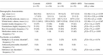
There were no significant differences among the groups in age, sex, handedness or whole-brain volume, as shown in Table 1. Current rates of antisocial personality disorder and multiple (⩾2) anxiety disorder were statistically comparable across groups. However, differences were found in the omnibus test at the 0.10 level for social class, race, medication status at the time of scanning, and lifetime history of substance use disorders. We also found a significant association between diagnostic status and IQ estimate [omnibus test: F(3, 93)=6.5, p<0.001]. We then assessed the association between each of these variables and the ROIs under study. We found associations at the 0.10 level for three regions in both the left and right hemisphere (a total of six associations): the middle frontal gyrus (IQ estimate), cerebellum cortex (IQ estimate), and amygdala (IQ estimate and race).
Demographic characteristics comparing ADHD v. Controls v. BBPD v. ADHD+BPD

ADHD, Attention deficit hyperactivity disorder; BPD, bipolar disorder; df, degrees of freedom; s.d., standard deviation; SES, socio-economic status.
a Social class as measured by the Hollingshead scale.
b Presence of disorder in the past month. Because of missing data, sample sizes for the BPD and ADHD+BPD groups are 8 and 26 respectively.
c Lifetime history of alcohol or drug abuse or dependence.
Omnibus multivariate tests
The multivariate test of the omnibus association between ADHD status and BPD status on the eight ROIs was statistically significant on both the left [F(16, 92)=2.28, p=0.007] and the right [F(16, 92)=1.78, p=0.046]. We therefore proceeded to estimate the effect of ADHD and BPD on each ROI individually.
Predicted effects of BPD and ADHD on brain volumes
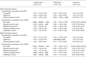
We tested the interaction between ADHD and BPD status in a series of models with each volume as the dependent variable. None of the interaction terms were significant (all p values >0.05). The main effects for the hypothesized ROIs are presented in Table 2.
Magnetic resonance imaging volumes in total sample of four groups of subjects (n=98)Footnote a, predicted by ADHD and BPD

ADHD, Attention deficit hyperactivity disorder; BPD, bipolar disorder; CI, confidence interval.
Bold text indicates statistical significance (p<0.05).
a Controls, n=23; ADHD, n=26; BPD, n=18; ADHD+BPD, n=31.
b Adjusted for bipolar status and structure-specific covariates (as indexed by Arabic numeral superscripts).
c Adjusted for ADHD status and structure-specific covariates (as indexed by Arabic numeral superscripts).
d Predicted volume given mean covariate values and the absence of both ADHD and BPD.
e Adjusted for age, sex and total cerebrum.
f Neocortex minus superior and middle frontal gyri, adjusted for age, sex and whole brain.
g Adjusted for age, sex and total cerebellum.
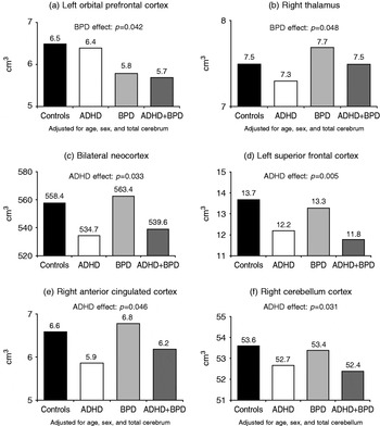
BPD adults had significantly smaller left orbital prefrontal cortex (adjusted mean difference of 10.8%) and significantly larger right thalamic volume (adjusted mean difference of 2.7%), independently of ADHD. Although the amygdala volumes in BPD were 8.7% smaller than in the controls, this difference was not significant. There were no other significant differences in lobar volumes for BPD.
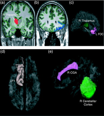
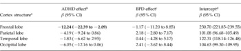
Adults with ADHD had significantly smaller overall left and right non-DLPFC neocortex (adjusted mean differences of 4.3% and 4.4% respectively), independently of BPD (see Table 2). Adults with ADHD also had significantly smaller right cerebellar gray matter volumes (adjusted mean difference of 1.8%) volumes, independently of BPD. In addition, adults with ADHD had significantly smaller left superior frontal cortex (adjusted mean volume difference of 11.0%), and right ACC volume (adjusted mean difference of 10.0%), independently of BPD (Fig. 1). Adults with ADHD also had significantly smaller overall frontal lobe volumes, independent of BPD status, age, sex and total cerebrum (adjusted mean difference of 5.3%; see Table 3).
Significant volumetric effects associated with bipolar disorder (BPD) and attention deficit hyperactivity disorder (ADHD). (a) 2D representation of the significant volumetric increases associated with BPD, depicted in red, in the thalamus. (b) 2D representation of the significant volumetric decreases associated with BPD, depicted in blue, in the frontal orbital cortex (FOC). (c) 3D representation of volumetric effects associated with BPD localized in the thalamus (green) and FOC (pink). (d) 3D representation of volumetric effects associated with ADHD in the superior frontal gyrus (F1). The letters and numbers in parentheses (e.g. ‘F1’) are codes used in the parcellation system (Caviness et al. Reference Caviness, Meyer, Makris and Kennedy1996b). (e) 3D representation of volumetric effects associated with ADHD in the cerebellar cortex, and anterior cingulate gyrus (CGA).

Magnetic resonance imaging neocortex (cortical gray matter) volumes in adults, predicted by ADHD and BPD

ADHD, Attention deficit hyperactivity disorder; BPD, bipolar disorder; CI, confidence interval.
Bold text indicates statistical significance (p<0.05).
a Adjusted for age, sex and total cerebrum.
b Adjusted for bipolar status and structure-specific covariates (as indexed by Arabic numeral superscript).
c Adjusted for ADHD status and structure-specific covariates (as indexed by Arabic numeral superscript).
d Predicted volume given mean covariate values and the absence of both ADHD and BPD.
We then reanalyzed any ROIs that: (1) exhibited significant associations with ADHD or BPD status as displayed in Table 2; and (2) were associated with potentially confounding demographic measures: only the right cerebellum cortex met both these criteria. With additional statistical adjustment for estimated IQ, ADHD status remained a statistically significant predictor of the right cerebellum cortex [β=−0.91, 95% confidence interval (CI) −1.80 to −0.01, p=0.048].
To further demonstrate the statistically significant effects of ADHD and BPD on the ROIs under study, we calculated adjusted mean volumes across the four diagnostic groups (controls, ADHD, BPD and ADHD+BPD; see Fig. 2: this figure is just for illustrative purposes, and does not signify that pairwise comparisons between the four groups were conducted; all analyses of ROIs were carried out using the model described above). As shown, ADHD and BPD are associated with separate alterations. In addition, the volumes of the co-morbid group are distinguished by the additive effects of each disorder.
Adjusted mean volumes for significant regions of interest (ROIs) across the four diagnostic groups.

Finally, in exploratory analyses, there were no significant differences at the p=0.05 level between any groups in volumes of the parietal cortex or of the striatal structures, including the caudate, pallidum, putamen or accumbens.
Discussion
Although prior morphometric studies have helped to clarify the neurobiological substrates of ADHD and BPD, there has been heterogeneity that remains unexplained. In the present work, we used a new strategy to address heterogeneity by examining cases with and without co-morbidity with the other disorder.
Our results showed that ADHD was selectively associated with smaller neocortical (including superior prefrontal cortex and ACC) volumes and cerebellar gray matter volume and BPD was associated with significantly larger thalamic volumes, and smaller left orbital prefrontal volumes, independently of the co-morbidity with the other disorder. The combined group manifested elements of each disorder's neuroanatomical deviations. This pattern of syndrome-congruent neuroanatomical findings suggests that ADHD and BPD contribute relatively selectively to brain volume alterations. To our knowledge, this is the first morphometric MRI study to address the effect of co-morbidity of ADHD and BPD.
Our findings are consistent with the literature on morphometric studies in children with ADHD that have implicated the cortical attentional network, especially DLPFC, the corpus callosum, caudate and the cerebellum (Seidman et al. Reference Seidman, Valera and Makris2005). Moreover, with this larger sample, we now show a significant volumetric reduction in cerebellar gray matter, which was not significant with a smaller sample (Seidman et al. Reference Seidman, Valera and Makris2006). The current results support the hypothesis that ADHD is associated with a neuroanatomical substrate implicating the networks subserving attention and executive functions.
Our hypothesis predicting an association between ADHD and the middle frontal gyrus was not supported by these data, although an ADHD effect was found for the superior frontal gyrus. While these results may suggest that our parcellation procedures did not correctly delineate the landmarks used to separate the superior and middle frontal gyri, this is highly unlikely because the landmark that defines the border between these gyri is the superior frontal sulcus, which is prominent and present in all humans. Although there is documented variability in its morphology, it is less than that of other sulci (Ono et al. Reference Ono, Kubik and Abarnathey1990), and highly trained technicians can reliably identify it as we documented in the methods section. Nevertheless, we recognize that the superior and middle frontal gyri are only proxies for DLPFC, as morphometric analyses are only estimates of underlying cytoarchitectonic features.
The reduced cerebellar gray matter in adults with ADHD is noteworthy because previous studies of children have demonstrated smaller cerebellar volumes. Clinical observations, experimental testing and neuroimaging studies have implicated the cerebellum in both cognitive (Schmahmann et al. Reference Schmahmann and Sherman1998; Cabeza et al. Reference Cabeza and Nyberg2000) and affective (Schmahmann et al. Reference Schmahmann and Sherman1998) behaviors. Furthermore, the identification of reciprocal cerebro-cerebellar connections provides an anatomical substrate for interactions between the cerebellum and prefrontal regions of the cortex (Middleton et al. Reference Middleton and Strick2001). Findings of reduced cerebellar volume in ADHD are therefore consistent with a hypothesis of an abnormal network contributing to cognitive deficits observed in ADHD (Valera et al. Reference Valera, Faraone, Biederman, Poldrack and Seidman2005).
By contrast, BPD was associated with abnormalities in brain regions involved in processing emotion including smaller orbital prefrontal cortex and larger thalamic volumes. Although our thalamic findings are consistent with some studies (Dupont et al. Reference Dupont, Jernigan, Heindel, Butters, Shafer, Wilson, Hesselink and Gillin1995; Strakowski et al. Reference Strakowski, DelBello, Sax, Zimmerman, Shear, Hawkins and Larson1999; Adler et al. Reference Adler, Delbello, Jarvis, Levine, Adams and Strakowski2007), the literature on thalamic volumes in BPD has been equivocal (Strakowski et al. Reference Strakowski, Delbello and Adler2005). These contradictory findings could be due to the neuroanatomical heterogeneity of the thalamus; only approximately 40% of the thalamus is involved with limbic networks and processing (Makris et al. Reference Makris, Meyer, Bates, Yeterian, Kennedy and Caviness1999). Thus, although our thalamic finding is consistent with theoretical models of BPD, they should be viewed as preliminary until confirmed in refined analyses of the limbic components of the thalamus (i.e. medial dorsal and anterior nuclei).
Our hypothesis regarding significant volume differences in the amygdala was not confirmed, but it was smaller by 8.7% in BPD than in control subjects. The literature shows large degrees of variability for the amygdala (McDonald et al. Reference McDonald, Zanelli, Rabe-Hesketh, Ellison-Wright, Sham, Kalidindi, Murray and Kennedy2004; Strakowski et al. Reference Strakowski, Delbello and Adler2005). These discrepancies could be due to the heterogeneity of BPD, including co-morbid ADHD and age of BPD onset, as well as differences in neuroimaging methodology. Moreover, while our inter-rater reliability estimate for the amygdala is adequate (0.84), this structure is difficult to measure and the modest reliability may also contribute to increased variability of the volumes.
Considering that ADHD is a relatively common co-morbidity in adults with BPD (Sachs et al. Reference Sachs, Baldassano, Truman and Guille2000), our findings may begin to explain some of the discrepant neuroimaging findings in adults with BPD. For example, in studies of adult BPD showing volumetric reductions in the frontal regions, including regions close to or inclusive of the DLPFC, co-morbid ADHD was not an exclusion (Sax et al. Reference Sax, Strakowski, Zimmerman, DelBello, Keck and Hawkins1999; Lopez-Larson et al. Reference Lopez-Larson, DelBello, Zimmerman, Schwiers and Strakowski2002). In fact, Sax and colleagues found significant associations between prefrontal volume and performance on the Continuous Performance Test, a test that is impaired in subjects with ADHD (Seidman, Reference Seidman2006). Therefore, co-morbid ADHD rather then the BPD itself could have accounted for reported volumetric reductions in some frontal regions. These findings stress the importance of attending to the co-morbidity with ADHD to disambiguate causes of structural heterogeneity in studies of bipolar subjects (McDonald et al. Reference McDonald, Zanelli, Rabe-Hesketh, Ellison-Wright, Sham, Kalidindi, Murray and Kennedy2004) and vice versa.
Our findings support the hypothesis that the co-occurrence of ADHD and BPD is a true co-morbidity and less likely to be a misdiagnosis of the other (Milberger et al. Reference Milberger, Biederman, Faraone, Murphy and Tsuang1995), and is consistent with family studies of ADHD and BPD (Faraone et al. Reference Faraone, Biederman, Mennin, Wozniak and Spencer1997, Reference Faraone, Biederman and Monuteaux2001), which found specificity in the familial aggregation of these disorders. This issue is of particular clinical relevance considering that BPD and ADHD require very different treatments.
Our findings should be viewed in light of some methodological limitations. Because our patient samples were referred, our results cannot be generalized to non-referred samples. Another limitation to generalizability is that our sample was skewed toward Caucasian participants and was not representative of lower socio-economic strata. The diagnoses of adult ADHD relied on self-report, and there is substantial evidence for the validity of such diagnoses (Shaffer, Reference Shaffer1994; Faraone et al. Reference Faraone, Biederman, Feighner and Monuteaux2000). Moreover, it is possible that different medications had effects on brain volumes. Studies of bipolar patients are particularly difficult to conduct in medication-naïve subjects as the seriousness of symptoms usually necessitates medication. Similarly, the effects of prior substance use disorders cannot be estimated in this sample, although the rate of lifetime substance abuse or dependence in our control group (30%) is consistent with nationally representative epidemiological data of the same outcome (26.6%) (Kessler et al. Reference Kessler, McGonagle, Zhao, Nelson, Hughes, Eshleman, Wittchen and Kendler1994). Although our parcellation procedures have demonstrated reliability, there are limitations inherent in the current technology of MRI that prevent more detailed examinations of neuroanatomy. Future studies will benefit from advances in MRI technology. Finally, although there were IQ differences between the groups, none of the significant effects in the ROIs could be accounted for by IQ. It should also be noted that the mean IQ of the ADHD group was somewhat higher that what might be expected, indicating that our sample may represent the upper end of the ADHD IQ distribution.
Despite these considerations, our findings suggest that ADHD co-morbid with BPD manifests elements of each disorder's neuroanatomical deviations. The syndrome-congruent findings identified suggest that ADHD and BPD may contribute selectively to brain volume alterations and support the hypothesis that individuals with both disorders are truly co-morbid.
Acknowledgments
National Alliance for Research on Schizophrenia and Depression (NARSAD) Distinguished Investigator Award, and Janssen Pharmaceuticals (J. B.); NIMH MH/HD 62152 and MH 63951, the Stanley Medical Research Institute; NARSAD, the March of Dimes Foundation, and the Mental Illness and Neuroscience Discovery (MIND) Institute (L. J. S.); National Research Service Award (NIMH F32 MH065040-01A1), Peter Livingston Fellowship through the Harvard Medical School Department of Psychiatry, and the Clinical Research Training Program Fellowship in Biological and Social Psychiatry MH-16259 (E. M. V.); MH-56956 (J. M. G.), the Stanley Medical Research Institute (S. B.); NIMH MH 57934 (S. V. F.), The National Center for Research Resources (P41RR14075).
We thank L. Barker, A. Brown, J. Burbridge, M. Cirillo, L. Denny, J.-A. Donatelli, C. Fetterer, J. Frazier, S. Hodge, K. Kelkar, M. Landi, E. Mick, N. Peace, A. Peters, S. Rauch, M. Schiller, J. Schlerf, T. Spencer, W. S. Stone and T. Wilens.
Declaration of Interest
J. B. has received research support from, has been a speaker for, or has been on the advisory board for: Shire, Eli Lilly, Pfizer, McNeil, Abbott, Bristol-Myers-Squibb, New River Pharmaceuticals, Cephalon, Janssen, Novartis, UCB Pharma, Astra-Zeneca, Forest Laboratories, Glaxo-SmithKline and Neurosearch. Other sources of research support are: the Stanley Medical Institute, Inc., the Lilly Foundation, and the Prechter Foundation. E. M. V. has been a speaker for Shire Pharmaceuticals. G. B. has received research support from, has been a speaker for, or has been on the advisory board for: Eli Lilly and Company, Pfizer, Inc., Shire U.S. Inc., Novartis Pharmaceuticals, Janssen Pharmaceuticals, McNeil Pharmaceuticals, and Johnson & Johnson. He has no financial interest in any of these entities. S. V. F. has received research support from, has been a speaker for, or has been on the advisory board for: Shire, Eli Lilly, McNeil, and Novartis. L. J. S has received research or educational support from, and has been a speaker in industry-sponsored symposia for: Shire, Eli Lilly, and Janssen.