INTRODCTION
Neospora caninum is an obligate intracellular protozoan parasite. The parasite has a worldwide distribution and causes neosporosis in a wide range of species including dogs, cattle, sheep, goats, horses and deer (Dubey and Lindsay, Reference Dubey and Lindsay1996; Dubey et al. Reference Dubey, Schares and Ortega-Mora2007). Neosporosis is one of the major causes of perinatal calf mortality and abortion in cattle and represents a serious threat to the dairy and beef industry, which can cause significant economic losses (Dubey, Reference Dubey1999). Considering the economic and agricultural impact of neosporosis, there is an urgent need to develop control measures to prevent its transmission and infection. Because no suitable treatment is available, effective vaccination is regarded as an economically promising option (Hasler et al. Reference Hasler, Regula, Stark, Sager, Gottstein and Reist2006a, Reference Hasler, Stark, Sager, Gottstein and Reistb).
Since N. caninum was first reported as a pathogenic parasite in animals (Dubey et al. Reference Dubey, Hanel, Lindsay and Topper1988), an increasing number of functional proteins have been identified, including proteins of microneme, rhoptry, and dense granule, as well as membrane proteins. The identified proteins are functional in different stages of the N. caninum life cycle, involved in adhesion, host-cell invasion, intracellular development, parasitophorous vacuole formation and/or acquiring nutrients from host cells. Some of the identified proteins have been investigated as vaccine candidates against neosporosis (Baszler et al. Reference Baszler, Shkap, Mwangi, Davies, Mathison and Mazuz2008; Ellis et al. Reference Ellis, Miller, Quinn, Ryce and Reichel2008; Debache et al. Reference Debache, Guionaud, Alaeddine, Mevissen and Hemphill2008; Nishikawa et al. Reference Nishikawa, Zhang, Ikehara, Kojima, Xuan and Yokoyama2009; Monney et al. Reference Monney, Debache and Hemphill2011), but not all proteins are good vaccine candidates.
A large number of functional proteins of N. caninum are still unidentified, and these unidentified proteins may contain some potential antigens for the development of effective vaccines to control N. caninum infection. Immune mapped protein 1 (IMP1) was a newly discovered protein in Eimeria maxima (Blake et al. Reference Blake, Billington, Copestake, Oakes and Quail2011), and was demonstrated to be immunogenic and conferred protection against E. maxima infection. Considering that IMP1 is a highly conserved gene of apicomplexan, it has the potential to be used as a novel antigen for other apicomplexan parasites. Although the IMP1 gene was also identified in the genome of N. caninum (NcLIV_000430) (Blake et al. Reference Blake, Billington, Copestake, Oakes and Quail2011), the gene has not been sequenced and extensively characterized, and its role is not completely understood. In this study, we reported the cloning, localization and initial characterization of the NcIMP1 gene of N. caninum Nc1 strain.
MATERIALS AND METHODS
Parasite culture and preparation
Neospora caninum tachyzoites (Nc1 strain) were maintained in African green monkey kidney (Vero) cells cultured in a nutrient Dulbecco's Modification of Eagle's Medium (DMEM) supplemented with L-glutamine, 8% heat-inactivated fetal bovine serum (FBS), penicillin (100 U/ml) and streptomycin (100 μg/ml) at 37°C in a 5% CO2 environment. Parasites were harvested and purified as described previously (Zhang et al. Reference Zhang, Compaore, Lee, Liao, Zhang and Xuan2007a). Tachyzoites were isolated by washing in cold phosphate-buffered saline (PBS), centrifugation, re-suspension in cold PBS, syringing 3 times through a 27-gauge needle, filtering through a 5·0 μm pore filter (Millipore, USA), washing twice with PBS, and finally centrifuging at 300 g for 10 min.
Cloning and sequencing of the NcIMP1 gene
Total RNA was extracted from Nc-1 tachyzoites (∼107) with the Trizol reagent (Invitrogen, USA). cDNA was synthesized using oligo-dT and a RevertAid First Strand cDNA synthesis kit (TransGen, China). The cDNA was used as the template for NcIMP1 cloning.
The NcIMP1 gene was amplified by PCR using the Phusion High-Fidelity DNA polymerase kit (NEB, USA) with IMP1-specific primers P1 and P2 (Table 1). The primers correspond to the respective ATG start site and TAG stop codon of the NCLIV_000430 gene sequence, which was predicted as the IMP1 gene of the NcLIV strain. The RT-PCR amplicon of the expected size of 1·2 kb was gel-recovered, cloned into the pEASY-Blunt cloning vector (TransGen, China). The sequence was determined by a commercial sequencing service (Invitrogen, Beijing).
Primers used in this study

| Name of primers (forward primer/reverse primer) | Forward primer sequence 5′−3′ | Reverse primer sequence 5′−3′ |
| P1/P2 | ATGGGAGGCGTTTGCTCTAAAAG | CTATTTCTTGCCGTCGAGGATGAG |
| P3/P4 | cgGGATCCATGGGAGGCGTTTGCTCTAAAAG | ccgCTCGAGTTTCTTGCCGTCGAGGATGAG |
| P5/P6 | gtcGATATCATGCATGGCGTTTGCTCTAAAAG | ccaATGCATTTTCTTGCCGTCGAGGATGAG |
| P5m/P6 | gtcGATATCATGGCAGGCGTTGCATCTAAAAG |
Sequence analysis
Sequence and structure analyses were performed using a series of bioinformatics tools. The NcIMP1 coding sequence was submitted to the Sanger database (http://www.Sanger.ac.uk) and Toxoplasma database (http://www.toxodb.org) for full cDNA sequence searching and structure analysis. Potential signal peptide cleavage sites were identified with SignalIP3.0 (http://www.cbs.dtu.dk/services/SignalP/) (Bendtsen et al. Reference Bendtsen, Nielsen, Von-Heijne and Brunak2004). The potential transmembrane region was predicted with TMHMM2.0 and the ProtScale tool on the Expasy server (http://expasy.org/tools/protscale.html). Functional protein motifs were identified using the Scan Prosite Tool (http://www.expasy.org/tools/scanprosite/) and online CSS-Palm predicted software (http://csspalm.biocuckoo.org/online3.php) (Zhou et al. Reference Zhou, Xue, Yao and Xu2006; Ren et al. Reference Ren, Wen, Gao, Jin, Xue and Yao2008).
Expression and identification of recombinant NcIMP1
To determine whether the putative NcIMP1 gene encoded a functional protein, the cDNA of NcIMP1 was amplified by PCR using primers P3 and P4 with introduced BamHI and XhoI sites (Table 1). The product was cloned into the prokaryotic expression vector pET-28a (Novagen, USA) and then transformed into E. coli for expressing. The recombinant NcIMP1 (rNcIMP1) fused to a (His)6-tag was expressed and purified using HisTrap FF purification columns (Novagen, USA). The purified protein was verified by SDS-PAGE and immune-blotting using sera from mice immunized with N. caninum Nc1 tachyzoites as previously described for Toxoplasma tachyzoites (Liao et al. Reference Liao, Xuan, Huang, Shirafuji, Fukumoto, Hirata and Fujisaki2005).
Production of anti-rNcIMP1 sera
BALB/c female mice (6 weeks old, Academy of Military Medical Sciences Animal Center, Beijing, China) were immunized subcutaneously with 100 μg of purified rNcIMP1 in an equal volume of Freund's complete adjuvant (Sigma, USA) for the first injection. The immunization was boosted twice with 50 μg of the antigen in Freund's incomplete adjuvant (Sigma, USA) at 2 and 4 weeks post-primary injection. Anti-rNcIMP1 sera were obtained 2 weeks after the last immunization and the antibody titre was assayed by ELISA as previously described (Zhang et al. Reference Zhang, Huang, Boldbaatara, Battura, Battsetsega and Zhang2010). Briefly, 96-well microtitre plates (Nunc, Denmark) were coated with rNcIMP1 (200 ng/well). Non-specific binding sites were blocked with 4% bovine serum albumin (BSA) in PBS. Mouse sera (1:100, diluted in PBS containing 3% skim milk) were added to the wells and incubated, followed by goat anti-mouse IgG-HRP (Proteintech Group Inc., Chicago, USA) conjugate as the secondary antibody. After incubation in a substrate solution, the absorbance was measured at 450 nm with an ELISA reader (Bio-TekEL_680, USA). All samples were run in triplicate. The anti-rNcIMP1 sera were sterilized by filtration through a 0·20 μm filter (Millipore, USA), and stored at −20°C.
Detection of native NcIMP1 protein by Western blotting
Neospora caninum tachyzoites were harvested and purified as described above. Purified Nc1 tachyzoites (1×10⁸) were re-suspended in 200 μl of cold PBS, lysed by 3 freeze-thaw cycles in liquid nitrogen, and then sonicated in an iced water bath. The protein concentration of the lysate was determined by a BCA protein assay kit (CoWin Biotech Co., Ltd, Beijing, China). The crude lysate was boiled in the SDS-PAGE loading buffer (Beyotime Institute of Biotechnology, China) for 10 min and then separated using a 12% (w/v) SDS-PAGE gel. Following electrophoresis, separated proteins were transferred onto polyvinylidene fluoride (PVDF) membranes (Millipore, USA). The PVDF membrane was blocked with 5% (w/v) skim milk diluted in PBS for 1 h at room temperature before incubation with mouse anti-rNcIMP1 serum (dilution 1:750). After washing in PBST, the PVDF membrane was incubated with 1:10 000 diluted goat anti-mouse IgG HRP-labelled secondary antibodies (Sigma, USA) for 1 h. Labelled NcIMP1 proteins were visualized with ECL chemiluminescence reagents (CoWin Biotech Co., Ltd., Beijing, China).
Localization of NcIMP1 in N. caninum tachyzoites by immunofluorescence analysis (IFA)
Extracellular and intracellular tachyzoites grown in Vero cells were analysed by IFA as previously described for Toxoplasma tachyzoites (Liao et al. Reference Liao, Xuan, Huang, Shirafuji, Fukumoto, Hirata and Fujisaki2005). For extracellular tachyzoites, 5×10⁴ were mounted on slides, dried and fixed with methanol for 20 min at −20°C. Intracellular tachyzoites were obtained by cultivation of tachyzoites in Vero cells attached to cover-slips at 37°C with 5% CO2 for 24–48 h. The culture was washed 3 times in PBS, fixed with methanol for 20 min at −20°C, and then treated with 0·2% Triton-100 in PBS for 5 min to permeabilize the plasma membrane of both Vero cells and parasites. Extracellular and intracellular preparations were blocked with PBS-BSA 1% for 30 min at room temperature, before incubation with mouse anti-rNcIMP1 serum as the primary antibody (1:75 in PBS-BSA 1%), and fluorescein isothiocyanate (FITC)-conjugated anti-mouse IgG (Proteintech, USA) diluted at 1:100 in PBS-BSA 1% as the secondary antibody. Finally, the parasites were observed under a fluoresence microscope IX71 (Olympus, Japan).
Localization of NcIMP1 protein through mutation of glycine-2 and cystenine-5
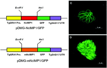
The full ORF sequence of NcIMP1 was amplified by PCR using primers P5 and P6 with introduced EcoRV and NsiI sites. The product was inserted into the plasmid pDMG (kindly provided by Professor Xuenan Xuan, National Research Center for Protozoan Diseases, Obihiro University of Agriculture and Veterinary Medicine, Inada-cho, Obihiro, Hokkaido, Japan) between the EcoRV and NsiI sites. The resulting plasmid as a localization transfer vector was designated as pDMG-NcIMP1/GFP. The stop codon of the NcIMP1 gene was excised before NcIMP1 was fused with the reporter gene GFP. The fused NcIMP1-GFP gene is under the control of T. gondii GRA1 promoter.
In order to investigate whether the localization of NcIMP1 is determined by the N-myristorylation site (glycine-2) and palmitoylation site (cystenine-5), the glycine-2 and cystenine-5 of NcIMP1 were both replaced with alanine by site mutation PCR with the primers P5 m (site mutation primer) and P6. The mutant PCR product was inserted into the pDMG plasmid between the EcoRV and NsiI sites. The mutant plasmid was designated as pDMG-mNcIMP1/GFP.
The recombinant plasmids were transfected to N. caninum tachyzoites by electroporation as described previously (Sibley et al. Reference Sibley, Messina and Niesman1994). After electroporation, the parasites were allowed to recover for 20 min at room temperature and then inoculated into Vero cells. The transfected parasites were examined using a laser confocal scanning microscope (Leica TCS SP5 II, Germany). The pictures were analysed by software Leica LAS AF.
In vitro host cell invasion assay
To investigate the role of NcIMP1 in host cell invasion by N. caninum tachyzoites, an in vitro invasion assay was performed as described previously (Zhang et al. Reference Zhang, Compaore, Lee, Liao, Zhang and Xuan2007a). The tachyzoites (2×10⁵) expressing GFP, made by Yoshifumi Nishikawa (kindly provided by Professor Xuenan Xuan, Japan), were suspended in 500 μl of DMEM containing mouse anti-rNcIMP1 antibodies diluted to 1:500, 1:250, 1:100, 1:50 and 1:25, and incubated for 1 h at 37°C/5% CO2 before infection of Vero cells. Untreated parasites were used as controls. Thereafter, all the treated and untreated parasites were added to the monolayers of Vero cells and incubated for 1 h at 37°C/5% CO2. Unbound parasites were removed by rinsing with DMEM. After another 30 h of culture, the percentage of fluorescent parasite-infected cells was determined using a flow cytometry (BD Accuri C6, USA), and 5×10³ cells were counted in each well sample. The percentage of invaded tachyzoites was calculated by the following formula: [1−the percentage of fluorescent parasites in the tested IgG treatment wells/the percentage of fluorescent parasites in control wells]×100%, which was described by Zhang et al. (Reference Zhang, Lee, Liao, Compaore, Zhang and Kawase2007b).
RESULTS
Cloning and sequence analysis of the NcIMP1 gene
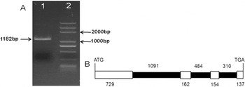
RNA was extracted from purified Nc1 strain tachyzoites and amplified by RT-PCR to obtain the open reading frame (ORF) of the IMP1 gene. A 1182 bp PCR product was amplified (Fig. 1A) and found to encode a protein of 393 amino acids. The sequence has been deposited in the GenBank database (Accession number JN651092). Analysis results of the Sanger database and toxoDB revealed that the full-length DNA sequence of NcIMP1 was 3067 bp, and the gene was interrupted with 3 introns (Fig. 1B).
(A) Gel electrophoresis of a 1182 bp amplification product of NcIMP1 from Nc1 strain tachyzoites (Lane 1) and molecular weight markers (Lane 2). (B) Schematic view of the NcIMP1 gene. White boxes indicate exons, and black bold lines indicate introns. The numbers indicate the number of nucleotides.

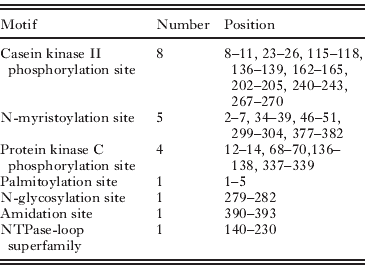
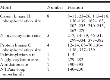
Analysis of the IMP1 sequence on identification/prediction platforms including SignalP3.0, TMHHM2.0 and online databases, showed the absence of a classical signal peptide or transmembrane region. Several kinds of functional protein motifs were identified, including phosphorylation sites, N-myristoylation sites, a palmitoylation site, a glycosylation site and an amidation site, as well as a p-loop NTPase superfamily (Table 2).
Motifs predicted of NcIMP1
| Motif | Number | Position |
| Casein kinase II phosphorylation site | 8 | 8–11, 23–26, 115–118, 136–139, 162–165, 202–205, 240–243, 267–270 |
| N-myristoylation site | 5 | 2–7, 34–39, 46–51, 299–304, 377–382 |
| Protein kinase C phosphorylation site | 4 | 12–14, 68–70,136–138, 337–339 |
| Palmitoylation site | 1 | 1–5 |
| N-glycosylation site | 1 | 279–282 |
| Amidation site | 1 | 390–393 |
| NTPase-loop superfamily | 1 | 140–230 |
Identification of NcIMP1 protein in N. caninum tachyzoites
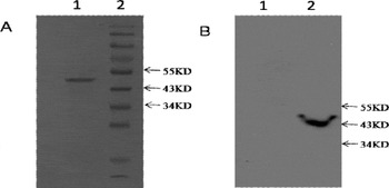
The recombinant NcIMP1 protein, comprising aa 1–393, was expressed as a (His)6-tagged fusion protein in E. coli and purified by Ni2+-affinity chromatography. The purified rNcIMP1 had a molecular weight of approximately 50 kD determined by SDS-PAGE (Fig. 2A), and was recognized by mouse serum raised against Nc1 tachyzoites. The specific anti-rNcIMP1 sera were obtained by immunizing mice with the recombinant protein according to a common procedure, and had a high titre (3·28×106) against rNcIMP1 as determined by ELISA. The specific sera were then tested by Western blotting against N. caninum tachyzoite lysates in which a ∼43 kDa protein was elicited (Fig. 2B). No parasite proteins were recognized by pre-immune mouse sera.
(A) SDS-PAGE of the recombinant NcIMP1, approximately 50 KD (Lane 1); molecular weight markers (Lane 2). (B) Native NcIMP1 was confirmed by Western blotting. No binding to pre-immune mouse serum as the primary antibody control (Lane 1); positive staining with mouse NcIMP1-specific antiserum as the primary antibody (Lane 2).

Localization of NcIMP1
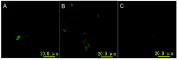
To determine the location of NcIMP1 on parasites, intracellular and extracellular parasites were labelled with mouse anti-rNcIMP1 serum and FITC-conjugated goat anti-mouse IgG. Green fluorescence was observed on the membrane of both intracellular and extracellular tachyzoites (Fig. 3). No fluorescence staining was observed in the culture incubated with serum from pre-immune mice. Our findings indicate that NcIMP1 is a membrane protein.
Location of NcIMP1 in Neospora caninum tachyzoites by IFAT. Immunofluorescence microscopy of extracellular (A) and intracellular (B) tachyzoites stained with mouse anti-rNcIMP1 antibodies and FITC-conjugated goat anti-mouse IgG, and tachyzoites stained with pre-immune mouse sera (C).

To further verify the localization of NcIMP1 in N. caninum tachyzoites, 2 plasmids, pDMG-NcIMP1/GFP (Fig. 4A) and pDMG-mNcIMP1/GFP (Fig. 4B), were constructed and transfected into N. caninum tachyzoites. pDMG-mNcIMP1/GFP was a plasmid with a site mutation at glycine-2 of the predicted N-myristoylation site GGVCSK and cysteine-5 of a predicted palmitoylation site MGGVC. In pDMG-NcIMP1/GFP transfected parasites, green fluorescence was observed on the membrane of the tachyzoites (Fig. 4C). In contrast, green fluorescence was dispersed throughout the parasites transfected with the mutated plasmid pDMG-mNcIMP1/GFP (Fig. 4D).
(A) Plasmid map of pDMG-NcIMP1/GFP. (B) Plasmid map of pDMG-mNcIMP1/pGFP (mNcIMP1 represents NcIMP1 with glycine-2 and cysteine-5 mutated). (C) Neospora caninum tachyzoites transfected with plasmid pDMG-NcIMP1/GFP. (D) N. caninum tachyzoites transfected with mutated plasmid pDMG-mNcIMP1/GFP.

Anti-NcIMP1 antibodies inhibited host-cell invasion by N. caninum
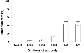
Treatment of N. caninum tachyzoites with antibodies against rNcIMP1 diluted to 1:25 or 1:50 reduced Vero cell invasion by 44±3·77% and 43±2·62% (P<0·05), respectively, compared with the untreated controls. Higher dilutions of the antibodies had no significant effect on cell invasion (Fig. 5).
In vitro inhibition of host-cell invasion. The value for the inhibition rate of Neospora caninum tachyzoites in the absence of antibodies was considered to be 0%. Statistically significant differences (P<0·05) are indicated by (**).

DISCUSSION
In this study, a highly conserved gene in N. caninum Nc1 strain, named NcIMP1, was identified and characterized by cloning, sequence analysis, expression, localization and initial functional investigation. The NcIMP1 gene was found to have an open reading frame of 1182 bp, encoding a 393 amino acid peptide. Sequence analysis suggested that NcIMP1 was a gene interrupted by 3 introns, similar to the exon/intron structure of EmIMP1. SignalIP3.0 and THMMH2.0 analyses revealed that neither a classical signal peptide nor a transmembrane region was present within the NcIMP1 sequence, and these findings were consistent with sequence structure of EmIMP1 (Blake et al. Reference Blake, Billington, Copestake, Oakes and Quail2011).
It is noteworthy that NcIMP1 has no predicted signal peptide but, interestingly, the protein was found to localize to the membrane of N. caninum tachyzoites. Previous work identified that N-myristoylation is a common form of co-translational modification of N-terminal glycine residues, and plays a vital role in protein membrane targeting and signal transduction in plants responding to environmental stress. This function of N-myristoylation was also found to exist in animals, fungi and viruses (Zha et al. Reference Zha, Weiler, Oh, Wei and Korsmeyer2000; Podell and Gribskov, Reference Podell and Gribskov2004; Martinez et al. Reference Martinez, Traverso, Valot, Ferro, Espagne and Ephritikhine2008). In many cases, protein N-myristoylation is required but is not sufficient for stable and permanent membrane anchoring (Peitzsch and McLaughlin, Reference Peitzsch and McLaughlin1993) and often occurs together with S-acylation of proximal cysteine residues or a polybasic amino acid domain next to the protein N-terminus (Resh, Reference Resh1999; Hof and Resh, Reference Hof and Resh2000). When N-myristoylation and palmitoylation concur the N-terminal end of a protein, dual acylation could direct protein location to the membrane (Cadwallader et al. Reference Cadwallader, Paterson, Macdonald and Hancock1994). Compared with the classical N-myristoylation site GXXX(S/T) (Maurer-Stroh et al. Reference Maurer-Stroh, Eisenhaber and Eisenhaber2002), one N-myristoylation site (GGVCSK) was found in the NcIMP1 sequence. Besides, there is a proximal cysteine residue formed palmitoylation site at the N-terminal end of NcIMP1. Therefore, we speculated that the membrane targeting function of N-myristoylation and palmitoylation is also effective in N. caninum. This was supported by our findings that N. caninum tachyzoites transfected with the pDMG-NcIMP1/GFP plasmid expressed the GFP reporter to the membrane of N. canium tachyzoites. In contrast, green fluorescence was dispersed throughout the parasite transfected with the plasmid pDMG-mNcIMP1/GFP with site mutations of the glycine-2 and cysteine-5. This result indicates that IMP1 is likely to be a membrane protein.
Usually, membrane proteins, such as NcSAG1 and NcSRS2 (Cannas et al. Reference Cannas, Naguleswaran, Muller, Eperon, Gottstein and Hemphill2003; Haldorson et al. Reference Haldorson, Stanton, Mathison, Suarez and Baszler2006; Pinitkiatisakul et al. Reference Pinitkiatisakul, Friedman, Wikman, Mattsson and Lundén2007), contribute to host cell invasion. The two major surface proteins, NcSAG1 and NcSRS2 have been studied as potential vaccine candidates, and shown to be capable of inducing protective immunity against N. caninum infection (Nishikawa et al. Reference Nishikawa, Xuan, Nagasawa, Igarashi, Fujisaki and Otsuka2001; Baszler et al. Reference Baszler, Shkap, Mwangi, Davies, Mathison and Mazuz2008). We demonstrated in an in vitro assay that anti-rNcIMP1 inhibited host cell invasion, suggesting NcIMP1 as a membrane protein may contribute to host cell invasion. However, further studies are still needed to determine the exact function of NcIMP1.
Although the cell-mediated immune response plays a vital role in N. caninum infection, the humoral immune response could also participate into the process of anti-N. caninum infection, especially for the membrane-specific antibody which could block parasite invasion (Nishikawa et al. Reference Nishikawa, Xuan, Nagasawa, Igarashi, Fujisaki and Otsuka2000). The study in which EmIMP1 was used as a subunit vaccine to immunize chickens, showed that the immunized chickens could be protected against challenge by the W strain with a 45% reduction in oocyst output (Blake et al. Reference Blake, Billington, Copestake, Oakes and Quail2011). In this study we showed that the NcIMP1 protein was a membrane protein of N. caninum, and that the NcIMP1-specific serum could inhibit the invasion of N. caninum tachyzoites into the host cell. Therefore NcIMP1 might be one potential vaccine candidate against neosporosis, but the immunogenicity of this protein in the ruminant system needs to be investigated in future studies.
In conclusion, in the present study, a novel protein IMP1 of N. caninum was identified and initially characterized. The results of this study indicate that NcIMP1 is likely to be a membrane protein of N. caninum. The N-myristoylation and palmitoylation sites contribute to the localization of NcIMP1. In addition, NcIMP1 may play an important role in parasite invasion. The identification of NcIMP1 opens another gateway for research on the parasite N. caninum.
ACKNOWLEDGEMENTS
This research was supported by the National Natural Science Foundation of China (Grant number: 30871861) and the Program for Cheung Kong Scholars and Innovative Research Team in University of China (No. IRT0866). We are grateful to Dr Jin Zhu (Therapeutic Goods Administration, Australia) for his assistance in the preparation of the manuscript. We declare that the experiments complied with the current laws of China where they were performed.