Introduction
The genus Labronemella was proposed by Andrássy (Reference Andrássy1985) with its type species, L. labiata, described. Three known species of the genus Labronema Thorne, Reference Thorne1939 and one of the genus Discolaimium Thorne, Reference Thorne1939 were also transferred into Labronemella (Andrássy, Reference Andrássy1985). With more subsequent studies, further species were described or transferred into Labronemella from other genera. Peña-Santiago et al. (Reference Peña-Santiago, Abolafia and Pedram2012) made a detailed compendium on Labronemella, provided new data on L. labiata Andrássy, Reference Andrássy1985, updated the list of assigned species and provided a key to all the nine species of the genus. Later, Peña-Santiago & Álvarez-Ortega (Reference Peña-Santiago and Álvarez-Ortega2014) transferred Labronemella and another seven genera (Labronema Thorne, Reference Thorne1939; Crassogula Andrássy, 1991; Crassolabium Yeates, 1967, Andrássy, Reference Andrássy1985; Nevadanema Álvarez-Ortega & Peña-Santiago, 2012; Scalpelus Ahmad, 2004; Skibbenema Van Reenen & Heyns, 1986; and Torumanawa Yeates, 1967) from Qudsianematidae to Dorylaimidae, based on morphological data, molecular analyses and observations of the post-embryonic development, and proposed a new subfamily, Labronematinae.
Nematodes extracted from soil samples collected from Qinghai Province, China, were studied. One population was identified as belonging to Labronemella as important diagnostic features of the genus were observed: a disc-like lip region with sunken oral field bearing six well-developed peri-oral liplets, slender odontostyle and a double guiding ring. Its detailed examination based on light microscopy suggested that it should be a new species and it is described here as Labronemella major n. sp. Scanning electron microscopy (SEM) observations are also included in the description. An improved key to species of the genus is also provided.
Materials and methods
Morphology and morphometrics
Nematodes were extracted by using the modified Baermann funnel method, heat killed at 62°C for 3 min, fixed in 4% formalin–glycerin (FG) fixative, dehydrated by using the glycerol–ethanol method and then mounted on permanent slides (Xie, Reference Xie2005). Some of the best-preserved specimens were observed, photographed and measured using a Zeiss Scope.A1 microscope equipped with a digital camera (AxioCam MRm, Zeiss, Jena, Germany) and specialized software (ZEN 2012, Zeiss), or a Nikon Eclipse 90i microscope equipped with a digitizing tablet (NIS-Elements BR 4.10) and digital camera (DS-RiI, Nikon, Tokyo, Japan). Drawings were scanned and then edited using Photoshop CS3.
For SEM observations, fixed nematodes were dehydrated by transferring them into a series of alcohol concentrations, critical point dried, mounted on a copper disc and sputtered with gold–palladium, as described by Abolafia & Peña-Santiago (Reference Abolafia and Peña-Santiago2005), and observed with a FEI XL-30-ESEM electron microscope (Philips, Amsterdam, The Netherlands) at 10 kV.
Results
Labronemella major n. sp.
Measurements
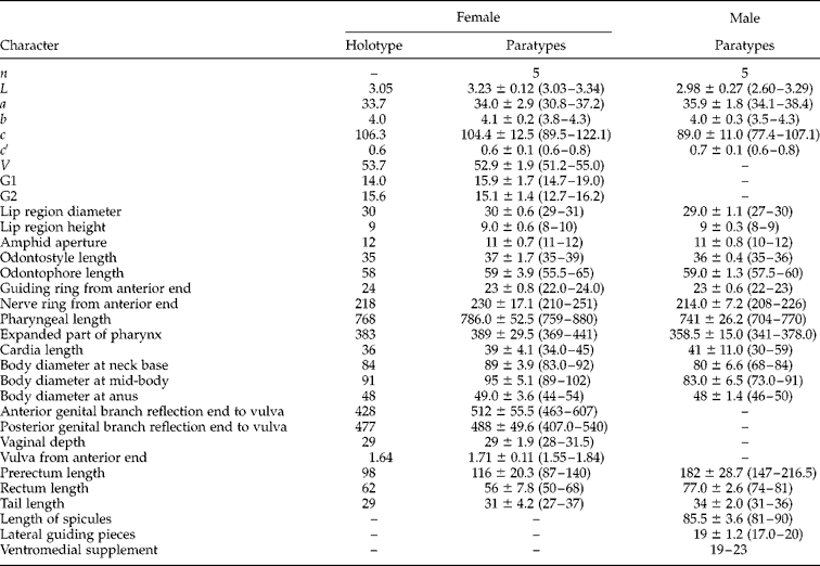
For measurements of L. major n. sp., see table 1.
Morphometric data of Labronemella major n. sp. All measurements are in the form: mean ± SD (range), and in μm (except for ‘L’ and ‘Vulva from anterior end’ in mm).

n, number of specimens observed; L, body length; a= L/maximum width; b= L/pharyngeal length; c= L/tail length; c′ = tail length/body diameter at anus; V= distance of vulva from anterior end × 100/L; G1 = anterior genital branch reflection end to vulva × 100/L; G2 = posterior genital branch reflection end to vulva × 100/L.
Description
Female
Body moderately slender, ‘C’-shaped following heat relaxation and fixation (figs 1–3). Cuticle two-layered, 4–5 μm thick in anterior body, 6–7.5 μm at mid-body and 10–11.5 μm on tail; the outer layer thin with fine transverse striations, the inner one bearing distinct radial striations. Lateral chord 21–27 μm wide at mid-body, about one-fifth to three-tenths of the body diameter at mid-body. Lip region well offset by a distinct constriction, about three times as wide as high or about 0.3 times as wide as body diameter at neck base. Under SEM, lip region completely amalgamated, forming a disc-like structure, with an almost regular hexagonal outer margin and rounded inner margin; six separated inner liplets protruding on oral field; lateral liplets trapezoidal, larger than the triangular subdorsal and subventral ones. Labial papillae slightly protruding, inner labial papillae located on inner margin of labial disc, outer labial papillae and cephalic papillae located on six angles of outer margin of labial disc. Amphid fovea funnel-shaped, bulging distinctly on body surface under SEM; aperture located at the labial constriction, about 0.4 times as wide as lip region width. Odontostyle straight with distinct lumen and opening, about 1.2–1.3 times longer than lip region width and its aperture about 39–47% of its length. Odontophore rod-like, 1.4–1.7 times as long as the odontostyle. Guiding ring double. Nerve ring situated at 25.7–32.8% of the neck length from the anterior end. Anterior part of pharynx slender and muscular, basal expanded part of pharynx up to half (48–50%) of the total neck length. Pharyngeal gland nuclei located as follows: D = 54–56, AS1 = 55–62, AS2 = 56–64, PS1 = 64–80, PS2 = 72–83. Cardia conoid to ligulate. Genital system amphidelphic, with both well-developed branches. Ovary reflexed, anterior 138.5–295 μm long and posterior 140.5–237 μm long, usually surpassing the junction of oviduct and uterus. Oocytes arranged in a single row, except within the germination zone. Oviduct consists of a long slender part and a well-developed pars dilatata which is usually filled with sperm; anterior 162–187 μm long and posterior 170–187 μm long. Sphincter present at the junction of oviduct and uterus. Uterus differentiated into three parts; with one slender and twisting proximal region, one shorter intermediate region with thick walls and one distal portion with thinner walls and distinct lumen; anterior 322–397 μm long, posterior 307–450 μm long. Vulva transverse, vagina extending inwards about one-third of the corresponding body diameter. Pars proximalis vaginae 28–31 × 19–22 μm with convex walls; pars refringens with two well-developed drop-shaped sclerotized pieces that are close to each other, measuring about 9 × 6 μm and with a combined width of about 15 μm; pars distalis relatively short, 3–5 μm long. One egg observed, measuring 124 × 50.5 μm. Prerectum 1.8–2.6 times and rectum 1.0–1.4 times in length compared to the body diameter at anus. Tail short, rounded to conoid.
Labronemella major n. sp. Female: (B) entire body; (C) anterior end; (D) amphid; (E) vulva; (G) posterior genital branch; (I) posterior end; (J) anterior region showing pharynx. Male: (A) entire body; (F) spicules; (H) posterior end.

Labronemella major n. sp. Female: (A) entire body; (C) posterior region, (D) amphid; (E, F) anterior ends; (G) anterior region showing pharynx; (H) genital system; (I, J) vulvas in lateral view. Male: (B) entire body; (K) posterior region showing ventromedial supplements; (L) spicules. Scale bars: A, B = 500 μm; C–L = 20 μm.

Labronemella major n. sp. Female: (A–C) anterior regions showing lip regions and amphids; (D) vulva; (E) posterior region. Male: (F, G) posterior regions. IL, inner labial papillae; LL, lateral liplet. Scale bars: A, C = 10 μm; B, D = 5 μm; E, G = 20 μm; F = 50 μm.

Male
Body similar to that of female, except that the posterior region of the body is more ventrally curved (figs 1–3). Ventromedial supplements 19–23, contiguous, the most posterior one lying 80–94.5 μm from the adanal one. Spicules dorylaimoid, slender with lateral guiding pieces, length 1.7–1.8 times the body diameter at anus. Prerectum relatively longer, 3.0–4.3 times, and rectum length 1.5–1.7 times the anal body width. Tail similar to that of female.
Taxonomic summary
Etymology
The new species is named as major (latin major= bigger, major) as it has the longest body length, odontostyle and spicules, and also the widest lip region, in the current known species of the genus.
Habitat and locality
Soil under the roots of grasses from Graham beach, Maqen, Guoluo Prefecture, Qinghai Province, China. GPS coordinate 100°12′37.2″ E, 34°28′0.2.4″ N.
Type material
Female holotype, five female and five male paratype specimens (slide numbers: QH8-1-(1) to (5); QH.8-1-(10) to (15)) are deposited in the Laboratory of Plant Nematology/Research Center of Nematodes of Plant Quarantine, South China Agricultural University, Guangzhou, Guangdong 510642, China.
Diagnosis and relationships
Labronemella major n. sp. is characterized by a body length of 3.03–3.34 mm; lip region wide, offset by a distinct depression, disc-like with six separated inner liplets; amphid fovea funnel-shaped, distinctly bulged on body surface in SEM; long and straight odontostyle with distinct lumen, aperture 39–47% of its length; odontophore rod-like and long; guiding ring double; pharyngeal basal expansion about half of the total neck length; uterus relatively long and tripartite; vulva transverse and sclerotized; spicules 81–90 μm long; ventromedial supplements 19–23; tail short, rounded to conoid.
The new species can be differentiated from all other species of the genus (Peña-Santiago et al., Reference Peña-Santiago, Abolafia and Pedram2012) by its relatively longer body (3.03–3.34 mm), odontostyle (35–39 μm), spicules (81–90 μm), and wider lip region (29–31 μm). Due to the distinct offset lip region and long (three or more times body diameter at mid-body) tripartite uterus, the new species is close to L. czernowitzensis (Micoletzky, 1922) Andrássy, 2002 and L. labiata Andrássy, Reference Andrássy1985. In addition to the morphological differences described here, the new species can also be distinguished from these species by the following characters. It differs from L. czernowitzensis (Andrássy, Reference Andrássy2009) by having a transverse vulva (vs. longitudinal) and lower a value (a= 30.8–37.2 vs. 40–43 in female; 34.1–38.4 vs. 43–47 in male). From L. labiata (Peña-Santiago et al., Reference Peña-Santiago, Abolafia and Pedram2012), it mainly differs by the amphid distinctly bulging from the body surface (vs. not bulging on body surface); longer rectum (50–68 μm vs. 41–48 μm) in female and more ventromedial supplements (19–23 vs. 14–17) in male.
Key to species of Labronemella (after Peña-Santiago et al., Reference Peña-Santiago, Abolafia and Pedram2012)
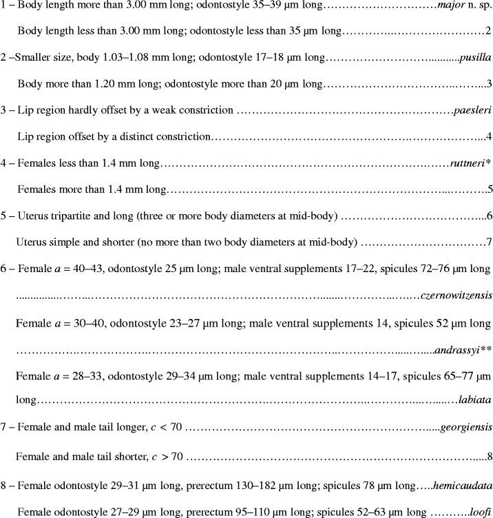
In the key to the species of the genus provided by Peña-Santiago et al. (Reference Peña-Santiago, Abolafia and Pedram2012), L. andrassyi was classified into the group having a body less than 2.00 mm long. As described by Choi (Reference Choi1999), the Korean populations of L. andrassyi have a body length that is more than 2.00 mm long (1.98–2.28 mm). As a result, the key provided by Peña-Santiago et al. (Reference Peña-Santiago, Abolafia and Pedram2012) needs to be revised. A new key to the species is presented in fig. 4.
Key to species of Labronemella (after Peña-Santiago et al., Reference Peña-Santiago, Abolafia and Pedram2012). References: *, (1) Thorne (Reference Thorne1939), (2) Andrássy (Reference Andrássy1985), (3) Peña-Santiago et al. (Reference Peña-Santiago, Abolafia and Pedram2012); **, (1) Choi (Reference Choi1999), (2) Peña-Santiago et al. (Reference Peña-Santiago, Abolafia and Pedram2012).

Financial support
This work was supported by a Special Project of Scientific and Technological Basis of the Ministry of Science and Technology of the People's Republic of China to H.X. (grant no. 2006FY120100).
Conflict of interest
None.