Implications
This observational study describes how the equilibrium between bone mobilisation and bone accretion changes throughout the reproductive cycle of sows, and particularly adds to the understanding of differences in bone metabolism between parities (gilts v. sows). The identification of the crucial periods in bone remodelling and the evaluation of bone markers allow targeted research to overcome bone metabolic problems.
Introduction
Increased mineral requirements at the end of gestation and during lactation change the mineral absorption and bone metabolism throughout the reproductive cycle of mammals (Liesegang et al., Reference Liesegang, Loch, Bürgi and Risteli2005 and Reference Liesegang, Risteli and Wanner2007; Counotte et al., Reference Counotte, Groenland and Salden2009). These processes are largely regulated by parathyroid hormone (PTH) and 1,25-dihydroxyvitamin D3 (1,25-(OH)2D3) hormone secretion (Peterson et al., Reference Prakobwong, Pinlaor, Yongvanit, Sithithaworn, Pairojkul and Hiraku2005; Liesegang et al., Reference Liesegang, Risteli and Wanner2006; Taylor et al., Reference Taylor, Knowlton, McGilliard, Swecker, Ferguson, Wu and Hanigan2008). The latter hormone increases an active calcium (Ca) and phosphorus (P) intestinal epithelial transport and mobilisation. The changes in bone metabolism stimulate the mobilisation of Ca and P from bone to enable foetal skeletal mineralisation during gestation and milk production during lactation (Fukuda and Iida, Reference Fukuda and Iida1993; Liesegang et al., Reference Liesegang, Loch, Bürgi and Risteli2005 and Reference Liesegang, Risteli and Wanner2006).
Although it is known that breeding sows have higher nutrient requirements during the end of gestation and lactation, scientific data on mineral requirements, especially Ca and P, is limited. Inadequate nutrition may lead to imbalanced mineral nutrition or even deficiency and may negatively impact bone quality (McDowell, Reference McDowell2003; Bonjour, Reference Bonjour2005; van Riet et al., Reference van Riet, Millet, Aluwé and Janssens2013). Changes in bone (de)mineralisation during gestation and lactation may affect other bone minerals. Although increased bone remodelling is conceivable in sows during reproduction as well as in other production animals, the effect of parity on bone mineralisation has not been studied in breeding sows. In other animal species, parity did influence bone mineralisation (cattle: Taylor et al., Reference Taylor, Knowlton, McGilliard, Swecker, Ferguson, Wu and Hanigan2008; Sato et al., Reference Sato, Onda, Kato, Ochiai, Kawai, Iriki, Kaneko, Yamazaki and Wada2011 and Reference Sato, Onda, Ochiai, Iriki, Yamazaki and Wada2013; sheep and goats: Liesegang et al., Reference Liesegang, Risteli and Wanner2007). Since primiparous sows are still growing and lack a preceding lactation period, we hypothesised that hormonal profiles differ between primiparous and multiparous sows.
Serum osteocalcin (OC), a marker for osteoblast activity during bone formation and serum crosslaps (CTX), indicating levels of epitopes of the carboxyterminal telopeptide released into the blood during bone resorption, are used as markers for bone remodelling (Liesegang et al., Reference Liesegang, Risteli and Wanner2006). Bone remodelling is a continuous process that serves to renew bone, maintain bone mineral content, and enhance bone strength (van Riet et al., Reference van Riet, Millet, Aluwé and Janssens2013). The fluctuation of hydroxyproline (HYP) and matrix metalloproteinase 2 (MMP2) during the reproductive cycle is unknown, whereas both molecules might contribute to understanding bone metabolism in sows. Hydroxyproline is important for collagen metabolism in bone and uterus and has been used in other studies as a marker for bone resorption (Klein et al., Reference Klein, Lafferty, Pearson and Curtiss1964; Smith and Nordin, Reference Smith and Nordin1964; van Mosel and Corlett, Reference van Mosel and Corlett1990). In those studies, HYP and bone resorption were positively correlated (Smith and Nordin, Reference Smith and Nordin1964; van Mosel and Corlett, Reference van Mosel and Corlett1990). MMP2 is important for collagen turnover and extracellular matrix remodelling (Prakobwong et al., Reference Percival2009).
In this study we evaluated the fluctuation of bone metabolism in both primiparous and multiparous sows throughout a reproductive cycle using bone markers and the CTX/OC ratio (measure for bone turnover) which may be involved in bone remodelling.
Material and methods
Animals and diets
The experiment was performed with 23 commercial hybrid sows (RA-SE Genetics) from the Institute for Agricultural and Fisheries Research (ILVO)-herd. One sow was excluded from the experiment due to the animals’ excessive stress during blood sampling. Two primiparous and two multiparous sows were not gravid and three primiparous sows aborted at day 58, 66 and 93 of gestation and therefore also did not contribute to the experiment. One sow developed an abscess under the cheek and required veterinary treatment. This sow remained in the experimental group, because her results were not considered outliers compared with other sows. The experiment therefore included ten multiparous and five primiparous sows that were followed one reproductive cycle. The multiparous sows (parity: 5.5±3.6 (SD), parity range: 2 to 11, number of sows per parity: parity 2 (n=2), parity 3 (n=2), parity 4 (n=2), parity 6 (n=1), parity 9 (n=1), parity 11 (n=2)) weighed 234±36 (SD) kg at weaning of the preceding lactation period (i.e. at the start of the study, day -5). The primiparous sows (gilts, 233±12 (SD) days old) weighed 142±7 (SD) kg at day -5.
The sows were housed individually during the first 4 weeks of gestation (gestation crates) and from 1 week before parturition until weaning (farrowing crates). The ventilation in the units was adapted to the temperature. The indoor temperature in the farrowing crate was set to 23°C. During mid- and end gestation the sows were housed as one group using an automated feeding system with individual sow recognition through an electronic transponder in the sow’s ear. The indoor temperature was set at 20°C.
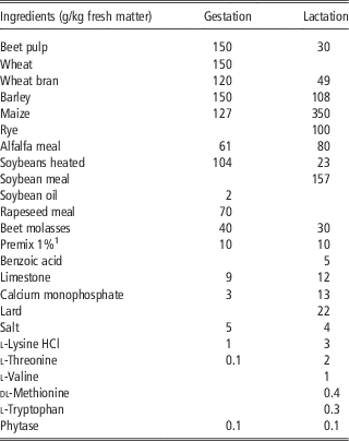
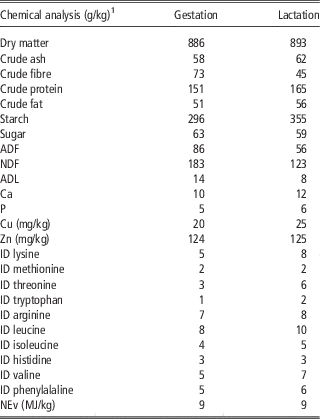
The sows were fed a gestation and lactation diet according to commercial dietary standards and nutrient requirements (National Research Council, 1998) (Tables 1 and 2). Feed samples of the gestation and lactation diet were subject to proximate analysis according to international standard methods accredited by ISO 17025 (2005). The gestation diet was provided from the start of the study until 1 week before parturition, whereas the lactation diet was provided 1 week before parturition until weaning. During the first 4 weeks of gestation, the sows were fed 2.3 kg per day, provided in two equal portions. During mid- and end gestation, the sows were fed 2.6 kg daily and during lactation twice daily, in total 3+0.25 kg per piglet (e.g. a sow with 10 piglets received 5.5 kg of feed daily). There were no feed leftovers. Throughout the experiment, all sows had ad libitum access to drinking water, except during the first 4 weeks of gestation. Drinking water was then automatically provided through nipple drinkers for 15 min every hour and 45 min while feeding in order to prohibit water spillage.
Ingredient composition of the gestation and lactation diet

1 Premix 1% included per kg diet: vitamin A (12 000 IU), vitamin D3 (2000 IU), vitamin E (75 mg), vitamin K3 (1 mg), vitamin B1 (2 mg), vitamin B2 (5 mg), vitamin B5 (18 mg), vitamin B6 (4 mg), vitamin B12 (0.03 mg), vitamin B3 (25 mg), vitamin B11 (3 mg), biotin (0.3 mg), choline (433 mg), FeSO4·H2O (Fe: 150 mg), CuSO4·5H2O (Cu: 16 mg), MnO (Mn: 50 mg), ZnO (Zn: 93 mg), Ca(IO3)2 (I: 2 mg), Na2O3Se (Se: 0.4 mg), Ca (1 mg), P (0.1 mg), Mg (0.2 mg), Na (3 mg), Cl (0.1 mg), K (0.1 mg), S (0.1 mg), lysine (0.04 mg), methionine (0.01 mg), threonine (0.03 mg), tryptophan (0.01 mg), butylhydroxytoluene (1 mg), ethoxyquin (1 mg), butylated hydroxy anisol (0.1 mg).
Analysed nutrient composition of the gestation and lactation diet

1 The chemical analyses of the ileal digestible (ID) amino acids and net energy for pigs (NEv) are according to CVB, 2007.
Experimental design
Blood samples were taken after overnight fasting (17.5 h) before feeding in the morning between 0800 to 1000 h, at fixed time intervals throughout the reproductive cycle: day -5 (multiparous sows’ weaning date of the preceding cycle; ‘start of the study in December 2011, winter season’), every 3 weeks during gestation (day 0 (insemination), 21, 42, 63, 84), around parturition (day 108, 112, 115 (parturition), 118), and during lactation (day 122, 129, 143 (weaning, end of the study in May 2012, spring season)). During gestation, the dates were determined based on the insemination date, whereas during lactation the sows were sampled based on the date of parturition. Blood samples were collected from the jugular vein, using stainless steel needles and plastic syringes, and were placed in a heparin and serum vacuum tube (Terumo Europe, Leuven, Belgium).
BW and backfat thickness were measured at the start of the study (i.e. weaning of the preceding lactation period for the multiparous sows (day -5)), at day 63, 84 and 108 of gestation, and at weaning (day 143). Backfat thickness was determined between the 3rd and 4th last rib, 7 cm at the left and right side of the vertebrae (P2). After P2 was lubricated, backfat measures by ultrasound were repeated three times, alternately at the left and right side (PIGLOG 105, SFK-Technology, Søborg, Denmark). The average thickness was used for further calculations. All experimental procedures involving these animals were approved by ILVO’s ethical committee for animal experiments (approval no. 2010/160, 16 August 2011).
Preparation and analysis of biomarkers
One ml heparinised blood was used for haematocrit (centrifuged at 2749×g, 30 min, 20°C). The vacuum serum tubes were centrifuged (1500×g, 10 min, 4°C) after resting overnight to clot at 4°C. Serum samples were equally divided into two disposable polystyrene tubes, stored for 24 h at −20°C and then stored at −80°C until analysis. The sows’ sera were analysed for OC, CTX, HYP and MMP2. MMP2 was only determined at day 84 of gestation, parturition (day 115), and at weaning (day 143).
Serum OC concentrations were determined using a radioimmunoassay (DiaSorin Inc., Stillwater, MN, USA). This test is based on a two-site immunoradiometric assay for the measurement of intact and N-terminal midregion OC fragment in serum (Liesegang et al., Reference Liesegang, Loch, Bürgi and Risteli2005 and Reference Liesegang, Risteli and Wanner2007). Serum CTX concentrations were determined using a one-step ELISA (Osteometer BioTech A/S, Herlev, Denmark). This test is based on two specific monoclonal antibodies against the amino acid sequence of the CTX. The second antibody is oxidase-conjugated (Liesegang et al., Reference Liesegang, Loch, Bürgi and Risteli2005 and Reference Liesegang, Risteli and Wanner2007). Serum HYP was determined using competitive ELISA (Porcine Hydroxyproline (HYP) Elisa kit, E07H0014; BlueGene Biotech CO., Shanghai, China). The product of the enzyme–substrate reaction forms a blue coloured complex and turns into yellow after the reaction stops, adding a stop solution. The intensity of the colour was measured spectrophotometrically at 450 nm (BEP 2000, Siemens AG, Munich, Germany). The HYP concentration was interpolated from the standard curve. Serum MMP2 was determined using quantitative sandwich ELISA (Pig Matrix Metalloproteinase 2/Gelatinase 2 Elisa kit, CBS-E12842p; Cusabio, Wuhan, China). Standards and samples were pipetted into the wells, which were pre-coated with antibody specific for MMP2. A biotin-conjugated antibody specific for MMP2 was added, and after washing, avidin conjugated Horseradish Peroxidase (HRP) was added. The wells were washed again and a substrate solution was added to colour the remaining amount of MMP2. The intensity of the colour was measured spectrophotometrically at 450 nm within 5 min after stopping the colour reaction (BEP 2000). Serum MMP2 concentrations below the detection limit (0.225 ng/ml) were corrected using the equation: detection limit/√2.
Statistical analyses
The effect of parity on BW, backfat thickness, reproduction and haematocrit was analysed using a linear model with performance as the outcome and parity as fixed effect. The evolution over time of BW, backfat thickness and haematocrit were analysed using a linear mixed model with performance as the outcome and parity as fixed effect. Sow was added as random effect to correct for repeated measures within sows.
The fluctuation over time of all markers was analysed using a linear mixed model for each phase (start of the study (day -5), gestation (day 0 to day 115) and lactation (day 118 to day 143)) with the marker as outcome and parity, haematocrit, time, interaction between parity and time as fixed effects. Based on the graphical exploration of the data, a quadratic time effect was also included in the model. The linear time component is dominant at the start of each phase while the quadratic component (deviation from linearity) becomes more dominant at the end of each phase. In case of repeated measures, a random effect for sow was introduced in the model. The analysed data were considered normally distributed, based on the graphical evaluation (histogram and QQ-plot) of the residuals. Observations for MMP2 were LOG transformed to obtain a normal distribution. In case of post hoc pairwise testing, P-values were corrected with the Tukey–Kramer adjustment for multiple comparisons. All analyses were performed using SAS 9.4 (SAS Institute Inc., Cary, NC, USA).
Results
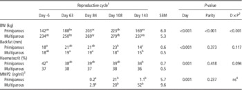
The BW increased during gestation and decreased after parturition (Table 3). The BW of multiparous sows decreased after parturition to similar values at weaning of the preceding cycle, whereas primiparous sows showed an increased BW at weaning compared to the start of the study. Backfat thickness decreased during lactation (P<0.001), independent of parity (P=0.373) (Table 3). Haematocrit tended to be lower at weaning (day 143) compared to the haematocrit at the start of the study (day -5) for primiparous sows (e.g. tendency for interaction between day and parity, P=0.094) (Table 3).
Mean BW, backfat thickness, haematocrit and serum matrix metalloproteinase 2 concentrations throughout the reproductive cycle of primiparous (n=5) and multiparous (n=10) sows

MMP2=matrix metalloproteinase 2.
a,b,cDifferent row superscripts indicate a significant difference between different days of the reproductive cycle (P<0.050).
x,yDifferent column superscripts indicate a significant difference between primiparous and multiparous sows for the specific day of the reproductive cycle (P<0.050).
1 Reproductive cycle represents day-5 (start of the study), day 63, 84, and 108 (gestation), and day 143 (weaning). The serum MMP2 concentration was determined at day 84, 115 instead of day 108, and 143.
2 Interaction between day of the reproductive cycle (D) and parity (P).
3 Serum MMP2 (ng/ml) concentrations below the detection limit (0.225 ng/ml) were corrected using the equation: detection limit/√2. Serum MMP2 concentrations were LOG transformed to obtain a normal distribution.
4 Interaction not significant between day (D) and parity (P) and therefore excluded from the final model.
No differences in reproductive performances (mean±SE) were observed between primiparous and multiparous sows for the number of piglets born alive (13±1.2 and 13±0.9, respectively, P=0.741), number of stillborn piglets (1±0.4 for both primiparous and multiparous sows, P=0.537), average BW of piglets born alive (1.3±0.1 and 1.5±0.1 kg, respectively, P=0.210), piglet mortality (13±5.8 and 10±2.4%, respectively, P=0.537), number of weaned piglets (11±0.9 and 12±0.9, respectively, P=0.480), and average BW of weaned piglets (8.9±0.2 and 8.4±0.2 kg, respectively, P=0.153).
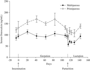
The mean OC concentrations in serum increased during the beginning of gestation (P=0.002) and decreased towards parturition (P=0.001) (Figure 1). Primiparous sows showed higher serum OC concentrations during gestation compared with multiparous sows (P=0.001). Serum OC concentration tended to decrease in the beginning of lactation (P=0.081) and increased towards the end of lactation (P=0.007). A significant parity effect was found during lactation (P=0.003), with primiparous sows showing higher OC concentrations. No parity effect was found at day -5 (P=0.103). Serum OC concentration was associated with haematocrit during gestation (P=0.006).
Mean and SE serum osteocalcin (ng/ml) concentrations throughout gestation and lactation of primiparous (n=5) and multiparous (n=10) sows. Gestation: serum OC increased during gestation (P=0.002) and decreased towards parturition (P=0.001). Lactation: serum OC increased towards the end of lactation (P=0.007). Primiparous sows have higher OC during gestation and lactation (P=0.001 and 0.003, respectively).

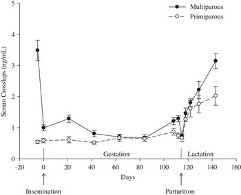
The serum CTX concentration decreased at the beginning of gestation (P=0.002) and increased towards parturition (P=0.001) (Figure 2). Primiparous sows showed lower CTX concentrations during gestation (P=0.005). During lactation, CTX concentration increased, although it increased less for primiparous sows (e.g. interaction between parity and day of lactation, P=0.013). At the start of the study, primiparous sows showed lower CTX concentration compared with multiparous sows (P<0.001). Serum CTX concentration was associated with haematocrit during gestation (P=0.037).
Mean and SE serum crosslaps (ng/ml) concentrations throughout gestation and lactation of primiparous (n=5) and multiparous (n=10) sows. Gestation: serum CTX decreased (P=0.002) and increased towards parturition (P=0.001). Lactation: serum CTX increased, though increased less for primiparous sows (P=0.013). Primiparous sows have lower CTX during gestation (P=0.005) and start of the study (day -5; P<0.001).

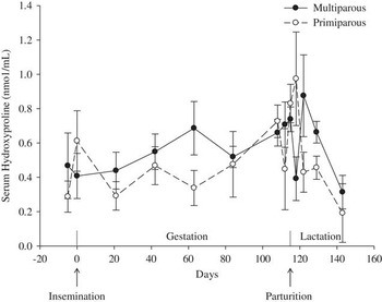
No significant changes in serum HYP concentration during the reproductive cycle were observed (P=0.765 and 0.569 for gestation and lactation, respectively) (Figure 3). No parity effect was found during gestation (P=0.430), lactation (P=0.797), and at start of the study (P=0.930).
Mean and SE serum hydroxyproline (nmol/ml) concentrations throughout gestation and lactation of primiparous (n=5) and multiparous (n=10) sows. Gestation and lactation: serum HYP did not fluctuated significantly (P=0.765 and 0.569, respectively). No parity effects found (P=0.430, 0.797, and 0.930 for gestation, lactation, and start of the study (day -5), respectively).

Between day 84 and 115, serum MMP2 concentration increased for both primiparous and multiparous sows (P=0.001), whereas during lactation no significant fluctuations were found (Table 3). No parity effects were found during gestation and lactation (P=0.237).
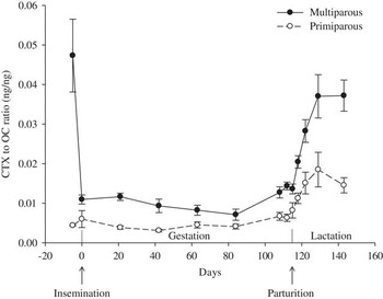
The bone remodelling rate (i.e. bone turnover rate), calculated as the ratio between CTX (bone resorption) and OC (bone formation) decreased at the beginning of gestation (P<0.001) and increased towards parturition (P<0.001) (Figure 4). Primiparous sows had a lower CTX/OC ratio during gestation compared with multiparous sows (P=0.008). During lactation, CTX/OC ratio increased at the beginning of lactation and a decrease or constant concentration was found until weaning, with a lower ratio for primiparous sows (e.g. interaction between parity and day of lactation, P=0.032). At start of the study primiparous sows had a lower CTX/OC ratio than multiparous sows (P=0.010).
Mean and SE serum crosslaps to serum osteocalcin ratio (ng/ml) throughout gestation and lactation of primiparous (n=5) and multiparous (n=10) sows. Gestation: CTX/OC decreased (P<0.001) and increased towards parturition (P<0.001). Lactation: CTX/OC increased at the beginning of lactation and decreased or remains constant until weaning, with a lower ratio for primiparous sows (P=0.032). Primiparous sows have lower CTX/OC ratio during gestation and start of the study (day-5; P=0.008 and 0.010, respectively).

Discussion
A clear parity effect in the concentration of some bone markers was observed, however fluctuations throughout the reproductive cycle followed a similar pattern for primiparous and multiparous sows. Although age, parity, and BW are unavoidably interrelated, which makes it impossible to differentiate between these factors, these results anyhow indicates differences in bone metabolism according to parity as a practical parameter.
Bone remodelling is a continuous process, in which bone formation (OC) and bone resorption (CTX) (processes of bone remodelling) show a parallel fluctuation if both processes are in equilibrium: if bone resorption increases, bone formation also increases (Eriksen, Reference Eriksen1986; Liesegang et al., Reference Liesegang, Risteli and Wanner2007). Although the time effect for OC and CTX during early- and mid-gestation (day 0 to 84), an equilibrium between bone formation and resorption was found (almost constant CTX/OC ratio), which is comparable to studies with goats and sheep where both decreased (Liesegang et al., Reference Liesegang, Risteli and Wanner2006 and Reference Liesegang, Risteli and Wanner2007) or in sows where both stayed constant (Peterson et al., Reference Prakobwong, Pinlaor, Yongvanit, Sithithaworn, Pairojkul and Hiraku2005).
Bone formation overrules bone resorption until day 84 of gestation (i.e. low CTX/OC ratio) for both parity groups. This low CTX/OC ratio may be caused by a changed nutrient requirement, influences of dietary intake (increased from beginning to mid-gestation: 2.3 to 2.6 kg), skeletal replenishment or bone mass compensation in multiparous sows, and the increased activity during group housing, because it seems that exercise increases bone formation as has been found in humans (Ischander et al., Reference Ischander, Zaldivar, Eliakim, Nussbaum, Dunton, Leu, Cooper and Schneider2007; Counotte et al., Reference Counotte, Groenland and Salden2009).
The continuing growth of primiparous sows explains the more marked bone formation and lower bone resorption in these animals during gestation compared to multiparous sows. This parity effect was also found in goats and sheep (Liesegang et al., Reference Liesegang, Risteli and Wanner2007) and in cows (van Mosel and Corlett, Reference van Mosel and Corlett1990; Liesegang et al., Reference Liesegang, Eicher, Sassi, Risteli, Kraenzlin, Riond and Wanner2000). Primiparous sows may require more nutrients for growth and development of muscle and skeletal tissues compared with multiparous sows (Mahan and Newton, Reference Mahan and Newton1995; Goodband et al., Reference Goodband, Tokach, Goncalves, Woodworth, Dritz and DeRouchey2013). However, the nutrient requirements for multiparous sows vary according to the weight loss of a preceding lactation period and concomitant required nutrient deposition.
The present study suggests that the equilibrium between bone formation and bone resorption (day 0 to 84) evolves to a state of net bone loss during late gestation and lactation (day 84 to 129). In the period between day 84 and 129, CTX increases and OC graphically decreases, except for a similar decrease of CTX and OC at parturition and a similar increase of CTX and OC until 3 days postpartum. At this reproductive phase, despite the increased absorption efficiency, the minerals absorbed from feed may not meet the increased nutrient requirements to facilitate Ca and P demands for foetal development and milk production (Liesegang et al., Reference Liesegang, Loch, Bürgi and Risteli2005; Counotte et al., Reference Counotte, Groenland and Salden2009; Donangelo and King, Reference Donangelo and King2012). In other studies, serum Ca concentrations decreases consequently and stimulates PTH and 1,25-(OH)2D3 secretion to support Ca transport and mobilisation (Liesegang et al., Reference Liesegang, Eicher, Sassi, Risteli, Kraenzlin, Riond and Wanner2000; Taylor et al., Reference Taylor, Knowlton, McGilliard, Swecker, Ferguson, Wu and Hanigan2008; Counotte et al., Reference Counotte, Groenland and Salden2009), thereby increasing bone resorption and decreasing bone formation (Halloran and DeLuca, Reference Halloran and DeLuca1980; Liesegang et al., Reference Liesegang, Loch, Bürgi and Risteli2005 and Reference Liesegang, Risteli and Wanner2006). In the present study, sows were fed close to ad libitum feed intake during lactation, which is in accordance with conventional practices. Still, it cannot be excluded that the results may have slightly differed when sows were fed ad libitum. This is in accordance with an increased CTX concentration in young adults (Villareal et al., Reference Villareal, Fontana, Das, Redman, Smith, Saltzman, Bales, Rochon, Pieper, Huang, Lewis and Schwartz2015). However, a study in sows did not observe differences in OC and CTX concentration, except at day 112, between feed restricted or ad libitum fed sows (Cools et al., Reference Cools, Maes, Decaluwé, Buyse, van Kempen, Liesegang and Janssens2014). In cows, OC concentration and pattern over time was also not different in ad libitum fed multiparous cows with varying dietary Ca intake (Taylor et al., Reference Taylor, Knowlton, McGilliard, Seymour and Herbein2009). Therefore, the influence of feed intake is probably minimal. The fluctuations of bone formation and resorption observed in the present study are in accordance with fluctuations around parturition found in cows (Naito et al., Reference Naito, Shindo, Sato and Murakami1990; Peterson et al., Reference Prakobwong, Pinlaor, Yongvanit, Sithithaworn, Pairojkul and Hiraku2005; Ekelund et al., Reference Ekelund, Spörndly and Holtenius2006), sows (Liesegang et al., Reference Liesegang, Loch, Bürgi and Risteli2005; Counotte et al., Reference Counotte, Groenland and Salden2009; Lauridsen et al., Reference Lauridsen, Halekoh, Larsen and Jensen2010; Cools et al., Reference Cools, Maes, Decaluwé, Buyse, van Kempen, Liesegang and Janssens2014), and goats and sheep (Farrugia et al., Reference Farrugia, Fortune, Heath, Caple and Wark1989; Liesegang et al., Reference Liesegang, Risteli and Wanner2006 and Reference Liesegang, Risteli and Wanner2007).
After 2 weeks of lactation (day 129), bone resorption further increased and bone formation started to increase until weaning, resulting in a constant and a decreased CTX/OC ratio for multiparous and primiparous sows, respectively. In other studies on goats, sheep and cows, decreased bone resorption at the end of lactation was observed (Liesegang et al., Reference Liesegang, Eicher, Sassi, Risteli, Kraenzlin, Riond and Wanner2000, Reference Liesegang, Risteli and Wanner2006 and Reference Liesegang, Risteli and Wanner2007). Those studies suggested that the mobilised bone mineral content is reversed towards the end of lactation when milk production decreases, a time in which animals replenish their skeletal reserves by increasing bone formation over bone resorption to form new bone (Liesegang et al., Reference Liesegang, Eicher, Sassi, Risteli, Kraenzlin, Riond and Wanner2000, Reference Liesegang, Risteli and Wanner2006 and Reference Liesegang, Risteli and Wanner2007). Bone resorption tremendously decreased after weaning in the present study, reflecting the high milk production in the 4-week lactation period and the still outweighed nutritional demands over the amount of nutrients absorbed. After weaning, the milk production stops, reducing the nutrient demands, facilitating the tremendous decrease in bone resorption. This decreased bone resorption after weaning indicates that milk production is the main trigger for bone resorption (Liesegang et al., Reference Liesegang, Risteli and Wanner2006) and that the sow may replenish her skeletal reserves mainly after weaning. Further, the observations of the present study compared with the results of other studies may suggest that replenishment of skeletal reserves is species dependent, depending on the length of the lactation period. In cows and humans bone formation and resorption markers are not directly related to milk yield, which may indicate not only that the high Ca demands for milk production initiate bone turnover, but also that high oestrogen concentration at the end of gestation influences bone metabolism (Holtenius and Ekelund, Reference Holtenius and Ekelund2005). Furthermore, nutrient intake of vitamins and minerals are also important factors associated with bone metabolism (New et al., Reference New, Robins, Campbell, Martin, Garton, Bolton-Smith, Grubb, Lee and Reid2000; Palacios, Reference Palacios2006; van Riet et al., Reference van Riet, Millet, Aluwé and Janssens2013).
The parity effect, unavoidably confounded with BW changes, during gestation was continued during lactation, where differences between parity groups depended on day of lactation for CTX. The higher OC concentrations in primiparous sows compared to multiparous sows were in agreement with the expectations, because OC reflects osteoblast activity in bone formation and these decreased with age (Liao et al., Reference Liao, Ozono, Sone, McDonnell and Pile1990), and were in accordance with other species such as goats and sheep (Liesegang et al., Reference Liesegang, Risteli and Wanner2007) and cows (van Mosel and Corlett, 1990; Kamiya et al., Reference Kamiya, Kamiya, Tanaka and Shioya2005; Taylor et al., Reference Taylor, Knowlton, McGilliard, Swecker, Ferguson, Wu and Hanigan2008). Though, the concentration of bone markers may vary independently of bone metabolism, as reported in Sato et al. (Reference Sato, Onda, Ochiai, Iriki, Yamazaki and Wada2013). Further research is warranted.
Hydroxyproline, important for collagen metabolism in collagen type II (articular cartilage) and collagen type III tissues (bone and uterus) (Garnero and Delmans, Reference Garnero and Delmans1996; Percival, Reference Prasadam, Friis, Shi, van Gennip, Crawford and Xiao1997; Liesegang et al., Reference Liesegang, Eicher, Sassi, Risteli, Kraenzlin, Riond and Wanner2000), was used in other studies as a marker for bone resorption (Klein et al., Reference Klein, Lafferty, Pearson and Curtiss1964; Smith and Nordin, Reference Smith and Nordin1964; van Mosel and Corlett, Reference van Mosel and Corlett1990), with a positive correlation between HYP and bone resorption (Smith and Nordin, Reference Smith and Nordin1964; van Mosel and Corlett, Reference van Mosel and Corlett1990). MMP2 is important for collagen turnover and extracellular matrix remodelling (Prakobwong et al., Reference Percival2009). It is expected that MMP2 expression increases if tissues are damaged (including bone), resulting in reactive oxygen species (ROS) formation (Knittel et al., Reference Knittel, Mehde, Grundmann, Saile, Scharf and Ramadori2000; Siwik et al., Reference Siwik, Pagano and Colucci2001; Prakobwong et al., Reference Percival2009). Therefore, HYP and MMP2 may reflect bone remodelling (Lajeunesse and Reboul, Reference Lajeunesse and Reboul2003; Prasadam et al., Reference Peterson, Orth, Goff and Beede2010; Sharma et al., Reference Sharma, Jagga, Lee and Nam2013). However, in the present study HYP and MMP2 as potential bone markers did not provide added value to the already established markers OC and CTX. Although HYP has been used as a bone remodelling marker before, the main limitation of HYP seems to be the high variability. The same applies to MMP2, indicating that these markers may not be specific enough to express changes in bone metabolism only. According to Liesegang et al. (Reference Liesegang, Eicher, Sassi, Risteli, Kraenzlin, Riond and Wanner2000), HYP may even originate from the uterus.
Conclusion
The fluctuation of bone formation and bone resorption markers, OC and CTX, follows a similar pattern for both primiparous and multiparous sows. However, primiparous sows have a higher bone formation and lower bone resorption throughout the reproductive cycle compared with multiparous sows. The results of this study demonstrate that the period between late gestation and the end of lactation (weaning) is the most intensive period for bone metabolism.
Based on the measured HYP and MMP2 concentrations, the added value of HYP and MMP2 in understanding bone remodelling seems limited.
Acknowledgements
This study was supported by the Institute for Promotion of Innovation through Science and Technology in Flanders (IWT, grant number 090938), and co-funded by Orffa, VDV Beton, Boerenbond, AVEVE, INVE and Boehringer. The authors thank the technicians M. van Yperen and T. Martens, animal caretakers of the ILVO experimental farm, and colleagues who were ‘sow sitting’ for their much appreciated assistance and support. Thanks also to Miriam Levenson for English-language editing. This study is dedicated to the first author’s grandfather.