Introduction
The Leishmaniases complex are divided into three distinct forms of clinical presentation. The major factor that determines the development of each form of the disease is the specie of parasite associated with the host's specific immune responses (not all those infected by the parasites will develop the disease). In the most common form of the disease, cutaneous leishmaniasis (CL), the parasites remain only at the site of the sand fly bite and cause localized long-term ulceration with no systemic symptoms. In some cases, inadequate treatment of a CL lesion may lead to later development of mucocutaneous leishmaniasis (ML). The third clinical form is visceral leishmaniasis (VL) that is the most pathogenic in which parasites migrate from the inoculation site to multiply in the host's liver and spleen macrophages and bone marrow, causing immunosuppression and death if not treated (Peacock, Reference Peacock2007; Campos et al., Reference Campos2008; David and Craft, Reference David and Craft2009; Bifeld and Clos, Reference Bifeld and Clos2015; Van Der Auwera and Dujardin, Reference Van Der Auwera and Dujardin2015). The control of leishmaniases is presently a serious problem due to the high death rates involved and the economic losses resulting from morbidity. In addition, the disease is strongly linked to poverty in the tropical and subtropical areas and with ever increasing cases worldwide each year. In total 98 countries are officially considered endemic for leishmaniases and estimates show that more than 58.000 VL and 220.000 CL cases are notified per year. More than 90% of global VL cases occurring in six countries – India, Bangladesh, Sudan, South Sudan, Brazil and Ethiopia – and around 75% of global CL cases occurring in ten countries – Afghanistan, Algeria, Colombia, Brazil, Iran, Syria, Ethiopia, North Sudan, Costa Rica and Peru (Alvar et al., Reference Alvar2012; De Vries et al., Reference De Vries, Reedijk and Schallig2015).
Rapid methods for the effective leishmaniases diagnosis and species identification are urgently needed, either by prevention and control of leishmaniases in endemic areas or treatment of infected patients. The definitive diagnosis of all suspected Leishmania infections should be performed in an association of clinical symptoms, and parasitological and immunologic findings. Several diagnostic methods have been described to detect the presence of the Leishmania parasites, with a huge variation in diagnostic accuracy, including direct parasitological examination by microscopy, histopathology and/or parasite culture, indirect testing by serology and molecular diagnostics. The demonstration of the presence of the parasite by parasitological diagnosis is still considered the gold standard in leishmaniases diagnosis because of its high specificity. However, the sensitivity of the direct examinations is low for the diagnosis of CL and ML, with a range from 15 to 70%, whilst in case of VL the sensitivity varies depending on the tissue used, with a range from 93 to 99% for spleen, 53 to 86% for bone marrow and 53 to 65% for lymph node aspirates. Moreover, parasite culture in the culture media is difficult, requires technical skills, is prone to contamination, and is time-consuming. As all other parasitological method for Leishmania detection, the parasite culture does not allow Leishmania species determination. The sensitivity of cultures depends on the parasite quantity but is estimated to be between 60 and 85% (Bhargava and Singh, Reference Bhargava and Singh2012; Rezvan, Reference Rezvan2014; De Vries et al., Reference De Vries, Reedijk and Schallig2015; Georgiadou et al., Reference Georgiadou, Makaritsis and Dalekos2015; Paiva-Cavalcanti et al., Reference Paiva-Cavalcanti2015).
Alternative and complementary diagnosis techniques also include the evaluation of indirect parameters. The Montenegro skin test has been successfully used in the diagnosis of cutaneous and mucocutaneous forms of the disease. Its sensitivity range from 86.4%, but it is not very reliable for detecting the presence of parasites in recent lesions, in diffuse forms of disease and in immunosuppressed patients, as well as not differentiating between past and present infection (Goto and Lindoso, Reference Goto and Lindoso2012; De Vries et al., Reference De Vries, Reedijk and Schallig2015; Handler et al., Reference Handler2015; Paiva-Cavalcanti et al., Reference Paiva-Cavalcanti2015).
Immunological diagnoses, another important indirect method for detecting leishmaniases, are based on the detection of either Leishmania antigens or anti-Leishmania antibodies in the host serum samples. The optimal test for serologic diagnosis is one that is easy to use, cheap to make and has both a high sensitivity and specificity. Most of the immunological techniques for the detection of anti-Leishmania antibodies have been based on the Enzyme-Linked Immunosorbent Assay (ELISA) technique and the sensitivity and specificity of this method depend on the antigen employed. Considering the variations of the individual immune responses to the infection, several antigens have been identified for potential use in the diagnosis of leishmaniases (Goto and Lindoso, Reference Goto and Lindoso2012; Maia et al., Reference Maia2012; Sánchez-Ovejero et al., Reference Sánchez-Ovejero2016).
Oligosaccharides are broadly present on cell surfaces arranged as glycan arrays, responsible for regulating the interaction between cells. They can be useful for disease diagnosis and also helpful in identifying new disease-causing microbial cell markers. The interactions among pathogens and their host cells are guided by different cell-surface glycans and glycan-binding receptors at each stage of the infectious process (Fernández-Tejada et al., Reference Fernández-Tejada, Cañada and Jiménez-Barbero2015). Glycoinositol phospholipids (GIPLs) are the main family of low molecular weight glycolipids synthesized by Leishmania parasites. It is expressed in abundance on promastigote and amastigote cell surfaces and is not linked to proteins, forming a protective surface revetment that provides essential host–parasite interactions. However, each parasite stage is already known to have a different glycoconjugate attached on its surface beyond GIPLs. Biochemical analyses reveal that, unlike amastigotes, the procyclic promastigotes surface coat expresses two other glycoconjugates which are less abundant than GIPLs and are responsible for protecting parasites from hydrolytic enzymes in the sandfly gut: (1) glycosylphosphatidylinositol (GPI) anchored to macromolecules such as metalloprotease and; (2) protein-free lipophosphoglycan complex (LPG), a GPI-anchored macromolecules underlying layer composed of densely, free-packed glycolipids. Altogether these molecules create an effective barrier which protects promastigotes from cell death processes like lysis mediated by complement system, oxygen radicals and hydrolases in the mammalian and insect host environments (Ilgoutz and McConville, Reference Ilgoutz and McConville2001; Mukhopadhyay et al., Reference Mukhopadhyay, Bandyopadhyay and Mandal2006; Barreto-Bergter and Vermelho, Reference Barreto-Bergter and Vermelho2010; Galili, Reference Galili2013a).
High levels of antibody produced against GIPLs have been reported in people infected with trypanosomatid parasites (Ávila et al., Reference Ávila, Rojas and Garcia1988, Reference Ávila, Rojas and Galili1989, Reference Ávila, Rojas and Acosta1991; Almeida et al., Reference Almeida1994). Remarkably, anti-α-Gal recognizes specifically the unique glycosphingolipids structures known as the GPI-anchored mucins in the trypomastigote stage of Trypanosoma spp (Galili, Reference Galili1993; Macher and Galili, Reference Macher and Galili2008; Schocker et al., Reference Schocker2016). However, the parasites escape through the antibodies’ action by penetrating tissues and continue to produce and release GIPLs and LPG with α-Gal epitopes, stimulating the immune system to continuously produce anti-α-Gal antibodies at high titres (Galili, Reference Galili2013a). Quantities of anti-α-Gal antibodies constantly produced can also represent a continuous immune response to Galα1-3 Gal structures found in various gastrointestinal bacteria, confirming the polyreactive nature of these antibodies in human serum (Galili, Reference Galili2013b; Galili et al., Reference Galili1984, Reference Galili1987).
The major GIPLs found in these parasites’ cell membranes include tetraglycosylinositol, pentaglycosylinositol and hexaglycosylphosphatidylinositol and sugar analysis of L eishmania mexicana and L eishmania braziliensis GIPLs revealed monosaccharide composition of Manose (Man), Galactose (Gal), Glucosamine (GlcN) and inositol (Ávila et al., Reference Ávila, Rojas and Acosta1991; McConville and Ferguson, Reference McConville and Ferguson1993).
Anti-α-Gal is the most abundant natural circulating human antibody and, since it binds specifically to the Galα1–3Gal glycosphingolipids (Galili, Reference Galili1993; Macher and Galili, Reference Macher and Galili2008; Schocker et al., Reference Schocker2016), it is assumed that the majority of the anti-α-Gal binding sites expressed in the cell membranes of many organisms have this non-reducing terminal structure (Galili, Reference Galili1993, Reference Galili2013a, Reference Galili2013b; Galili and Andrews, Reference Galili and Andrews1995; Galili et al., Reference Galili1988). However, quantities of anti-α-Gal antibodies constantly produced can also represent a continuous immune response to Galα1-3 Gal structures found in various gastrointestinal bacteria (Galili, Reference Galili2013b; Galili et al., Reference Galili1984, Reference Galili1987), confirming the polyreactive nature of these antibodies in human serum (Satapathy and Ravindran, Reference Satapathy and Ravindran1999).
The α-Gal has the potential for many clinical uses once anti-Gal antibodies are widely produced in healthy humans and α-gal epitopes can be easily synthesized by several methods. This study describes a standardized ELISA used to measure serum levels of anti-α-Gal antibodies in individuals with CL from different endemic regions in Brazil and compares them with those from healthy individuals living in the same endemic areas and non-endemic areas using different neoglycoproteins (NGPs) from the antigen.
Materials and methods
Sample collection
A total of 149 serum samples from females and males of different age groups were collected from CL endemic and non-endemic regions in Brazil. The main inclusion criterion was the proven diagnosis of CL by parasite presence on the direct examination of lesion smears obtained from the edge of the active lesion with a punch biopsy tool and/or positivity in the parasite culturing. The samples were divided into seven groups (Table 1): (1) H1 (23): healthy individuals from non-endemic areas; (2) H2 (37): healthy individuals from endemic areas; (3) CL1 (12): CL patients with active lesion and no treatment; (4) CL2 (5): CL patients with active lesion under the treatment; (5) CL3 (13): CL patients that had finished treatment and were under observation for the subsequent 3 months; (6) OD (54): serum reagent patients for other diseases such as Hepatitis B and C, Syphilis and truly positive patients for Tuberculosis; (7) CD (5): serum reagent patients for Chagas disease (CD).
Demographic data of the study cohort

H1, healthy individuals from non-endemic areas; H2, healthy individuals from endemic areas; CL1, CL patients with active lesion and no treatment; CL2, CL patients with active lesion under the treatment; CL3, CL patients that had finished treatment and were under observation for the subsequent 3 months; OD, serum reagent patients for other diseases such as Hepatitis B and C, Syphilis (diagnosis performed by a blood bank) and truly positive patients for Tuberculosis; CD, serum reagent patients for Chagas disease (diagnosis performed by a blood bank).
Individuals from H2 group were medically examined to discard any previous CL infection. CL patients were treated according to the Brazilian Healthy Ministry guideline, with meglumine antimoniate and amphotericin B. Patients with other diseases such as CD were also studied to evaluate the chance of cross-reactivity in these tests.
Ethical approval was obtained from the Universidade Federal do Paraná Ethical Committee under number 684.244.
Enzyme-linked immunosorbent assay
To determine whether levels of anti-α-Gal antibodies in human serum, both non-infected and infected with CL and other diseases can identify Leishmania α-Gal epitopes, optimal ELISA conditions were established, such as dilutions of sera (1:100, 1:200, 1:400 and 1:800), conjugate (1:5000, 1: 10 000 and 1: 20 000) and antigens (0.1, 0.5 and 1 µg well−1). Once the best condition was established, polystyrene microtiter plates with 96 wells (NUNC C96 Immuno Plate Maxisorp Surface, Thermo Scientific) were coated overnight at 4 °C with 100 µL of 0.1 µg well−1 of two Galα1-3Gal neoglycoprotein series with 3-atom spacer (Galα1-3Galβ1-4GlcNAc conjugated with human albumin serum, identified as NGP 2334; and Galα1-3Gal conjugated with human albumin serum, identified as NGP 2203 - Dextra Laboratories) and one Galα1-3Gal analogue neoglycoprotein with 3-atom spacer (Galα1-3Galβ1-3GlcNAc conjugated with human albumin serum, identified as NGP 2333 – Dextra Laboratories), diluted in carbonate–bicarbonate buffer (pH 9.6). In addition, the β-Gal NGP β1-4-Galactosyl-Galactose conjugated with bovine serum albumin and with a 3-atom spacer (NGP 0204, Dextra Laboratories) was also included in the study as the ‘β-control’ and soluble proteins from the crude extract from L. (Viannia) braziliensis culture (strain MHOM/BR/84/LTB300) as a positive control. Human albumin serum was chosen for the generation of NGPs because of its absence of immunological reaction with human serum and its adequacy as a carrier protein.
The following day, the plates were washed twice with 200 µL well−1 of washing solution (NaCl 0.9% + Tween 20 at 0.05%), then the wells were blocked with 120 µL of blocking solution (Pierce Protein – Free T20 – PBS Blocking Buffer, Thermo Scientific) for 1 h at 37 °C, and after were washed twice again with 200 µL well−1 of washing solution. Immediately after, serum samples were diluted to 1:800 in a solution of PBS −0.25% casein +0.5% Tween 20 and were added to their respective wells and incubated at 37 °C for 1 h. Then the plates were washed four times with 200 µL well−1 of washing solution and polyclonal rabbit anti-human IgG HRP conjugate (1.3 g L−1, Dako) and was added to each well for 1 h at 37 °C. Finally, the reaction was developed by adding 100 µL of SureBlue TMB™ Microwell Peroxidase Substrate to each well at room temperature for 15 min, avoiding light, and to interrupt the reaction 20 µL of a solution 1:20 of H2SO4 was added. Plates were read in the Infinite F200 PRO multimode reader (Tecan) at 450 nm and values were expressed in absorbance. Each sample was measured in triplicate, including the ‘no serum’ control for preventing the background from the secondary antibody (blank control), and the whole assay described above was performed in duplicate. The absorbance readings were performed subtracting the mean of blank control from the mean of the unknown samples in triplicate.
Determination of α-Gal specific activity
Based on Al-Salem et al. (Reference Al-Salem2014) CBAG treatment protocol to determine the α-Gal specific activity, 0.1 µg well−1 of each NGP previously tested, except for NGP 2204, were treated overnight at 28 °C with 0.04 U well−1 of α-galactosidase from green coffee beans (Sigma). After incubation, the plates were washed five times with washing solution and the ELISA was performed as described above. A pool of seven serums from CL1 and CL2 and 10 from H1 individuals was also used as positive and negative controls, respectively.
Statistical analysis
The homogeneity of variance analysis and the Kolmogorov–Smirnov test for the normal condition of variables evaluation were performed using the Statistica 7 and MedCalc 16.1 software, respectively. Once data showed non-parametric distribution, a Kruskal–Wallis one-way analysis with a Dunn post-test was performed to validate the significant difference among groups. The Mann–Whitney analysis was used to compare the means of each group between antigens and positive and negative controls. All non-parametric analyses were performed using GraphPad Prism 6 software, assuming the significant level of P < 0.05. Finally, receiver operating characteristics (ROC) curve were performed based on the logistic regression model, considering the classification of the samples (presence or absence of the disease) as a dependent variable and each antigen as an independent variable. The logistic regression model, ROC curve and sensitivity and specificity analyses were performed using R software (R CORE TEAM, Version 3.4.0, 2017) with auxiliary pROC system (Robin et al., Reference Robin2011) and a significant level of P < 0.05 was adopted.
Results
Enzyme-linked immunosorbent assay
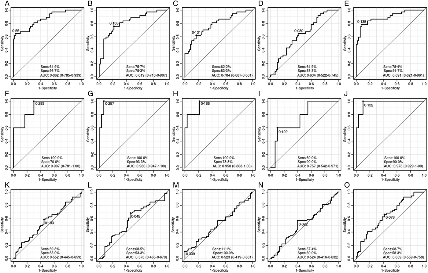
All serum samples collected were analysed on two occasions. First, absorbance values of samples from H1 and CL1 groups were used to perform the ROC curve analysis, sensitivity, specificity and odds ratio of all antigens. The results obtained were compared with the same parameters of the same sample groups for the positive control (Fig. 1 and Table 2). The cut-off values for CL were: 0.030 for NGP 0204, 0.135 for L. braziliensis crude extract and NGP 2333, 0.131 for NGP 2203 and 0.550 for NGP 2334; whilst sensitivity and specificity ranged from 62.2% (NGP 2203) to 78.4% (L. braziliensis crude extract), and 58.3% (NGP 0204) to 96.7% (NGP 2334), respectively.
ROC curve analysis and sensitivity and specificity for NGP 2334, NGP 2333, NGP 2203 and NGP 0204 antigens compared with the ROC curve analysis for the positive control (L. braziliensis) for CL, Chagas disease and other diseases, performed using R software with auxiliary pROC system. NGP 2334: Galα1-3Galβ1-4GlcNAc-HAS, NGP 2333: Galα1-3Galβ1-3GlcNAc-HAS, NGP 2203: Galα1-3Gal-HAS, NGP 0204: β1-4-Galactosyl-Galactose-BSA, L. braziliensis: soluble proteins from crude extract from Leishmania (Viannia) braziliensis culture (strain MHOM/BR/84/LTB300), (A) NGP 2334 for CL, (B) NGP 2333 for CL, (C) NGP 2203 for CL, (D) – NGP 0204 for CL, (E) L. braziliensis crude extract, (F) NGP 2334 for Chagas disease, (G) NGP 2333 for Chagas disease, (H) NGP 2203 for Chagas disease, (I) NGP 0204 for Chagas disease, (J) L. braziliensis crude extract for Chagas disease, (K) NGP 2334 for other disease, (L) NGP 2333 for other disease, (M) NGP 2203 for other disease, (N) NGP 0204 for other disease and (O) L. braziliensis crude extract for other disease.

Odds ratio for all antigens and diseases, considering H1 and CL1 groups as truly negative and positive groups, respectively.

NGP 2334: Galα1-3Galβ1-4GlcNAc-HAS, NGP 2333: Galα1-3Galβ1-3GlcNAc-HAS, NGP 2203: Galα1-3Gal-HAS, NGP 02040: β1-4-Galactosyl-Galactose-BSA.
The P values with an asterisk show a significant difference between groups (P < 0.05).
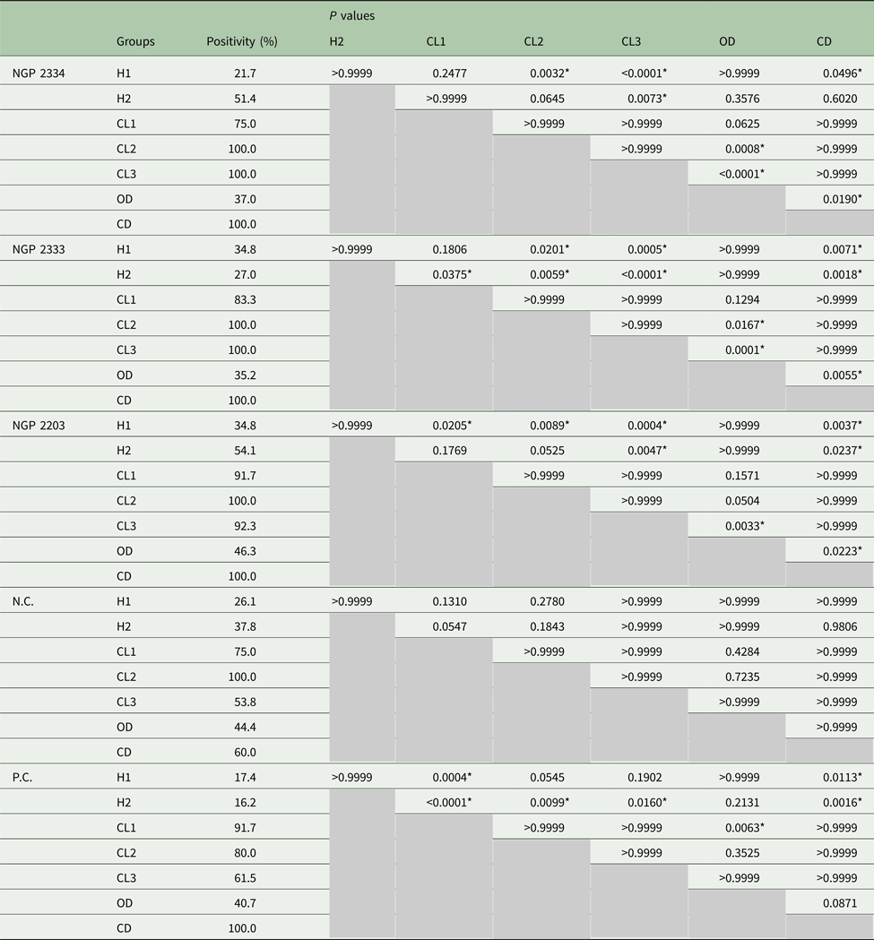
The presence of Leishmania anti-α-Gal antibodies was determined by comparing anti-α-Gal levels in CL patients and healthy individuals living in the same endemic area, non-endemic area and individuals with other diseases (Table 3). It was found that means of anti-α-Gal antibody titres of only CL2 and CL3 groups were significantly higher (P < 0.05) than the H1 group for NGP 2234 and NGP 2333. For NGP 2203, all CL groups presented significantly higher means of antibody levels (P < 0.05) compared with H1 group. No cross-reaction between the NGPs and antibodies from patients with other diseases was detected, except for the CD, since the aetiological agent of this disease is another trypanosomatid parasite.
Kruskal–Wallis statistical analyses of levels of anti-α-Gal antibodies found in individuals from Cutaneous Leishmaniasis endemic and non-endemic areas
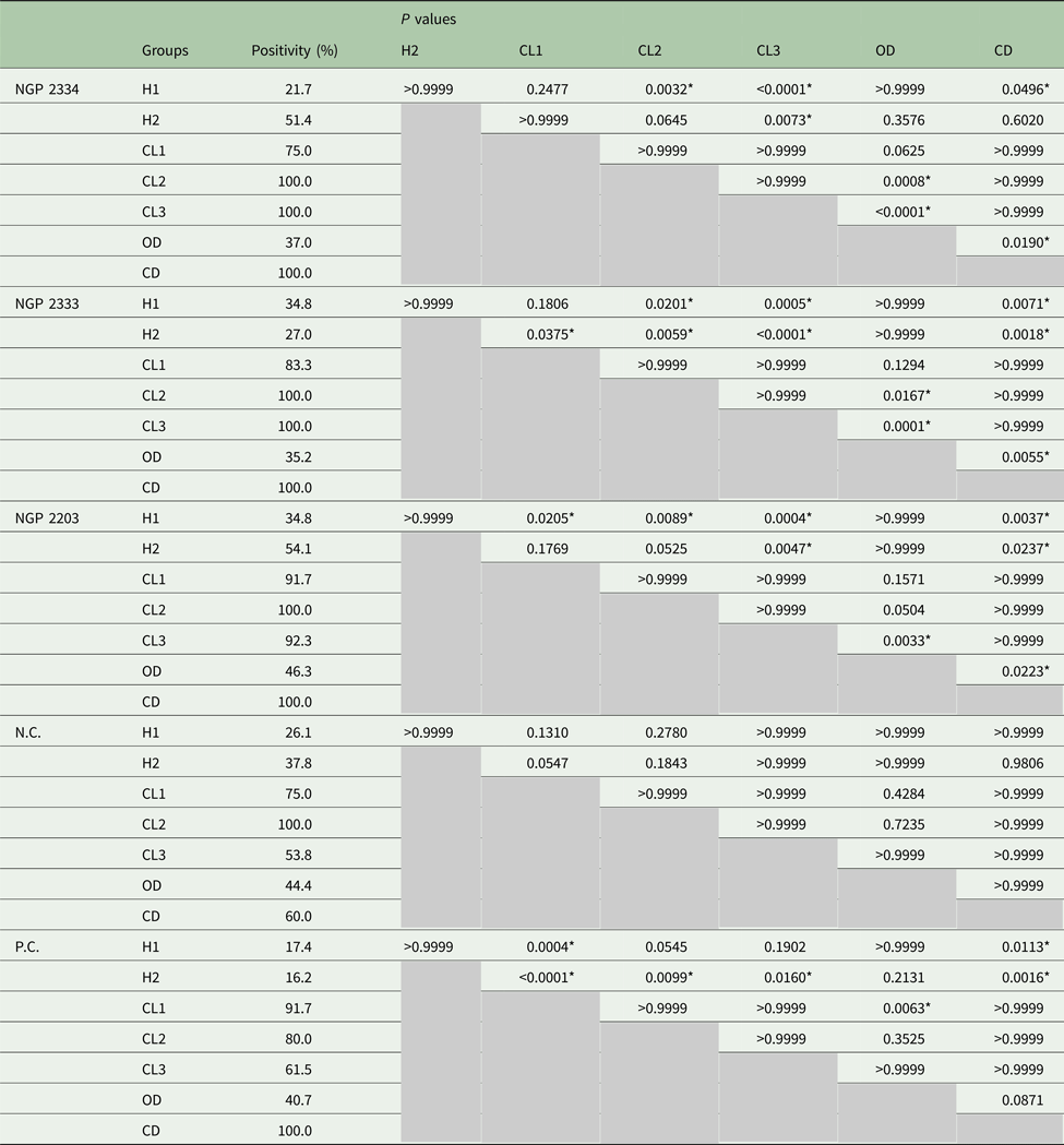
NGP 2334: Galα1-3Galβ1-4GlcNAc-HAS, NGP 2333: Galα1-3Galβ1-3GlcNAc-HAS, NGP 2203: Galα1-3Gal-HAS, N.C.: β1-4-Galactosyl-Galactose-BSA (NGP 02040); P.C., soluble proteins from crude extract from Leishmania (Viannia) braziliensis culture (strain MHOM/BR/84/LTB300); H1, healthy individuals from non-endemic areas; H2, healthy individuals from endemic areas; CL1, CL patients with active lesions and no treatment; CL2, CL patients with active lesions under the treatment; CL3, CL patients that had finished treatment and were under observation for the subsequent 3 months; OD, serum reagent patients for other diseases – such as Hepatitis B and C, Syphilis and A3B2 truly positive patients for Tuberculosis; CD, serum reagent patients for Chagas disease.
The P values with an asterisk show a significant difference between groups (P < 0.05).
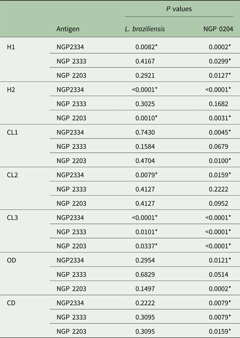
When comparing each sera group for each antigen singly (Table 4), means of antibody titres of CL1 group from all NGPs showed no significant difference compared with the same group from L. braziliensis. However, means of antibody titres of CL2 from NGP 2334 and CL3 groups from all NGPs showed a higher difference compared with the same groups L. braziliensis. Also, means of antibody titres of CL3 and CD groups from NGP 0204 showed lower significant differences compared with all NGPs, indicating that the β-Gal NGP is not able to be detected by antibodies of CD patients and/or CL patients after treatment.
Mann–Whitney statistical analysis between groups of individuals from CL endemic and non-endemic areas and α-Gal antigens
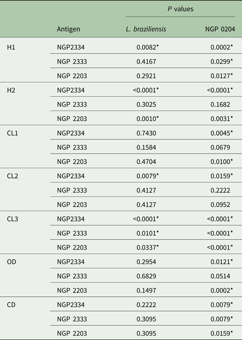
NGP 2334: Galα1-3Galβ1-4GlcNAc-HAS, NGP 2333: Galα1-3Galβ1-3GlcNAc-HAS, NGP 2203: Galα1-3Gal-HAS, NGP 0204, β1–4-Galactosyl-Galactose-BSA; L. braziliensis, soluble proteins from crude extract from Leishmania (Viannia) braziliensis culture (strain MHOM/BR/84/LTB300); H1, healthy individuals from non-endemic areas; H2, healthy individuals from endemic areas; CL1, CL patients with active lesions and no treatment; CL2, CL patients with active lesions under the treatment; CL3, CL patients that had finished treatment and were under observation for the subsequent 3 months; OD, serum reagent patients for other diseases – such as Hepatitis B and C, Syphilis and truly positive patients for Tuberculosis; CD, serum reagent patients for Chagas disease.
The P values with an asterisk show a significant difference between groups (P < 0.05).
Determination of α-Gal specific activity
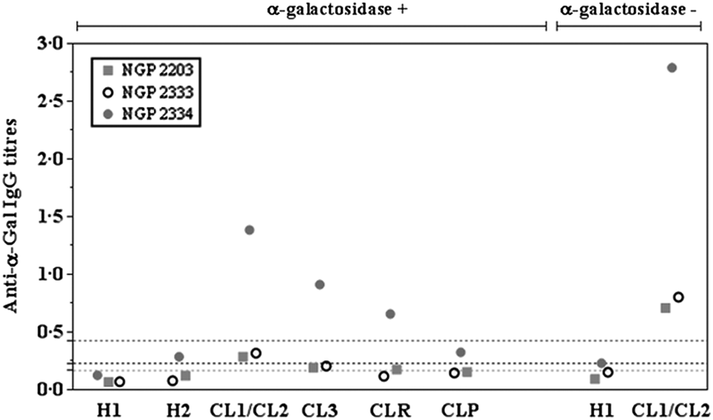
To determine the specific immunogenic activity of the α-Gal residues, all NGPs, except for NGP 2204, were treated with α-galactosidase enzyme from green coffee beans, which specifically unlinks Galα1-3Gal into two parts – Galα1 and 3Gal – and, consequently, abrogates the recognition of α-Gal epitope by anti-α-Gal antibodies (Fig. 2). Although it is clear there is still an antigen–antibody reaction detected in the assay performed with the presence of the α-galactosidase enzyme, compared with the controls, patients from the CL1/CL2 and CL3 groups used in enzymatic treatment assay with NGP 2334 showed a 2- and 3-fold decrease in the antibody titres than positive control, respectively. The same result was found for the other antigens: a 2.5- and 4-fold decrease in the antibody titres for CL1/CL2 and CL3 groups for NGP 2333 antigen, respectively; and 2.5- and 4-fold for the CL1/CL2 and CL3 groups NGP 2203 antigen, respectively. H2 and H1 groups used in enzymatic treatment also presented a decrease in the antibody titres when compared with the positive and negative controls from non-enzymatic treatment.
Levels of anti-α-Gal antibodies detected in individuals from CL endemic and non-endemic areas in Brazil after cleavage specific with α-galactosidase enzyme from Green Coffee beans on α-galactosylated NGPs antigen. NGP 2334: Galα1-3Galβ1-4GlcNAc-HAS, NGP 2333: Galα1-3Galβ1-3GlcNAc-HAS, NGP 2203: Galα1-3Gal-HAS, H1: healthy individuals from non-endemic areas, H2: healthy individuals from endemic areas, CL1: CL patients with active lesions and no treatment, CL2: CL patients with active lesions under the treatment, CL3: CL patients that had finished treatment and were under observation for the subsequent 3 months.

Discussion
Different NGP sequences were able to be better detected by anti-α-Gal antibodies from CL patients under treatment and those that were under observation post-treatment compared with active CL patients, pointing out that the increase in the anti-α-Gal levels could be used as a potential biomarker for detecting the presence of CL. However, means of antibody titres of CL patients with active lesions showed no difference compared with the same group for positive control, meaning that all NGPs tested were able to detect anti-Leishmania antibody levels in CL patients with active lesions at the same level of the crude extract from L. braziliensis. Previous studies also suggest that sera from patients with active Leishmania lesions and cured individuals were able to recognize α-Gal epitopes (Ávila and Rojas, Reference Ávila and Rojas1990; Ávila et al., Reference Ávila, Rojas and Galili1989; Al-Salem et al., Reference Al-Salem2014).
When Leishmania spread through the mammalian host cells, the parasites are rarely exposed directly to the humoral immune response and the presence of antibodies is induced by the complement system when in contact with the Leishmania membrane (Bifeld and Clos, Reference Bifeld and Clos2015). Levels of anti-α-Gal antibodies increase as the disease progresses (Ávila and Rojas, Reference Ávila and Rojas1990), also giving support in explaining why few truly infected individuals had presented anti-α-Gal levels below the cut-off line. Either total parasite clearance can take several years to complete or the treatment could allow a parasite cell lysis, releasing more α-Gal epitopes to the host immune system.
No cross-reaction between the NGPs and antibodies from patients with other diseases was detected, except for the CD, since the etiological agent of this disease is another trypanosomatid parasite. Ashmus et al. (Reference Ashmus2013) also showed that chagasic anti-α-Gal antibodies strongly recognize saccharides containing the non-reducing terminal disaccharide Galα1-3Galβ moiety.
The presence of different antibodies binding to similar epitopes, either in CL or patients with chronic Trypanosoma infections can indicate the strong presence of highly immunogenic oligosaccharide antigens linked to phosphatidylinositol in trypanosomatid parasites (Ávila et al., Reference Ávila, Rojas and Garcia1988, Reference Ávila, Rojas and Acosta1991). Anti-α-Gal antibodies specifically interact with glycoconjugates that have Galα1-3Galβ1-4GlcNAc residues but do not interact with Galα1-4Galβ1-4GlcNAc, β-galactosyl, or glycoconjugates with other carbohydrate residues. The α-Gal binding site has a size corresponding to the free trisaccharide Galα1-3Galβ1-4GlcNAcα- and it exhibits 7-fold higher in affinity of human anti-α-Gal than that to the disaccharide Galα1-3Gal, and much more strongly than α-Gal alone (Galili et al., Reference Galili1985; Galili, Reference Galili1989, Reference Galili2013b; Obukhova et al., Reference Obukhova, Rieben and Bovin2007; Schocker et al., Reference Schocker2016). A decrease of antibody titres from CL patients was noticed when the α-Gal residues specific immunogenic activity protocol was applied. The same results were observed by Galili et al. (Reference Galili1984) and Ávila and Rojas (Reference Ávila and Rojas1990); Ávila et al. (Reference Ávila, Rojas and Garcia1988, Reference Ávila, Rojas and Galili1989, Reference Ávila, Rojas and Acosta1991), suggesting that GPIs oligosaccharide chains have only terminal α-galactose residues. It can lead to the conclusion that the disaccharide Galα1-3Galβ is the immunodominant saccharide in Leishmania cell surface and is the unique non-reducing terminal glycosphingolipids structure recognized by anti-α-Gal.
Acknowledgements
The authors would like to thank both Universidade Federal do Paraná (UFPR), Brazil and Lancaster University (LU), UK for the use of the facilities of the Molecular Biology Laboratory of the Bioprocess and Biotechnology Engineer Department at UFPR and all the laboratories of Biomedical and Life Sciences Department at LU in order to accomplish the objectives of this study.
Financial support
This study was supported by grants from Coordenação de Aperfeiçoamento de Pessoal de nível Superior (CAPES, Brazil – Process number 99999.008072/2014-00) which had no influence in study design or interpretation of the results and Lancaster University, UK.
Declaration of interest
None of the authors has any financial or personal relationships with other people or organizations that could inappropriately influence (bias) the work.
Ethical standards
This study was conducted in accordance with the International Ethical Guidelines for Biomedical Research in Human Beings. In addition, ethical approval was obtained from the Universidade Federal do Paraná Ethical Committee under number 684.244 and we respected the low of the Southern Common Market Treaty (Mercosur), Resolution No. 129/96.