Congenital bronchopulmonary vascular malformations represent a spectrum of different developmental disorders of three systems, namely the airway, arteries, and veins, of the lung. The nomenclature for this pathology is varied in the literature and includes pulmonary sequestration, variants of scimitar syndrome, malinosculation, and broncho-arteriovenous malformations.Reference Sade, Clouse and Ellis 1 – Reference Irodi, Prabhu and John 4
Scimitar syndrome is characterised by a hypoplastic lung, which is drained by an abnormal vein into the systemic venous system. Other accompanying anomalies are hypoplasia of the right – and rarely the left – lung and right pulmonary artery, dextraposition of the heart, a characteristically curved anomalous right pulmonary vein that drains from the lower lobe to the inferior caval vein, which resembles the curved Turkish sword “scimitar”. Sometimes an anomalous systemic arterial supply with or without sequestrated lung parenchyma, abnormal bronchial supply, horseshoe lung, bronchogenic cyst, or diaphragmatic defects may also be seen. The scimitar vein’s connection to the caval vein may be above or below the level of diaphragm.Reference Cicek, Arslan and Ugurlucan 5
Embryologically, pulmonary venous drainage is the product of an initial vasculogenesis from the splanchnopleuric mesoderm followed by angiogenesis during the development of the intra-acinar region. Primary blood supply changes from the postbranchial descending aorta to the portion of the sixth aortic arch, which becomes the pulmonary artery. Any injury during this period can cause persistence of systemic arterial supply and the underdevelopment of the right pulmonary artery and right lung.Reference Hall, Hislop and Haworth 6
Standard chest radiographs are usually diagnostic, and performing a cross-sectional imaging technique is necessary to confirm the diagnosis and associated anomalies. Indications for surgical treatment of patients are being symptomatic or having increased pulmonary blood flow and signs of right heart chamber dilation.
The treatment of scimitar syndrome is not standard. Intra-atrial baffle repair, reimplantation of the scimitar vein on the left atrium, and pneumonectomy or lobectomy can be preferred depending on the patient. Ligation or embolisation of aberrant vessels is only feasible when there is dual arterial supply or venous drainage of the related parenchyma.Reference Fuchigami, Gabe and Takahashi 7 , Reference Guneyli, Cinar and Bozkaya 8 Transcatheter therapy has some limitations: it is unsuitable for vessels that are too large or too small, and its effect may be palliative. Thoracoscopic resection of pulmonary sequestration is widely used; on the other hand, so far, there is only one article in the literature on employing a thoracoscopic approach for an anomalous pulmonary venous connection in a child.Reference Fuchigami, Gabe and Takahashi 7
We report the first case of scimitar syndrome with both systemic arteriovenous connections to be treated via the thoracoscopic approach.
Patient and surgical technique
Patient
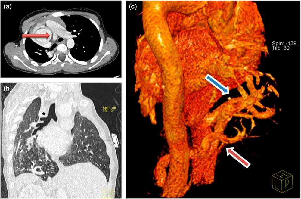
Routine chest radiography was carried out for a 10-year-old boy to investigate mild exertion dyspnoea and cardiac dextraposition. Echocardiographic examination revealed dextroversion, dilatations of the right atrium, left ventricle – left ventricle end-diastolic diameter of 5.8 mm, z score of +3.44 – and aorta – diameter of 3.4 cm, z score of +7.55 – bicuspid aortic valve and mild aortic failure, atrial septal defect of ostium secundum, and hypoplasia of the right pulmonary artery with a peak gradient of 20 mmHg and pulmonary arterial pressure of 30 mmHg and an ejection fraction of 50%±5%. Thoracic CT-scan also revealed a blind ending in the right upper bronchus, a hypoplastic right lung with two lobes, normal pulmonary arterial supply, and venous drainage of the lobes. A third cone-shaped pulmonary vein with a diameter of 7–10 mm draining from basal segments of the right lower lobe to the inferior caval vein, and an additional anomalous artery with a diameter of 7–9 mm branching from the coeliac artery and extending trans-diaphragmatically into the basal segments of the lower lobe of the right lung were detected (Fig 1). Catheter angiography showed the left-to-right shunt along with right ventricle pressures of 23/5 mmHg – systolic or end-diastolic – left ventricle pressures of 79/5 mmHg, systolic pulmonary artery pressures of 22/9/18 mmHg – the main, right or left pulmonary artery – pulmonary vascular resistance of 1.16 U/m2, and pulmonary to systemic blood flow ratio (Qp/Qs) of 1.6. Application of a vascular plug was ruled out because of a high migration risk.
( a ) Thorax CT showing dilated main pulmonary artery with normal left pulmonary artery and hypoplastic right pulmonary artery indicated with a red arrow. ( b ) Coronal reformatted CT image demonstrates the blind-ending right upper lobe bronchus (black asterisk). ( c ) Virtually reconstructed three-dimensional image from the posterior side, showing the anomalous arterial supply (red arrow) and venous drainage (blue arrow).

Operation
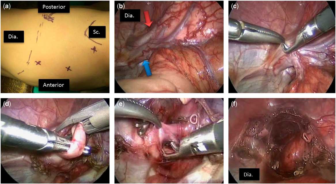
In the left lateral decubitus position, exposure was maintained via three 5-mm ports without the need for single lung intubation. The right lung consisted of two lobes. The scimitar vein and aberrant artery were exposed with their branches that were meticulously dissected, sealed, clipped, and cut. There was minimal bleeding with no complication. Ventilation and perfusion of both lobes were normal at the end of the procedure. The operative time was 110 minutes (Fig 2).
( a ) Positions of the trocars, ( b ) scimitar vein (blue arrow) and anomalous arterial supply (red arrow), ( c – e ) meticulous dissection and ligation of anomalous venous and arterial vessels, ( f ) operative view at the end of the procedure. Dia=diaphragm; Sc=scapula.

Postoperative follow-up
Early postoperative echocardiographic control revealed a significant increase in ejection fraction from 50±5% to 75±8%. The patient had a better effort capacity, and his pulmonary arterial pressure decreased from 30 to 20 mmHg on echocardiography, and his left ventricle end-diastolic diameter decreased from 5.8 cm (z score: +3.44) to 2.8 cm (z score: −3.84) on echocardiography at the 6th month of follow-up.
Discussion
Scimitar syndrome is frequently associated with various cardiovascular anomalies. The incidence is around 1–3:100,000 live births, with female gender predominance (2:1). There are two forms of the syndrome: infantile and paediatric or adult. The first is more severe and fatal, whereas the latter form may vary from less-severe to asymptomatic. Our case represents a rare variant of this syndrome with all aspects of the malformation.
Three-dimensional reconstruction of images obtained using cross-sectional imaging techniques allow for a non-invasive method of diagnosis with a good definition of the anatomy, as it was in our case. Following an elaborate cardiological evaluation, interdisciplinary collaboration is mandatory for an accurate depiction of abnormal vascular anatomy and for planning individualised treatment options.Reference Katre, Burns and Murillo 9
In a multicentric study with 68 patients who underwent intra-atrial baffle repair, reimplantation of the scimitar vein on the left atrium, or pneumonectomy or lobectomy for scimitar syndrome, morbidity and mortality were significantly higher in the pneumonectomy or lobectomy group, in which patients were younger.Reference Katre, Burns and Murillo 9 The researchers of this study found hospital mortality to be associated with pulmonary arterial hypertension, preoperative congestive heart failure, and severe respiratory symptoms; they also found late mortality to be related to right ventricle failure. In our case, the patient had elevated pulmonary arterial pressure, right atrium dilatation, increased left ventricle diameter, and decreased ejection fraction rate, which were considered signs of increased volume of the left ventricle, necessitating closure of the left-to-right shunt.
In a review of the outcome of coil embolisation of anomalous arterial supply in scimitar syndrome, an additional surgical intervention was required in 7 of 16 patients.Reference Uthaman, Abushaban and Al-Qbandi 10 Because of the large calibre of the systemic connection, transcatheter therapy was not preferred for the patient in our case. Thoracoscopic ligation of additional systemic arterial supply from the coeliac artery and systemic venous drainage to the inferior caval vein was feasible as the right lower pulmonary lobe had its own normal pulmonary arterial and venous circulation. The thoracoscopic approach maintained perfect exposure and precise ligation of anomalous arteries and veins. Significant improvement in ejection fraction and exercise capacity and progressive decrease in left ventricle end-diastolic diameter confirmed the efficiency of the procedure.
Conclusion
Scimitar syndrome necessitates multidisciplinary detailed evaluation for associated anomalies with the possible coexistence of a broad range of variations. Treatment options may differ individually, and minimally invasive techniques may safely and efficiently be performed in selected cases.
Acknowledgements
The authors thank Sevinc Kalin for reproducing three-dimensional images, Yasemin Ozdemir Sahan for echocardiographic evaluation, and Timur Mese for angiographic assessment.
Financial Support
This research received no specific grant from any funding agency, commercial, or not-for-profit sectors.
Conflicts of Interest
None.
Ethical Standards
The authors assert that all procedures contributing to this work comply with the ethical standards of the relevant national guidelines on human experimentation and with the Helsinki Declaration of 1975, as revised in 2008, and has been approved by the institutional committees.