Introduction
The family Echinostomatidae Looss, 1899 comprises a globally distributed group of digeneans characterised by complex life cycles involving multiple hosts. These parasites exploit a broad range of final vertebrate hosts, with a particularly high diversity in birds. According to phylogenetic evidence (Tkach et al. Reference Tkach, Olena Kudlai and Kostadinova2016), freshwater pulmonate gastropods typically serve as the first intermediate hosts. A wide variety of invertebrates and vertebrates, with molluscs and amphibians being the most studied, act as second intermediate hosts, in which the encysted metacercaria constitutes the predominant stage (Keeler and Huffman Reference Keeler, Huffman, Fried and Toledo2009). The extensive host diversity and broad geographic distribution help explain the remarkable taxonomic richness observed in the group, which includes over 40 recognised nominal genera (Kostadinova Reference Kostadinova, Jones, Bray and Gibson2005; Tkach et al. Reference Tkach, Olena Kudlai and Kostadinova2016). These characteristics, combined with their complex life cycles, underscore the value of echinostomes for studying evolution, ecology, and systematics.
In South America, more than 80 species belonging to 25 genera of the family Echinostomatidae have been recorded, primarily from Brazil, followed by Argentina, where around 30 species mainly parasitise birds and mammals (Fernandes and Kohn Reference Fernandes and Kohn2014; Fernandes et al. Reference Fernandes, Justo, Cardenas and Cohen2015). A similar number of larval stages of this family have been described in Argentina (Merlo et al. Reference Merlo, Parietti, Fernández, Flores and Davies2022); however, the vast majority remain unlinked to their corresponding adult forms, highlighting a significant gap in our understanding of life cycle connections. The principal obstacle to resolving these associations lies in the fact that digenean alpha taxonomy is fundamentally based on adult morphological traits, particularly reproductive structures, which are absent or rudimentary in larval forms. Although ideally, species descriptions should include larval stages (Blasco-Costa and Poulin Reference Blasco-Costa and Poulin2017), this requires fieldwork targeting intermediate hosts and life cycle elucidation.
Within the family Echinostomatidae, the number and arrangement of collar spines, especially in the genus Echinostoma, are shared by both adults and larvae, which offers a useful approach for identifying cercariae based on adult traits (Kanev et al. Reference Kanev, Fried and Radev2009). However, these morphological characters alone are not sufficient to link larval and adult stages, even more so considering the existence of cryptic species, which can only be distinguished through molecular data (Georgieva et al. Reference Georgieva, Selbach, Faltýnková, Soldánová, Sures, Skirnisson and Kostadinova2013; Valadão et al. Reference Valadão, Alves, López Hernández, Assis, Coelho, Geiger and Pinto2023). Although experimental infections can contribute to life-cycle elucidation, they are often difficult to perform due to the specificity and availability of the hosts involved, require ethical consideration, and do not necessarily provide a clear understanding of their true range of hosts in nature. In this context, molecular data obtained from larval trematodes support the formal description of larval trematodes (Hechinger Reference Hechinger2023) and provide an alternative means of linking the larval stages of parasites to known adults (Assis et al. Reference Assis, Lopez-Hernández, PulidoMurillo, Melo and Pinto2019; Bagnato et al. Reference Bagnato, Gilardoni, Pina, Rodrigues and Cremonte2016; Locke et al. Reference Locke, McLaughlin, Lapierre, Johnson and Marcogliese2011; Nolan and Cribb Reference Nolan and Cribb2004; Pina et al. Reference Pina, Russell-Pinto and Rodrigues2007; Soldánová et al. Reference Soldánová, Georgieva, Rohácˇová, Knudsen, Kuhn, Henriksen, Siwertsson, Shaw, Kuris, Amundsen, Scholz, Lafferty and Kostadinova2017). However, this requires expanding the available database in GenBank and sequencing the same genomic regions for comparisons.
In Argentina, only 4.7% of the larval digenean species described to date have genetic sequences available in GenBank (Merlo et al. Reference Merlo, Parietti, Fernández, Flores and Davies2022). This study provides morphological and molecular data on three echinostomatid morphotypes found in the freshwater snail Biomphalaria aff. straminea (Dunker 1848) from a rice field in Corrientes, Argentina. The ecological and reproductive traits of Biomphalaria, including broad habitat tolerance and self-fertilisation (Gérard et al. Reference Gérard, Carpentier and Paillisson2008; PAHO 1968; Rumi Reference Rumi1986; Tuan et al. Reference Tuan, Ohlweiler, Palasio, Dalla Zanna, Guimarães and Rokni2012), promote its persistence and high prevalence in rice agroecosystems. In South America, Biomphalaria species serve as intermediate hosts in the transmission of Schistosoma mansoni, the causative agent of human schistosomiasis; among them, B. straminea, native to the region, has recognised epidemiological importance, particularly in Brazil. Considering the ecological characteristics of the host, the present study provides new data on echinostomatid trematodes that expand current knowledge of freshwater parasitic fauna and contribute molecular information useful for elucidating their life cycles.
Materials and methods
Sample collection
The molluscs were collected in an agricultural area with cultivated rice plots connected with the Paraná River basin, located approximately 40 km South of Corrientes city, in Corrientes province, Argentina (27°49′09.91″S, 58°46′09.47″W) (Figure 1).
(A) Map of South America showing the location of the study area, and an enlarged map of Corrientes Province (Argentina) indicating the agricultural sampling site in the northwestern region; (B) detailed view of the rice plantation and the sampled channel; the arrow indicates the sampled site.

Figure 1. Long description
Panel A consists of two maps. On the left is a continental map of South America with a black dot in the South marking Argentina. Dashed lines extend from this dot to an enlarged map on the right showing Corrientes Province. This provincial map is bordered by Chaco and Paraguay to the North, Misiones and Brasil to the East, Uruguay to the Southeast, and Entre Rios to the South. A location pin icon in the Northwest region of Corrientes, near the Rio Parana, identifies the specific sampling site. Panel B is a color photograph of an agricultural landscape under a blue sky with scattered clouds. In the foreground, a dirt path runs alongside a narrow water channel filled with aquatic vegetation. A white arrow points downward into this channel. In the background, a vast green rice plantation extends to a distant tree line on the horizon.
Sampling was carried out along the edges of cultivated plots and irrigation channels during flooding periods, from sowing to shortly after rice harvesting, between January 2017 and February 2018. The channels studied (n = 2) were approximately 2 m wide and 250 m long (Figure 1). Collection was performed by two people in the morning for 1.5 hours, using simple mesh nets, locally named “copos” (25 cm frame diameter). The molluscs were transported to the laboratory, where they were enumerated and maintained individually in vials containing 20 ml of dechlorinated water. They were then alternately exposed to artificial photostimulation and darkness for five days, and subsequently evaluated for the presence of cercariae using a Leica M60 stereomicroscope (Leica Microsystems, Germany). Snails that were not releasing cercariae (shedding) were dissected to check for other larval intramolluscan stages (sporocysts, rediae, and metacercariae). For morphological identification of snails, we examined the genital anatomy following Rumi (Reference Rumi1986).
Morphological study
Cercariae were studied alive, with and without vital dyes (0.01% aqueous solutions of neutral red and Nile blue sulphate). Drawings of cercariae were made using a Leica microscope DM 2500 with the aid of a camera lucida (Leica
Microsystems, Germany). Measurements given in the description of each cercaria are based on heat-killed formalin-fixed specimens and expressed in micrometres (μm), with the range followed by the mean ± SD in parentheses. The arrangement of collar spines was analysed following Kostadinova (Reference Kostadinova, Jones, Bray and Gibson2005). Cercariae fixed in hot 4% formalin were preserved in vials with 70% ethanol, and deposited at the Helminthological Collection of the Centro de Ecología Aplicada del Litoral (CECOAL), Corrientes, Argentina, and Museum of Southwestern Biology, University of New Mexico, USA.
Photographs were taken with a Leica DFC 295 camera mounted on a Leica DM 2500 microscope. Specimens to be studied by scanning electron microscopy (SEM) were dehydrated in an ethanol series, dried using the critical point technique, coated with gold-palladium, and examined under a Zeiss Evo 15 Scanning Electron Microscope.
The overall prevalence was calculated following Bush et al. (Reference Bush, Lafferty, Lotz and Shostak1997).
DNA extraction, PCR amplification, and Sanger sequencing
We sequenced a partial portion of the 28S rDNA gene because for many of the echinostomes listed in GenBank, 28S is one of the more well-represented markers in the database for echinostomes (Tkach et al. Reference Tkach, Olena Kudlai and Kostadinova2016) and provides a broader taxonomic comparison of our specimens into genera. Additionally, a partial portion of the nicotinamide adenine dinucleotide dehydrogenase subunit 1 (nad1) was sequenced by polymerase chain reaction (PCR) to discern potentially closely related species (Laidemitt et al. Reference Laidemitt, Brant, Mutuku, Mkoji and Loker2019). One or two cercariae were used for DNA extraction. Genomic DNA was extracted using the QIAamp DNA Micro Kit following the manufacturer’s instructions, with a final elution volume of 35 μl (Qiagen, Valencia, CA). The 28S gene was amplified using forward primer dig12 (5′-AAG CAT ATC ACT AAGCGG-3′) and reverse primer 1500R (5′-GCT ATC CTG AGGGAA ACT TCG-3′) (Tkach et al. Reference Tkach, Littlewood, Olson, Kinsella and S´widerski2003). The volume of each PCR reaction was 25 μl with 1 μl of 50 ng of DNA, 0.8 mM/l dNTPs, 2.5 mM/l MgCl2, 0.25 units of Ex Taq DNA (Clontech, Mountain View, CA), and 0.25 μM/l of each primer. PCR cycles were performed on Eppendorf Mastercycler epigradient machine which were programmed as follows: 2 min denaturation at 94°C; 94°C for 1 min, 60°C for 30 s, and 72°C for 1 min for three cycles; 94°C for 1 min, 59°C for 30 s, and 72°C for 1 min for three cycles; 94°C for 1 min, 58°C for 30 s, and 72°C for 1 min for three cycles; 94°C for 1 min, 57°C for 30 s, and 72°C for 1 min for 20 cycles, and followed by an extension step for 7 min at 72°C. The nad1 gene was amplified using forward primer NDJ11 (Morgan and Blair, Reference Morgan and Blair1998) (5′ -AGA TTCGTA AGG GGC CTA ATA-3′) and the reverse primer NDJ2a (5′-CTT CAG CCT CAG CAT AAT-3′) (Kostadinova et al. Reference Kostadinova, Herniou, Barrett and Littlewood2003). The volume of each PCR reaction was 25 μl with 1 μl of 50 ng of DNA, 0.8 mM/l dNTPs, 2.5 mM/l MgCl2, 0.4 units of ExTaq DNA (Clontech, Mountain View, CA), and 0.25 μM/l of each primer. PCR cycles were performed on Eppendorf Mastercycler epigradient machines which were programmed as follows: 2 min denaturation at 94°C; 94°C for 1 min, 54°C for 30 s, and 72°C for 1 min for three cycles; 94°C for 1 min, 53°C for 30 s, and 72°C for 1 min for three cycles; 94°C for 1 min, 52°C for 30 s, and 72°C for 1 min for three cycles; 94°C for 1 min, 51°C for 30 s, and 72°C for 1 min for 20 cycles, and followed by an extension step for 7 min at 72°C.
PCR fragments were separated by agarose gel electrophoresis and visualised with 0.5% GelRed™ Nucleic acid gel stain (Biotium, Hayward, CA, USA). PCR products were purified using VWR® ExoCleanUp FAST (USA). Both strands were sequenced using an Applied Biosystems 3130 automated sequencer and BigDye terminator cycle sequencing kit Version 3.1 (Applied Biosystems, Foster City, CA). DNA sequences were verified by aligning reads from the 5′ and 3′ directions using Sequencher 5.1 and manually corrected for ambiguous base calls (Gene Codes, Ann Arbor, Michigan). The obtained DNA sequences have been deposited in GenBank under the accession numbers: PZ149699-PZ149702 (28S) PZ546940-PZ546942 (nad 1).
Phylogenetic analysis
28S and nad1 sequences were used in phylogenetic analyses using Maximum Likelihood (ML) analysis. The analysis included 114 Echinostomatidae specimens from NCBI-GenBank for 28S comparisons. We additionally compared 40 nad1 sequences from NCBI-GenBank to compare Drepanocephalus, 147 Echinostoma, and 12 Echinostomatidae nad1. Non-redundant sequences were aligned by eye and edited in MEGA12 (Kumar et al. Reference Kumar, Stecher, Suleski, Sanderford, Sharma and Tamura2024). A total of 1,134 positions were used for 28S alignment and 404 bases (for Echinostoma sp. 20), 430 bases (for Drepanocephalus sp.), and 508 bases (for Echinostomatidae gen. sp. 21 sp.) for nad1 alignments. MEGA12 was used to find the best fit model of substitution for both genes. Heuristic searches were utilised for ML analyses, and 1000 bootstrap replicates were run for each dataset. Uncorrected pairwise distance values (p-distance) were calculated in MEGA12 (Kumar et al. Reference Kumar, Stecher, Suleski, Sanderford, Sharma and Tamura2024). We followed other studies in using a p-distance value >5% in mtDNA markers to provisionally designate our specimens as distinct species (Vilas et al. Reference Vilas, Criscione and Blouin2005).
Results
A total of 3,913 Biomphalaria aff. straminea (Dunker, 1848) were examined; 83 were infected with echinostome larval stages (overall prevalence 2.12%). The following echinostomatid morphotypes were identified: Cercaria Echinostoma sp. 20 (1.63%); Cercaria Echinostomatidae gen.sp. 21 (0.25%); and Cercaria Drepanocephalus sp. (0.23%). Additionally, metacercariae of Echinostoma sp. 20 were detected in 81 snails (2.07%). The morphological and molecular data obtained from the new echinostomatid morphotypes are described below.
Morphological description
Family Echinostomatidae Looss, 1899
Cercaria Echinostoma sp. 20
Voucher specimens: CECOAL 17082922; MSB:PARA: 53269.
Redia (Figure 2)
Echinostoma sp. 20. (A) cercarial body; (B) tail; (C) redia; (D) detail of head collar with collar spines; (E) metacercaria of Echinostoma sp. 20 with detail of head collar with collar spines; (F) detail of cyst. Scale bars: 50 μm.

Figure 2. Long description
A multi-panel line drawing showing the morphology of Echinostoma species 20.
* Panel A shows the cercarial body in a ventral view. It features a prominent oral sucker at the anterior end and a ventral sucker at the center. Internal structures include a branched excretory system marked by small arrowheads and numerous cystogenous glands filling the lateral fields. A scale bar indicates 50 micrometers.
* Panel B depicts the long, slender tail of the cercaria with a visible central canal and lateral fin folds.
* Panel C illustrates an elongated redia containing multiple developing cercariae at various stages of maturation within its body cavity.
* Panel D provides a high-magnification detail of the head collar. It shows the oral sucker surrounded by a collar of sharp, pointed spines arranged in a specific pattern.
* Panel E shows the metacercaria stage, highlighting the head collar with its characteristic spines and the underlying muscular pharynx and esophagus.
* Panel F displays the metacercaria encysted within a spherical, multi-layered cyst. The coiled larva is visible inside, showing the oral sucker and a cluster of excretory granules.
Scale bars representing 50 micrometers are present in panels A, B, C, D, E, and F to provide physical dimensions.
Body lightly orange-brown pigmented 1,750–2,500 (2,104 ± 254)/250–337.5 (277 ± 27) long/wide, containing larger germinal cells and cercaria; muscular pharynx 38–60 (47 ± 27)/34–50 (40 ± 5); intestinal caeca 300–510 (374 ± 62) long, occupying less than 1/3 of redial length; a collar on anterior end, and a pair of appendages near anterior and posterior end.
Light micrographs and scanning electron micrograph of Cercaria Echinostoma sp. 20. (A) Ventral view of cercarial body with refractile granules in the main excretory tubes and cystogenous cells with granule-shaped contents; (B) anterior end with detail of dorsal spines arranged in double row; (C) detail of angle spines; (D) ventral view of cercarial body showing spines and sensory papillae arranged between oral sucker and ventral sucker; (E) ventral view of tail showing the ventral large lobulated fin-folds; (F) detail of papillae in ventral sucker; (G) detail of head collar-spines.

Figure 3. Long description
A multi-panel figure containing seven micrographs labeled A through G.
* Panel A is a light micrograph showing the ventral view of the cercarial body. It features clusters of dark, refractile granules within the main excretory tubes on both sides of the midline. A scale bar indicates 40 micrometers.
* Panel B is a high-magnification light micrograph of the anterior end, revealing a double row of dorsal spines. A scale bar indicates 20 micrometers.
* Panel C is an S E M micrograph showing a close-up of the angle spines near the oral sucker. A scale bar indicates 10 micrometers.
* Panel D is an S E M micrograph of the full ventral body. It shows the oral sucker at the top and the ventral sucker in the lower third, with sensory papillae and spines distributed between them. A scale bar indicates 30 micrometers.
* Panel E is an S E M micrograph of the long, slender tail. A white arrow points to the large, lobulated ventral fin-folds. A scale bar indicates 40 micrometers.
* Panel F is a high-magnification S E M micrograph of the ventral sucker, showing the circular arrangement of papillae around the opening. A scale bar indicates 10 micrometers.
* Panel G is an S E M micrograph focusing on the head collar-spines, showing their arrangement around the anterior tip. A scale bar indicates 10 micrometers.
Measurements based on 10 specimens. Body 250–330 (272 ± 20) long and 125–165 (142 ± 12) wide. Tegument thick with spines and sensory papillae arranged between oral sucker and ventral sucker. Tegument with spines and 10 pairs of sensory hairs on lateral margins of body: 3 pairs at level of oral sucker and 7 pairs between pharynx and ventral sucker. Head collar well developed, with 37 spines: 17 dorsal spines in double row, 6 lateral spines in single row, and 4 angle spines on each side, longer than marginal spines. Oral sucker 36–43 (40 ± 2) long and 36–43 (39 ± 3) wide. Prepharyngeal body absent. Prepharynx present, pharynx muscular, 17–24 (21 ± 2) long and 17–24 (19 ± 3) wide. Oesophagus long, intestine bifurcating just anterior to ventral sucker and caeca reaching level of excretory vesicle. Ventral sucker post-equatorial, 38–52 (45 ± 4) long and 36–49 (45 ± 4) wide. Numerous large cystogenous cells with granule-shaped contents, between pharynx and end of body. Excretory system stenostomate, with main tubes extending from anterior wall of excretory vesicle to prepharyngeal level; main tubes dilated between pharynx and anterior border of ventral sucker, with 36–46 (38 ± 3) refractile granules, ranging in size between 1 and 5 in diameter, the smaller ones near pharynx. Flame cells present in at least 16 pairs; excretory vesicle divided into two unequal chambers. Caudal duct of excretory system enters anterior portion of tail, bifurcates into 2 branches ending on lateral body margins. Tail 500–550 (522 ± 16) long and 35–50 (45 ± 4) wide, with 4 fin-folds: one ventral large lobulated, one dorsal large lobulated which occupy the entire length of the tail, and 2 small fin-folds near end of the tail.
Metacercariae (Figure 2)
Measurements based on 10 specimens. Cysts spherical, on average 145.2 ± 5.5 in diameter; cyst walls composed of 2 layers, the outer layer transparent, 11.3 ± 1.2 thick, and the inner layer opaque, thin, 4.1 ± 0.7 μm thick. The metacercariae could not be excysted entirely; only one specimen retained an intact anterior end. The tegument covered with large spines and the collar-spines with 37 spines, arranged in a configuration consistent with that observed in Echinostoma sp. 20.
Remarks. Considering geographical proximity, two cercariae with 37 collar spines have been described: Cercaria Echinostomatidae N° 2 Martorelli, Alda, La Sala and Montes 2013 from Biomphalaria tenagophila in Boquerón de Franquía, Uruguay (adjacent to Corrientes province) and Echinocercaria sp. XIV Fernández, Hamann and Ostrowki de Núñez 2014 from B. straminea and Biomphalaria occidentalis in Corrientes province. In contrast to the present cercaria, Cercaria Echinostomatidae N° 2 has the collar-spines arranged in simple row, further has a larger body (413 ± 3 vs. 272 ± 20) and suckers (oral 55 ± 4 vs. 40 ± 2, ventral 70 ± 8 vs. 45 ± 4), smaller tail (337 ± 63 vs. 522 ± 16), a greater number of refractive granules (59–65 vs. 36–46), the body has no spines or sensitive hairs in the cuticle, and different number of fin-fold in the tail. Echinocercaria sp. XIV differs from present Cercaria Echinostoma sp. 20 by the arrangement in two rows of dorsal and lateral collar-spines, further has more refractile granules in main tubes of excretory system (84–112 vs. 36–46) and has cystogenous cells with bar-shaped contents.
The characteristics of the collar-spines of the present echinocercaria (well-developed, reniform, with collar-spines not sharply pointed, numbering 31–55, arranged in a single row laterally and a double row dorsally, and with the angle collar spines longer than the marginal spines) are consistent with the genus Echinostoma (Kostadinova, Reference Kostadinova, Jones, Bray and Gibson2005).
The life cycle of only one species of the genus Echinostoma, E. parcespinosum (Martorelli, Reference Martorelli1987), is known in Argentina; however, it does not match the characteristics of the collar of spines of the present Cercaria Echinostoma sp. In Brazil, the life cycles of seven Echinostoma species bearing 37 collar spines have been described (Pinto and Melo Reference Pinto and Melo2013). The Cercaria Echinostoma sp. reported here closely resembles that of Echinostoma barbosai Lie and Basch, Reference Lie and Basch1966, obtained from Biomphalaria glabrata collected near Recife, Brazil (Lie and Basch Reference Lie and Basch1966). Both share the same number and arrangement of collar spines, the presence of cystogenous cells with granule-shaped contents, refractile granules of similar size, and a comparable configuration of fin folds in the tail. However, E. barbosai differs in having a larger body (335–423/169–196 vs. 250–330/125–165), larger suckers (oral 40–52 vs. 36–43 ventral 53–65 vs. 38–52), and a larger pharynx (23–52/19–24 vs. 17–24/17–24).
The molecular analysis suggests that this is a unique species of Echinostoma and it does not group with any other Echinostoma in GenBank. We compared our specimen to 147 different Echinostoma nad1 GenBank sequences (>404 bp) and it did not group with any other specimen or clade (p-distance <5%) (Supplementary Figure 1). The closest specimen was greater than 18% (p-distance value). Additionally, our specimen did not group with any other Echinostoma 28S sequences from GenBank (Figure 4).
Maximum Likelihood tree of Echinostoma sp., Echinostomatidae gen. sp. 21 and Drepanocephalus sp., including 114 GenBank specimens within the family, Echinostomatidae. The ML analysis was based on 1,136 bp of the 28S rDNA gene. A discrete Gamma distribution was used to model evolutionary rate differences among sites (5 categories (+G, parameter = 1.6055)). The rate variation model allowed for some sites to be evolutionarily invariable ([+I], 50.25% sites). Support values are indicated above or below the branches. Nodes that were >90% supported are shown. Sequences highlighted in bold correspond to those collected from this study.

Figure 4. Long description
A Maximum Likelihood tree with a scale bar of 0.05 at the bottom left. The tree branches from left to right.
* The top major clade includes various Echinostoma species such as E. revolutum, E. miyagawai, and E. paraensei.
* Highlighted in blue is Echinostoma sp. Argentina.
* Highlighted in green is a sister clade containing two sequences of Echinostomatidae sp. 21 Argentina with a support value of 100.
* Below this is a clade containing Neoacanthoparyphium echinatoides and the Patagifer Clade.
* Further down is the Echinoparyphium Clade, represented by a large black triangle, and species like Moliniella anceps.
* A middle section contains Ribeiroia, Cathaemasia, and Rhopalias.
* A distinct clade follows containing Drepanocephalus species. Highlighted in orange is Drepanocephalus sp. Argentina, nested with D. spathans and D. mexicanus with a support value of 100.
* The lower section includes Euparyphium capitaneum, Chaunocephalus ferox, and a large group of Petasiger and Neopetasiger species.
* Near the bottom is a clade of Isthmiophora species and a large black triangle representing the Petasiger Clade.
* The tree is outgrouped at the very bottom by Caballerotrema sp. K T 9 5 6 9 4 1.
* Support values are printed at the nodes, with values such as 92, 98, 94, 97, 99, and 100 visible throughout the structure.
Cercaria Echinostomatidae gen. sp. 21
Voucher specimens: CECOAL 1802052; MSB:Para:53268; MSB:Para:53277.
Redia (Figure 5): Orange-brown pigmented body 1,120–2,720/234–365 long/wide, with muscular pharynx 46–58/42–55, and intestinal caeca 242–567 long occupying less than 1/3 of redial length. Two pairs of appendages, 1 anterior and 1 posterior.
Cercaria Echinostomatidae gen. sp. 21. (A) cercarial body; (B) tail; (C) detail of head collar with collar spines; (D) redia. Cercaria Drepanocephalus sp.; (E) cercarial body; (F) cercaria and tail ventral view; (G) detail of head collar with collar spines; (H) redia. Scale bars: 50 μm.

Figure 5. Long description
A multi-panel line drawing showing two species of trematodes. Panels A through D represent Cercaria Echinostomatidae gen. sp. 21. Panel A shows the cercarial body with a prominent oral sucker at the anterior end, a ventral sucker at the center, and complex internal excretory and digestive systems. Panel B shows the elongated tail with fin-like membranes. Panel C provides a close-up of the head collar, featuring a ring of sharp collar spines surrounding the oral sucker. Panel D shows the redia, an elongated larval stage containing multiple developing cercariae. Panels E through H represent Cercaria Drepanocephalus sp. Panel E shows the cercarial body with a large ventral sucker and distinct internal glands. Panel F shows a ventral view of the entire cercaria including the tail. Panel G illustrates the head collar with a specific arrangement of collar spines. Panel H shows the redia stage with several internal embryos. Vertical scale bars representing 50 mu m are placed next to each specimen to indicate physical dimensions.
Cercaria (Figures 5 and 6): The measurements are provided in Table 1. Tegument without spines, with sensory papillae arranged between oral sucker and ventral sucker and sensory hairs on lateral margins of body. Head collar well developed, collar with 37 sharply pointed spines, 29 dorsal and lateral spines in double row, 4 angle spines on each side slightly longer than the lateral and dorsal spines. Oral sucker subterminal, prepharyngeal body present. Pharynx muscular, oesophagus long, intestine bifurcating just anterior to ventral sucker, caeca dorsal to excretory tubes, reaching level of excretory vesicle. Ventral sucker situated posterior to midbody. Numerous cystogenous cells with bar-shaped contents, between pharynx and end of the body. At least 3 pairs of penetration glands observed in the central body between main collecting tubes and oesophagus. Excretory system stenostomate, with main tubes extending from anterior wall of excretory vesicle to prepharyngeal level; main collecting tubes dilated between pharyngeal and ventral sucker levels and filled with 81–113 refractile granules, the smaller ones near pharynx. Flame cells arranged in at least 12 pairs, up to 8 ciliary patches in common collecting ducts. Caudal duct of excretory system enters anterior portion of tail, bifurcates into 2 branches ending on lateral body margins. Tail with 7 fin-folds: 2 dorsal, 2 ventral, 2 lateral, and 1 ventral small lobulated fin-fold at level of the 2 lateral fin-folds.
Light micrographs of Echinostomatidae gen. sp. 21. (A) cercarial body showing main tubes of excretory system with numerous refractile granules; (B) anterior end with detail of dorsal spines arranged in double row; (C) detail of angle spines; (D) detail of prepharyngeal body showing rosette-shaped form. Light micrographs and scanning electron micrograph of cercaria Drepanocephalus sp. (E) ventral view of cercarial body showing refractile granules in the main excretory tubes; (F) latero-ventral view of cercarial body showing the ventral sucker with cup-shaped; (G) anterior end with detail of angle spines; (H) anterior end with detail of dorsal spines.

Figure 6. Long description
The figure consists of eight panels labeled A through H.
Panel A is a light micrograph of Echinostomatidae gen. sp. 21 showing a full cercarial body with a 100 micrometer scale bar. The main excretory tubes contain dark, refractile granules.
Panel B shows the anterior end with a double row of dorsal spines and a 20 micrometer scale bar.
Panel C provides a detail of the angle spines with a 20 micrometer scale bar.
Panel D shows the prepharyngeal body with a rosette-shaped form and a 20 micrometer scale bar.
Panel E is a light micrograph of Drepanocephalus sp. in ventral view, showing refractile granules in the excretory tubes and a 100 micrometer scale bar.
Panel F is an S E M micrograph in latero-ventral view showing a cup-shaped ventral sucker and a 50 micrometer scale bar.
Panel G is an S E M micrograph of the anterior end showing the oral opening and angle spines with a 10 micrometer scale bar.
Panel H is a light micrograph of the anterior end showing dorsal spines with a 10 micrometer scale bar.
Morphometric data of Echinocercaria XIV (voucher ID) and Echinostomatidae gen. sp. 21 found in B. aff. straminea from two rice fields of Corrientes Province, Argentina (in micrometers (μm) minimum–maximum and mean values ± SD provided in parentheses)

Table 1. Long description
The table compares two species across six anatomical categories: Body, Prepharyngeal body, Pharynx, Oral sucker, Ventral sucker, and Tail. For each category, Length L and Width W are provided as a range followed by the mean plus or minus standard deviation.
* Body: Echinocercaria sp. X I V has L 270 to 366 mean 310 plus or minus 25 and W 138 to 222 mean 187 plus or minus 22. Echinostomatidae sp. 21 has L 300 to 375 mean 324 plus or minus 24 and W 125 to 155 mean 142 plus or minus 11.
* Prepharyngeal body: Echinocercaria sp. X I V has L 14 to 21 mean 15 plus or minus 2 and W 18 to 23 mean 22 plus or minus 1. Echinostomatidae sp. 21 has L 13 to 17 mean 15 plus or minus 1 and W 18 to 19 mean 18 plus or minus 1.
* Pharynx: Echinocercaria sp. X I V has L 18 to 23 mean 21 plus or minus 2 and W 11 to 18 mean 15 plus or minus 2. Echinostomatidae sp. 21 has L 12 to 18 mean 15 plus or minus 2 and W 14 to 22 mean 16 plus or minus 2.
* Oral sucker: Echinocercaria sp. X I V has L 34 to 46 mean 41 plus or minus 4 and W 32 to 46 mean 39 plus or minus 5. Echinostomatidae sp. 21 has L 32 to 36 mean 35 plus or minus 1 and W 31 to 36 mean 34 plus or minus 2.
* Ventral sucker: Echinocercaria sp. X I V has L 44 to 62 mean 49 plus or minus 5 and W 46 to 69 mean 52 plus or minus 6. Echinostomatidae sp. 21 has L 40 to 44 mean 42 plus or minus 1 and W 42 to 48 mean 44 plus or minus 2.
* Tail: Echinocercaria sp. X I V has L 480 to 642 mean 566 plus or minus 37 and W 42 to 60 mean 49 plus or minus 5. Echinostomatidae sp. 21 has L 575 to 640 mean 621 plus or minus 20 and W mean 48 plus or minus 3.
* Measurements based on 10 specimens.
Remarks. Although the number of collar spines in the present cercaria is consistent with that reported for species of the genus Echinostoma, the arrangement of the collar spines differs from the fourth 37-spine model proposed by Kanev et al. (Reference Kanev, Fried and Radev2009). In the present cercaria, all collar spines are arranged in a double row, whereas in the model proposed by Kanev et al. (Reference Kanev, Fried and Radev2009) the additional spines are restricted to the latero-oral region. In contrast, the arrangement of all spines in a double row, with 29–45 sharply pointed spines, is characteristic of the genus Echinoparyphium Dietz, 1909, although in this genus the dorsal aboral spines are always noticeably longer than the dorsal oral spines (Kostadinova Reference Kostadinova, Jones, Bray and Gibson2005), a feature that is not clearly evident in the collar of spines of the present cercaria. Two species described within this genus possess cercariae with 35–37 collar spines, one of which bears a finfold on the tail (Yamaguti Reference Yamaguti1975). However, based on its morphological features and genetic identity, it was not possible to assign this cercaria to a specific genus.
Echinostomatidae gen. sp. 21 displays morphological characteristics consistent with those of Echinocercaria sp. XIV, as described by Fernández, Hamann, and Ostrowski de Núñez (2014), with measurements that closely correspond, except for a larger tail observed in the present specimen (Table 1). Both cercariae share the same intermediate host, Biomphalaria straminea, and were collected from rice fields located approximately 10 km apart. Given their marked morphological similarity, it is possible that they represent the same species; however, this could not be confirmed through molecular identification, as such data are not available for Echinocercaria sp. XIV.
To date, no life cycles have been reported in Argentina with cercariae possessing a 37-spine collar (Merlo et al. Reference Merlo, Parietti, Fernández, Flores and Davies2022). In Corrientes Province, Cercaria Echinostomatidae N° 2 was described by Martorelli et al. (Reference Martorelli, Alda, Marcotegui, La Sala and Montes2013), exhibiting a collar with 37 spines, but arranged in a single row.
Molecular analysis of Echinostomatidae gen. sp. 21 suggests that it is a species closely related to Patagifer, but likely not within the genus. There were differences in phylogenetic positioning between the 28S tree and the nad1 tree (Supplementary Figure 2). This is similar to what was found morphologically, where there are certain traits that are Patagifer-like (tail fins, and a prepharyngeal body present); however, the spine count of 37 spines is quite different from the genus. The p-distance value between Patagifer and Echinostomatidae gen. sp. 21 ranged from 6.4 to 12% (for nad1). Mitochondrial genome sequence comparison and continued species sampling will be needed for more accurate phylogenetic placement.
Cercaria Drepanocephalus sp.
Voucher specimens: CECOAL 18020524; MSB:Para:53277; MSB:Para:53270.
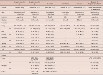
Redia (Figure 5): Body orange-yellow pigmented 863–1,175 (1,015 ± 99)/175–237 (197 ± 23) long/wide, containing larger germinal cells and cercariae; muscular pharynx 54–98 (73 ± 17)/40–78 (56 ± 13); intestinal caeca 450–650 (520 ± 78) long, occupying approximately half of the redial length; a collar in the anterior region and a pair of posterior appendages.
Cercaria (Figures 5 and 6): Measurements based on 10 specimens. Body 260–340 (292 ± 23) long and 100–160 (135 ± 17) wide. Spines and sensory hairs on lateral margins of body were not observed. Head collar falciform, not well differentiated from body with very shallow ventral incision and faint ventral ridge. Collar with 27 spines: 11 dorsal spines, of which 7 central spines are clearly arranged in a double row; 4 lateral spines in single row; and 4 angle spines on each side, larger than the marginal spines. Oral sucker, 36–47 (41 ± 3) long and 34–47 (41 ± 4) wide. Prepharyngeal body absent. Prepharynx present, pharynx muscular, 23–28 (25 ± 2) long and 16–24 (18 ± 3) wide. Oesophagus long, intestine bifurcating just anterior to ventral sucker and caeca reaching level of excretory vesicle. Ventral sucker cup-shaped with deep cavity (Figure 6F), post-equatorial, 32–60 (48 ± 9) long and 41–60 (52 ± 7) wide. Numerous cystogenous cells with granule-shaped contents, between pharynx and end of body. Excretory system stenostomate, with main tubes extending from anterior wall of excretory vesicle to prepharyngeal level; main tubes dilated between pharynx and anterior border of ventral sucker, with 11–20 (16 ± 3) refractile granules, ranging in size between 6 and 15.6 in diameter, the smaller ones near pharynx and ventral sucker. Excretory vesicle divided into two unequal chambers. Caudal duct of excretory system enters anterior portion of tail, bifurcates into 2 branches ending on lateral body margins. Tail 345–440 (388 ± 40) long and 35–50 (48 ± 5) wide, without fin-folds.
Remarks. The falciform collar with a very shallow ventral incision and a faint ventral ridge, the number of spines, the relatively larger angle spines compared with the dorsal and lateral spines, and the structure of the ventral sucker observed in the present echinocercaria correspond to the diagnostic characteristics of adult stages of the genus Drepanocephalus Dietz, 1909 (Kostadinova Reference Kostadinova, Jones, Bray and Gibson2005), with the exception of the arrangement in single row of spines in cephalic collar. In addition, the absence of a prepharyngeal body, a feature of cercariae of Drepanocephalus, is also observed in the material described herein. However, the present cercaria exhibits a double row arrangement of seven centrally located dorsal spines. Although this pattern does not correspond to the dorsal spine arrangement described for the genus (Kostadinova Reference Kostadinova, Jones, Bray and Gibson2005), it has been observed in other species of Drepanocephalus, including adults of D. mexicanus (see line drawings in Hernández-Cruz et al. 2017), metacercariae of Drepanocephalus sp. from Brazil (see light micrographs in Pinto et al. Reference Pinto, Griffin, Quiniou, Ware and Melo2016), and the cercaria of Drepanocephalus sp. from Salta, Argentina (Davies and Ostrowski de Núñez Reference Davies and de Núñez M2024).
In South America, cercariae of this genus have been described in Brazil and in Argentina. Pinto et al. (Reference Pinto, Griffin, Quiniou, Ware and Melo2016) described, using morphological and molecular data, two echinostomatid morphotypes of Drepanocephalus spp. parasitising B. straminea in Belo Horizonte, Minas Gerais, Brazil. Both species (D. auritus and Drepanocephalus sp.) possess oral and ventral suckers with cristae, a feature not observed in Cercaria Drepanocephalus sp. The present cercaria is similar to Drepanocephalus sp. in the number of refractile granules while D. auritus have more refractile granules (11–20 vs. 30). Comparably, cercariae and rediae of present Drepanocephalus sp. cercaria were smaller than larvae of Drepanocephalus spp. from Brazil (Table 2).
Comparative measurements of cercariae of the genus Drepanocephalus described from the Americas. Measurements are in micrometres (μm) (minimum–maximum and mean values ± SD in parentheses). L, length; W, width

Table 2. Long description
The table compares six datasets of Drepanocephalus species. The columns from left to right are:
1. Drepanocephalus sp. (Present study, Argentina, Host: Biomphalaria aff. straminea).
2. Drepanocephalus sp. (Pinto et al. 2016, Brazil, Host: Biomphalaria straminea).
3. D. auritus (Pinto et al. 2016, Brazil, Host: Biomphalaria straminea).
4. D. spathans (Griffin et al. 2012, USA, Host: Planorbella trivolvis).
5. D. auritus (Alberson et al. 2017, USA, Host: Biomphalaria havanensis).
6. Cercaria Cf. Drepanocephalus sp. (Davies and Ostrowski de Núñez 2024, Argentina, Host: Biomphalaria tenagophila).
Measurements for Cercaria include:
- Body Length: Ranges from 234 to 412 micrometres across studies. The present study shows 260 to 340 (mean 292 plus or minus 23).
- Body Width: Ranges from 94.6 to 216 micrometres.
- Oral Sucker Length/Width: Approximately 34 to 49 micrometres.
- Pharynx Length/Width: Approximately 13 to 34 micrometres.
- Ventral Sucker Length/Width: Approximately 32 to 83 micrometres.
- Tail Length: Ranges from 345 to 668 micrometres. The present study shows 345 to 440 (mean 388 plus or minus 40).
Measurements for Rediae (where available) include:
- Body Length: 1,204 to 3,112 micrometres.
- Pharynx Length: 35 to 108 micrometres.
- Caeca Length: 457 to 1,633 micrometres.
In Salta, Argentina, Davies and Ostrowski de Núñez (Reference Davies and de Núñez M2024) described, using morphological data, cercaria cf. Drepanocephalus sp. parasitising B. tenagophila. In contrast to present Cercaria Drepanocephalus sp., cercaria cf. Drepanocephalus sp. shows a slightly higher number of corpuscles (11–20 vs. 21–29), a larger body, suckers, and tail (Table 2), and possesses suckers provided with cristae. In addition, Cercaria Drepanocephalus sp. resembles Echinocercaria sp. 1 described by Fernández et al. (Reference Fernández, Hamann and Ostrowski de Núñez2016), which parasitizes B. occidentalis and B. tenagophila in Corrientes province, Argentina. Both echinocercariae have a cephalic collar with 27 spines, with dorsal spines in double row. However, in Echinocercaria sp. 1 the angle spines are not larger than the marginal spines, and exhibit cystogenous cells containing bar- and granule-shaped contents, a higher number of calcareous corpuscles (23–42), a slightly larger body size (270–396/132–192 vs. 260–340/100–160), and a longer tail (444–600 vs. 345–440). Additionally, the rediae of Echinocercaria sp. 1 are longer (1,200–1,785/180–285 vs. 863–1175/175–237), with the intestinal caecum extending to the posterior end of the body.
On the other hand, in Mississippi, USA, cercariae of Drepanocephalus auritus Kudlai, Kostadinova, Pulis and Tkach, 2015 from Biomphalaria havanensis L. Pfeiffer, 1839 and Planorbella trivolvis Say, 1817 (Alberson et al. Reference Alberson, Rosser, Buddenborg, Khoo, Loker, Richardson, Woodyard, Wise, Pote and Griffin2017), and cercariae of Drepanocephalus spathans Dietz, 1909 from P. trivolvis (Griffin et al. Reference Griffin, Khoo, Quiniou, O’Hear, Pote, Greenway and Wise2012) were molecularly identified. In both cases, morphological descriptions are limited, preventing direct comparison; however, morphometric data indicate that the present cercaria, Drepanocephalus sp., is smaller than larvae of D. auritus and larger than larvae of D. spathans (Table 2).
Molecular sequence data suggests the cercariae recovered from B. aff. straminea in this study represent a species of Drepanocephalus that falls within the same clade as Drepanocephalus specimens collected from cormorants collected in Mexico, from B. straminea collected in Brazil, and from Biomphalaria havanensis from the United States (p-distance values <1.6%) (Supplementary Figure 3). However, there is no consensus on species name in this clade as the adult samples collected from cormorants in Mexico were named D. spathans and the larvae from Brazil and the USA were named D. auritus. Similar problems occur in the sister clade where there is a mix of names used for Drepanocephalus adults and larvae. Additionally, based on a limited number of bp from GenBank (only 434 bp), the p-distance value between our specimen and the other clade was only 4.6%. This is lower than our species cut-off value of >5%. With more sequence data from different specimens, we could more accurately discern if these are two distinct species or not. GWAS studies and mitochondrial genomes are needed. Moreover, adult specimens of Drepanocephalus spathans from Nannopterum brasilianus in Mexico (Hernández-Cruz et al. 2018) and the cercaria described herein share a crescent-shaped head collar, but differ in the arrangement of the dorsal spines. Therefore, we retain the designation Drepanocephalus sp. for our material, as our material is represented only by larval stages, pending additional molecular data and the availability of adult specimens.
Discussion
In general, larval stages remain poorly known, likely less than half of one percent of the expected total number of trematode species (Hechinger Reference Hechinger2023). This represents a major gap in our understanding of trematode diversity and life cycles. In this study, morphological and molecular data are combined to characterise three echinostomatid morphotypes parasitising Biomphalaria aff. straminea in northeastern Argentina. The echinostomatid morphotypes presented here add to the 16 previously reported parasitising Biomphalaria spp. from various aquatic environments in Corrientes Province, Argentina (Fernández et al. Reference Fernández, Hamann and Ostrowski de Núñez2014, Reference Fernández, Hamann and Ostrowski de Núñez2016; Martorelli et al. Reference Martorelli, Alda, Marcotegui, La Sala and Montes2013; Ostrowski de Núñez et al. Reference Ostrowski de Núñez, Hamann and Rumi1990, Reference Ostrowski de Núñez, Hamann and Rumi1991, Reference Ostrowski de Núñez, Hamann and Rumi1997), and to the 36 records of larval digeneans of Echinostomatidae described in the country (Merlo et al. Reference Merlo, Parietti, Fernández, Flores and Davies2022). Although Echinostomatidae is the most represented family in terms of records of larval digeneans in Argentina, molecular data are currently available for the larval stages of only one species parasitising the apple snail Pomacea canaliculata (Martinez et al. Reference Martinez, Gilardoni, Medina, Cremonte and Etchegoin2024). Therefore, the sequences reported here substantially expand the scarce molecular data available for larval stages of this family, contributing to a better understanding of their diversity and host associations in the region.
Two of the echinocercariae described possess 37 collar spines, but with different spine arrangements. The cercaria Echinostoma sp., which presents lateral spines in single rows, was identified as belonging to the genus Echinostoma based on both molecular and morphological analyses. This genus is the most diverse within the family Echinostomatidae, comprising numerous species with a broad range of hosts and distributions (Kostadinova et al. Reference Kostadinova, Herniou, Barrett and Littlewood2003). The adult stage inhabits the intestinal tract of a wide range of aquatic birds and mammals. Available evidence indicates that their cercariae develop in pulmonate (Planorbidae, Lymnaeidae) and prosobranch (Viviparidae) gastropods, whereas metacercariae occur in a broad array of molluscan hosts (Gastropoda, Bivalvia), as well as in planarians and tadpoles (Kostadinova Reference Kostadinova, Jones, Bray and Gibson2005).
In South America, approximately 30 species of Echinostoma are known (Fernandes and Kohn, Reference Fernandes and Kohn2014; Fernandes et al. Reference Fernandes, Justo, Cardenas and Cohen2015; Lunaschi et al. Reference Lunaschi, Drago and Nunez2018; Valadão et al. Reference Valadão, López-Hernández, Alves and Pinto2022, Reference Valadão, Alves, López Hernández, Assis, Coelho, Geiger and Pinto2023), most of them parasitising birds. Brazil is the country with the highest number of records, and also the greatest number of Echinostoma life cycles described, many of which correspond to species bearing 37 collar spines. Cercaria Echinostoma sp. showed similarities to the cercaria of E. barbosai, whose adults have only been reported from experimental domestic bird hosts, including ducks (Anas platyrhynchos), pigeons (Columba livia domestica), and chickens (Gallus gallus domesticus) (Lie and Basch Reference Lie and Basch1966). Nevertheless, E. barbosai has so far only been reported from Brazil, and no molecular sequences of this species are currently available in GenBank for comparison with the present material.
In Argentina, species with 37 collar spines arranged in single lateral rows and a double dorsal row include Echinostoma revolutum (Frölich, 1802) in Myocastor coypus and Echinostoma revolutum ‘group’ in Sturnus vulgaris (Lunaschi and Drago, Reference Lunaschi and Drago2007; Valente et al. Reference Valente, Ibañez, Lorenti, Dafne Fiorini, Montalti and Diaz2014), as well as Echinostoma rodriguensis Hsu, Lie, and Basch, 1968 in Rattus norvegicus (Lunaschi and Drago, Reference Lunaschi and Drago2007). However, based on molecular data and the sequences currently available in GenBank, the Echinostoma cercaria described herein is not closely related to E. revolutum, and no sequences of E. rodriguensis are currently available in GenBank. Despite the high species diversity of the genus in South America, molecular studies involving Echinostoma species remain scarce (Maldonado et al. Reference Maldonado, Loker, Morgan, Rey and Lanfredi2001; Martínez et al. Reference Martinez, Gilardoni, Medina, Cremonte and Etchegoin2024; Valadão et al. Reference Valadão, López-Hernández, Alves and Pinto2022, Reference Valadão, Alves, López Hernández, Assis, Coelho, Geiger and Pinto2023) and none of the sequenced species show close affinity to the cercaria described in this study. Therefore, expanding the molecular database for this group is essential, especially considering that morphological similarity and overlapping diagnostic traits within this genus have led to taxonomic confusion, particularly within the 37-spined E. revolutum group, where integrative approaches have revealed substantial cryptic diversity.
Kostadinova (Reference Kostadinova, Jones, Bray and Gibson2005) characterised the genus Echinostoma by possessing a collar composed of 31–55 spines, with the lateral spines arranged in single row and dorsal spines in double row. This characterisation does not correspond to the observations made for Echinostomatidae gen. sp. 21, as the latter presents lateral spines that are arranged in double row. The molecular results further support this distinction, showing that Echinostomatidae gen. sp. 21 does not align with any of the Echinostoma sequences currently available in GenBank. However, Echinostoma mendax Dietz, 1909, has an arrangement of collar spines similar to Echinostomatidae gen. sp. 21 (dorsal and lateral spines in double row). This species has been reported in Argentina and Brazil parasitising anseriform birds of the family Anatidae (Lunaschi et al. Reference Lunaschi, Cremonte and Drago2007; Travassos et al. Reference Travassos, de Freitas JF and Kohn1969), with a wide distribution of some of them in the province of Corrientes (eBird, 2026a, b, c). Nevertheless, no molecular sequences of E. mendax are currently available in GenBank, preventing a direct comparison with Echinostomatidae gen. sp. 21.
The genus Drepanocephalus comprises a few species that infect the intestines of piscivorous birds, primarily cormorants, throughout North and South America. The species composition of the genus has had an unsettled taxonomic history. The most recent revision (Hernández-Cruz et al. 2017), which incorporated both phylogenetic and morphological analyses, recognises three species: Drepanocephalus mexicanus, previously Petasiger mexicanus (Tkach et al. Reference Tkach, Olena Kudlai and Kostadinova2016); Drepanocephalus spathans; and Drepanocephalus olivaceus (for which molecular data are still lacking). In addition, Drepanocephalus auritus was proposed as synonym of Drepanocephalus spathans. The ML phylogenetic trees recovered in the present study are consistent with this proposal, as sequences identified as D. spathans and D. auritus cluster within the same monophyletic lineage. Furthermore, sequences assigned to both nominal species are intermixed in two closely related sister clades, suggesting that the available molecular data do not clearly support their separation. However, more sequence data is needed to discern if these are two species or are distinct lineages of one species.
Drepanocephalus spathans Dietz, 1909 was described in Argentina parasitising Nannopterum brasilianum (Gmelin, 1789) in the provinces of Buenos Aires and Formosa (Drago et al. Reference Drago, Lunaschi and Schenone2011; Ostrowski de Núñez Reference Ostrowski de Núñez1968). Phylogenetic analyses based on the nad1 gene indicate that the sequences obtained from the present Drepanocephalus sp. cercaria cluster with those of D. spathans from Mexico, suggesting a close genetic relationship between them (<4.6% genetic divergence). Additionally, the other clade comprised mostly larvae and one adult from a vagrant Sula leucogaster from New Mexico. This pattern is consistent with previous reports of this species in the country and in nearby regions where this larval stage has been recorded. However, given the relatively small dataset available (434 bp), additional sequence data and sampling may help to clarify the boundaries between these taxa. To date, molecular data for the larval stages of Drepanocephalus in molluscan hosts have only been reported from Brazil and the United States (Alberson et al. Reference Alberson, Rosser, Buddenborg, Khoo, Loker, Richardson, Woodyard, Wise, Pote and Griffin2017; Griffin et al. Reference Griffin, Khoo, Quiniou, O’Hear, Pote, Greenway and Wise2012; Pinto et al. Reference Pinto, Griffin, Quiniou, Ware and Melo2016). The molecular identification of Drepanocephalus in B. aff. straminea from Argentina expands the geographic representation of the available molecular dataset for these parasites in South America. Molecular data from adult specimens of this genus are still needed in the region to allow comparisons between larval and adult stages.
In this context, molecular identification of both larval and adult stages is essential to establish reliable links between life-cycle stages and, together with morphological and ecological data, to improve species delimitation and clarify phylogenetic relationships in echinostomes species of South America. Echinostomes are important components of trematode communities because they are often competitively dominant within snail hosts (Hechinger et al. Reference Hechinger, Wood and Kuris2011) and can influence the structure of larval assemblages; thus, integrating morphological and molecular data is essential to distinguish species, clarify their interactions and life cycles, and better document their diversity.
Supplementary material
The supplementary material for this article can be found at http://doi.org/10.1017/S0022149X26101680.
Acknowledgements
We are grateful to the owners of the rice fields for granting us permission to carry out sampling on their properties and for generously providing accommodations during sampling. This study was funded by the Consejo Nacional de Investigaciones Científicas y Técnicas (CONICET), Argentina.
Financial support
This study was financed by the Consejo Nacional de Investigaciones Científicas y Técnicas (CONICET) through grant PIP 11220150100119. Technical assistance at the University of New Mexico Molecular Biology Facility was supported by the National Institute of General Medical Sciences of the National Institutes of Health under Award Number P30GM110907. The content for this paper is solely the responsibility of the authors and does not necessarily represent the official views of the National Institutes of Health.
Competing interests
All authors declare that there are no conflicts of interest.