Introduction
Infections of the respiratory tract have a significant economical impact on poultry production worldwide. Various pathogens have been known to cause respiratory diseases; acting either in a primary or secondary role. The avian metapneumovirus (aMPV), also known as turkey rhinotracheitis virus (TRTV), causes infection of the upper respiratory tract of turkeys, chickens and other birds. Related viruses include human, bovine, ovine and caprine respiratory syncytial viruses and pneumonia virus of mice (Arias et al., Reference ARIAS, MACHUCA and PETRUCCELLI2015). It is distributed across America, Europe, Asia and Africa where turkeys and chickens are raised, causing significant production losses. Turkeys and chickens of any age are known to be natural hosts of aMPV. The aMPV infection was first described in South Africa in the late 1970s in turkeys showing clinical signs of coughing, sneezing, nasal and eye secretions. Since then, it has been reported in most regions of the world (Buys and Du Perez, Reference BUYS and DU PEREZ1980). In the beginning, it was thought that SHS was produced by a combined corona virus and Escherichia coli infection (Morley and Thomson, Reference MORLEY and THOMSON1984) but the syndrome could not be reproduced experimentally using both agents. Later, aMPV infections were reported between 1985 and 1986 in turkeys in France (Giraud et al., Reference GIRAUD, BENNEJEAN, GUITTET and TOQUIN1986) and the United Kingdom (McDougall and Cook, Reference McDOUGALL and COOK1986) where the causative agent was isolated and identified as a pneumovirus (Cavanagh and Barrett, Reference CAVANAGH and BARRETT1988). Shortly after its appearance in France and England, aMPV was reported in other parts of Europe, the Middle and Far East and Latin America and was soon recognised as a major disease threat in both turkeys and chickens all over the world (Catelli et al., Reference CATELLI, CECCHINATO, LUPINI, SPERATI, PESENTE, PICCIRILLO, FRANCIOSI and NAYLOR2006; Cook, Reference COOK2009). In the US, it was reported for the first time in the year of 1996 in turkeys (Cook et al., Reference COOK, HUGGINS, ORBELL and SENNE1999). Serological evidence has suggested that aMPV is widespread throughout the world and still studies cited that Australia is the only region free from aMPV (Rivera-Benitez et al., Reference RIVERA-BENITEZ, MARTÍNEZ-BAUTISTA, RÍOS-CAMBRE and RAMÍREZ-MENDOZA2014).
In 1988, the causal agent was characterised as a virus belonging to the Paramyxoviridae family and initially categorised in the genus Pneumovirus (Jones, Reference JONES2001). It has now been reclassified into the genus Metapneumovirus. The virus is an important respiratory pathogen of poultry which causes impaired egg production in turkeys, chickens and ducks (McDougall and Cook, Reference McDOUGALL and COOK1986). Usually, respiratory symptoms have been observed in young birds with a drop in egg production in breeder flocks (Cook, Reference COOK2009). It can lead to significant economic losses, particularly when secondary pathogens (Escherichia coli, Bordetella and Pasteurella spp.) are involved (Giovanardi et al., Reference GIOVANARDI, LUPINI, PESENTE, ROSSI, ORTALI and CATELLI2014; Umar et al., Reference UMAR, ARIF, SHAH, MUNIR, AHMED and KHAN2015; Reference UMAR, SHAH and MUNIR2016a; Reference UMAR, SHAH, SUBHAN, TARIQ and MUNIR2016b).
Due to different clinical presentations the disease has different names. The disease observed in turkeys is called ‘turkey rhinotracheitis’ (TRT), while in chickens is called ‘swollen head syndrome’ or SHS, Generally, it is called ‘avian rhinotracheitis’ as it can affect other avian species. These presentations were named on the basis of clinical signs and lesions observed in different species (Jones, Reference JONES2001).Serological tests such as enzyme-linked immunosorbent assay (ELISA) and virus neutralisation (VN) test are the most commonly used methods to diagnose the aMPV infection. The VN test is performed in a variety of systems, including tracheal organ cultures, chicken embryo fibroblast or vero cells. However, ELISA and VN tests show similar sensitivity (Cook et al., Reference COOK, HUGGINS, ORBELL and SENNE1999) but the ELISA is the most commonly used assay (Seifi and Boroomand, Reference SEIFI and BOROOMAND2015). In most immunoprophylaxis programs, one-day-old turkey poults are administered with live attenuated vaccines against TRT by coarse spray to provide the earliest possible protection of the upper respiratory tract against aMPV infections. Despite vaccination, TRT outbreaks in the field are noted very frequently (Smialek et al., Reference SMIALEK, PESTKA, TYKALOWSKI, STENZEL and KONCICKI2015).Regarding the genetic similarity between human metapneumovirus (hMPV) and aMPV, it was found that hMPV may cause clinical disease in turkeys and aMPV can infect humans (Velayudhan et al., Reference VELAYUDHAN, NAGARAJA, THACHIL, SHAW, GRAY and HALVORSON2006).
This review summarises the currently available information about the aMPV and subsequent disease. Secondly, it addresses the possible pathogenesis mechanism in poultry. Finally, the future prospects of vaccination and other possible novel methods for the control of aMPV are discussed. The aim of this review is to facilitate veterinary practitioners for better control of aMPV infections in poultry.
Classification of the virus
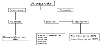
The aMPV, previously called avian pneumovirus (APV) or turkey rhinotracheitis virus (TRTV), is a member of the Paramyxoviridae family, Pneumovirinae subfamily, within the new genus Metapneumovirus (Lamb et al., Reference LAMB, COLLINS, KOLAKOFSKY, MELERO, NAGAI, OLDSTORE, PRINGLE and RIMA2000; Jones, Reference JONES2001).The subfamily Pneumovirinae comprises two genera, Pneumovirus, which are included respiratory syncytial virus (RSV) of mammals, and Metapneumovirus, including human and avian Metapneumovirus ( Figure 1 ).
Classification of Avian Metapneumovirus (aMPV).

There is only one single serotype recognised and four subtypes within that serotype; A, B, C and D on the basis of their antigenicity and diversity of genetic sequence. The highly variable attachment G gene, encoding a surface glycoprotein and serving as one of the major antigens of the metapneumoviruses, is a good candidate for type differentiation and for epidemiological studies (Homayounfar et al., Reference HOMAYOUNFAR, SHOUSHTARI, CHARKHKAR and BOZORGMEHRIFARD2015). The aMPV subgroup A and B were first described in England and France respectively and are highly genetically related. Mainly, aMPV of subtype A or B have been detected in Europe ( Catelli et al., Reference CATELLI, CECCHINATO, LUPINI, SPERATI, PESENTE, PICCIRILLO, FRANCIOSI and NAYLOR2006; Cecchinato et al., Reference CECCHINATO, LUPINI, MUNOZ POGORELTSEVA, LISTORTI, MONDIN, DRIGO and CATELLI2013; Listorti et al., Reference LISTORTI, LUPINI, CECCHINATO, PESENTE, ROSSI, GIOVANARDI, NAYLOR and CATELLI2014), Asia, and in other parts of the world (Rivera-Benitez et al., Reference RIVERA-BENITEZ, MARTÍNEZ-BAUTISTA, RÍOS-CAMBRE and RAMÍREZ-MENDOZA2014). Subtype C aMPV was first reported in turkeys in the USA and subsequently isolated from ducks in France, pheasants in South Korea, and more recently in China (Arias et al., Reference ARIAS, MACHUCA and PETRUCCELLI2015), while the subtype D has been reported only in France (Giraud et al., Reference GIRAUD, BENNEJEAN, GUITTET and TOQUIN1986). Based on nucleotide sequence of the glycoprotein G of aMPV subtypes, it was revealed that subtypes A and B are more closely related to each other than with subtype C while subtype D exhibits differences in sequence of the G gene with respect to the subtypes A, B and C. The genome organisation of aMPV (particularly type C) is quite similar to that of hMPV. The genomes of aMPV and hRSV closely resemble each other, excluding a few differences in the order of the genes and the absence of the non-structural genes from the aMPV genome ( Figure 3 ) (Cook et al., Reference COOK, HUGGINS, ORBELL and SENNE1999; Shin et al., Reference SHIN, NAGARAJA, MCCOMB, HALVORSON, JIRJIS, SHAW, SEAL and NJENGA2002; Gough and Jones, Reference GOUGH and JONES2008).
Molecular virology
The aMPV virion is pleomorphic in nature and its size varies from 150 nm to 600 nm. The genomic orientation of aMPV resembles other members of the Paramyxoviridae family. The virus genome is unsegmented and composed of single-stranded negative sense RNA of approximately 13.4 kilo bases. The genome consists of eight genes that encode for viral polypeptides of which two are glycosylated and three are non-structural virus-specified proteins. The viral polypeptide have been identified as nucleocapsid (N), phosphor protein (P), matrix (M), fusion (F), second matrix (M2), small hydrophobic (SH), surface glycoprotein (G), and RNA dependent RNA polymerase (L), organised as 3- leader-N-P-M-F-M2-SH-G-L-trailer-5′ (Figures 2 and 3). The RNA core is surrounded by M protein and covered by a lipid envelope. This envelope contains the three surface glycoproteins (F, SH, and G), in the form of spikes of approximately 13-17 nm. With the help of the G and F proteins, aMPV attaches and fuses to heparan sulphate receptors on the cell surface. After the fusion process, the viral nucleocapsid enters into the cytoplasm of the host cell and undergoes replication. The newly synthesised viral genome assembles with the viral P, N, L, and M2 proteins, and moves towards the host cell membrane. The virion now buds out of the cell, with the F, SH, and G proteins exposed on the outer side of the membrane ( Figure 4 ). The P protein acts as a co-factor to stabilize the L protein, allowing the formation of the virus ribonucleoprotein (RNP) complex during virus replication. The M protein plays a crucial role in virus assembly and budding by interacting with the RNP complex. The N protein encapsidates the viral genome and protects it from nuclease activity (Gough and Jones, Reference GOUGH and JONES2008).
Structure of aMPV. The viral envelope, containing two main surface glycoproteins: fusion protein and attachment protein, surrounds the single stranded RNA genome which is encapsidated by the nucleocapsid protein and bound by phosphoprotein and the large polymerase protein. Underlying the membrane is a layer of matrix proteins.

Genomic organisation of aMPV. N, nucleoprotein; P, phosphoprotein; M, matrix protein; F, fusion protein; SH, small hydrophobic protein; G, attachment protein; L, large polymerase protein.

Schematic illustration of the life cycle of aMPV. Transcription and replication of the viral genome occurs in the cytoplasm by the action of the viral RNA-dependent RNA polymerase. The newly synthesized viral components translocate to discrete sites at the infected cell plasma membrane where assembly and budding of infectious virus particles occur.

Susceptibility to chemical and physical agents
The susceptibility of aMPV has been studied using a strain isolated from turkeys in Minnesota. The virus remained viable in temperatures at -70 and -20°C for over 26 weeks, at 4°C for less than 12 weeks, 20°C for less than 4 weeks, 37°C for 48 hours and 50°C for less than six hours. The virus kept its activity for 12 cycles of freezing and thawing. It showed resistance to variable pH within the range of 5-9 for one hour. Several disinfectants were effective in reducing the viability of the virus, including ethanol, sodium hypochlorite, iodophos, a phenol derivative and quaternary ammonia. The virus showed surprisingly high resistance and could be recovered on cell culture after seven days of drying at room temperature. In general though, the virus is considered to be quite sensitive (Townsend et al., Reference TOWNSEND, HALVORSON, NAGARAJA and SHAW2000).
Transmission and pathogenesis
Chicken and turkeys are the primary hosts for aMPV infection but other avian species, such as guinea fowl, sparrows, ducks, geese, terns, gulls and pheasants can be infected by aMPV (Shin et al., Reference SHIN, NAGARAJA, MCCOMB, HALVORSON, JIRJIS, SHAW, SEAL and NJENGA2002; Gough and Jones, Reference GOUGH and JONES2008). Transmission of the virus mostly occurs due to direct contact but the rapid spread in many countries indicates that transmission must occur in other ways as well. Indirect spread through transports, equipment, contaminated water and airborne spread has been suggested. Although the virus can be detected in the reproductive tracts in laying birds, there are no evidence for vertical transmission (Gough and Jones, Reference GOUGH and JONES2008). The importance of transmission through wild birds has also been discussed and might explain the rapid spread of aMPV across the world since the first outbreak in South Africa (Cook and Cavanagh, Reference COOK and CAVANAGH2002).The US turkey outbreaks of APV displayed seasonal patterns with approximately 80% of the outbreaks occurring in spring (April to May) and autumn (October to December), suggesting that environmental factors such as migratory birds may be involved in the spread of virus. The detection of aMPV in wild birds is important for the understanding of the epizootology of aMPV in turkeys and chicken and, therefore, for the planning of biosecurity measures and vaccination protocols for poultry farms (Felippe et al., Reference FELIPPE, DA SILVA, SANTOS, SAKATA and ARNS2011).
This virus replicates mainly in the upper respiratory tract, but also in the lungs and air sacs (Gough and Jones, Reference GOUGH and JONES2008). The virus can be isolated from the nasal mucosa of birds up to 14 days post inoculation. PCR can detect genetic material up to 17 days post inoculation (Jones, Reference JONES2001). The aMPV associated with ciliated and non-ciliated cells of epithelioid nasal and tracheal, causing ciliostasis, ciliary destruction and necrosis, which favours colonisation by secondary bacterial agents (Majó et al., Reference MAJÓ, MARTÍ, O'LOAN, ALLAN, PAGÈS and RAMIS1996). The viraemic period is short, which makes isolation because when the birds begin to show clinical signs, the virus is no longer in the blood, and the agents that predominate are those who colonised by the ciliostasis, the ciliary epithelial destruction and necrosis caused by aMPV.
In addition to regulating viral transcription and replication, the M2 protein plays a major role in virulence by decreasing the host's innate immunity. The virus antagonises cellular responses by regulating pattern recognition receptors, such as toll-like receptors and retinoic acid-inducible gene-like receptors and other signalling molecules. Infection interferes with dendritic cell activity and reduces antigen-specific T cell activation. Thus, virus clearance remains incomplete and the chances of re-infection occurring increase (Jones, Reference JONES2001). It is capable of causing the disease with high morbidity and sometimes high rates of mortality in field conditions. However, the situation is very different in experimental conditions in which the birds are exhibiting signs of milder rhinotracheitis. It appears that the difference in severity of clinical signs would be related to the environment in which the birds live and the presence or absence of microorganisms that cause secondary infections. Several bacteria have been isolated during SHS in poultry including, Pasteurella multocida, Mycoplasma spp, E.coli and Staphylococcus spp, although it has been noted that the most important agent was E. coli (Seifi and Boroomand, Reference SEIFI and BOROOMAND2015). It has been noted that clinical signs are exacerbated during the coinfection of aMPV and E.coli indicating their synergistic effects on pathological pictures in poultry ( Majó et al., Reference MAJÓ, MARTÍ, O'LOAN, ALLAN, PAGÈS and RAMIS1996; Gough and Jones, Reference GOUGH and JONES2008).
Experimentally, turkeys infected with Chlamydia psittaci prior to inoculation with aMPV showed a significant increase in the severity of infection (Van Loock et al., Reference VAN LOOCK, LOOTS, VAN DE ZANDE, VAN HEERDEN, NAUWYNCK, GODDEERIS and VANROMPAY2006). Moreover, synergisms between aMPV and ORT agent have been reported to enhance disease severity in poultry (Marien et al., Reference MARIEN, DECOSTERE, MARTEL, CHIERS, FROYMAN and NAUWYNCK2005). The use of vaccines against aMPV reduced disease presentation (Cook, Reference COOK2000). At present, it is considered that aMPV is primary agent of the syndrome and then other bacteria are associated, complicating the picture, such as E. coli, Pasteurella, Ornithobacterium rhinotracheal (ORT) and Mycoplasma spp. (Jones, Reference JONES2001; Seifi and Boroomand, Reference SEIFI and BOROOMAND2015). Of course, as in all infectious diseases, environmental conditions play a crucial role and should be considered as an input in the presentation of the disease.
Clinical features
The clinical signs of aMPV infection are unspecific and characterised by respiratory and reproductive problems. The same clinical symptoms can result from infection with other organisms such as Newcastle virus, avian influenza virus, infectious bronchitis and avian paramyxovirus type 1. Secondary bacterial infections are not uncommon and the characteristic swollen head in SHS is usually caused by a secondary infection with E. coli (Gough and Jones, Reference GOUGH and JONES2008). The aMPV is of great importance in turkeys, where the virus causes a severe infection involving the upper respiratory tract (turkey rhinotracheitis). In younger turkeys common symptoms can be sneezing, nasal and eye discharge, conjunctivitis, submandibular oedema, swollen infraorbital sinus, snicking and rales. Secondary bacterial infections can dramatically increase the clinical signs. Young adults experience coughing and head shaking. Morbidity can be as high as 100%, with mortality ranging from 0.4% to 50%. The disease is associated with reproductive failure, which is of great importance to breeders. The decrease in egg production can be up to 70%. The quality of eggs can be affected with an increased incidence of thin-shells and egg-peritonitis. Severe coughing resulting from a lower respiratory tract infection can sometimes lead to prolapses of the uterus in breeding turkeys (Cook, Reference COOK2000; Gough and Jones, Reference GOUGH and JONES2008).
In chickens the role of aMPV as a primary pathogen was, until quite recently, questioned and infection may not always be associated with clinical signs. In layer breeders, the disease can affect the quality of eggs and cause a range of reproductive tract abnormalities, such as peritonitis, folded shell membranes in the oviduct, misshapen eggs and ovary and oviduct regression (Villarreal et al., Reference VILLARREAL, BRANDÃO, CHACÓN, ASSAYAG, MAIORKA, RAFFI, SAIDENBERG, JONES and FERREIRA2007).The clinical features of SHS is characterised by swelling of the infra-and periorbital sinuses, torticollis, cerebral disorientation, and opistotonus. The morbidity for SHS is often less than 4%, but respiratory signs are often widespread in the flock. The mortality is low, usually not more than 2% (Gough and Jones, Reference GOUGH and JONES2008).
Gross lesions include oedema and purulent exudate in the subcutaneous tissue of the head, neck and infraorbital sinusitis (Aung et al., Reference AUNG, LIMAN, NEUMANN and RAUTENSCHLEIN2008). Poor management conditions may result in more severe pathological lesions as aerosaculitis, peritonitis, pneumonia and peritonitis perihepatitis. Laying hen oviduct prolapse may occur due to intensive irritation and inflammation. In natural outbreaks, the picture tends to be complicated by the presence of associated pathogens, joining more severe lesions as aerosaculitis, peritonitis, pneumonia and perihepatitis (Jones, Reference JONES2001; Bennett et al., Reference BENNETT, LARUE, SHAW, YU, NAGARAJA, HALVORSON and NJENGA2005).
Microscopic lesions include hypertrophy and gland hyperplasia of the mucosa of the nares and loss of ciliated epithelium, along with hyperemia and mild nuclear mono infiltration. These lesions were observed in experimental cases, and may be more severe between four and 10 days after inoculation. Full recovery of the mucosa is observed 18 days after inoculation. Moreover, lesions can be observed in the trachea as in- thickening of the mucosa due to congestion, oedema and mononuclear infiltration (Majó et al., Reference MAJÓ, MARTÍ, O'LOAN, ALLAN, PAGÈS and RAMIS1996; Gough and Jones, Reference GOUGH and JONES2008). In conclusion, aMPV cause circumscribed lesions in the upper respiratory tract.
Diagnosis
Clinical signs are not pathognomonic for a diagnosis of aMPV. A diagnosis may be made by serology, polymerase chain reaction (PCR) or virus isolation. The diagnosis of this disease poses a challenge due to difference in clinical features and genome. The aMPV can be very difficult to isolate and the success rate might below. Moreover, viral isolation can be time consuming and requires additional investigation. Furthermore, for reasons that are not apparent, virus isolation from chickens is more difficult than from turkeys. However, virus isolation is the only method of choice if live virus is required for the research.
Ideally the virus is isolated from nasal secretion and tissue scraped from the sinuses of the affected birds. It has also been isolated from trachea, lung and viscera of affected poults. It is important to collect the samples from flocks that have not yet obtained severe clinical signs as the virus may not be present in sinuses and turbinates for more than six days. Secondary bacterial infections can be the cause of the severe clinical signs seen later and this can be the reason for the difficulties in isolating aMPV from chickens suffering from SHS (Cook and Cavanagh, Reference COOK and CAVANAGH2002; Gough and Jones, Reference GOUGH and JONES2008).
The methods of choice for primary isolation of aMPV are either the use of embryonated eggs (Gough and Jones, Reference GOUGH and JONES2008) or chicken tracheal organ cultures (TOCs) (Arias et al., Reference ARIAS, MACHUCA and PETRUCCELLI2015). Once the virus has been isolated in one of these two systems, it can be adapted to growth in cell cultures (Cook and Cavanagh, Reference COOK and CAVANAGH2002). A multiple approach should be applied to the method of isolation as experience has shown that different isolates can require different methods. To confirm the identity of the aMPV, electron microscopy or immunochemical methods can be used. When studied under the negative staining electron microscopy the aMPV virus has a highly pleomorphic appearance, the particles are enveloped and the sizes range from 80 nm to 500 nm (Gough and Jones, Reference GOUGH and JONES2008). There are three immunochemical methods used for detecting aMPV in turkeys; immunofluorescence (IF), immunoperoxidase (IP) and immunogold staining. For detecting aMPV in chickens IF and IP are being used. The value of these tests under field conditions has not been fully evaluated, nor have any scientific studies been undertaken to compare the sensitivity and specificity of IF and IP (Cook and Cavanagh, Reference COOK and CAVANAGH2002).
Diagnosis of aMPV infection in both turkeys and chickens are usually done serologically. ELISA is the most common serological way of diagnosing aMPV infection (Cook, Reference COOK2000). Other serological methods like VN and indirect immunofluorescence can be used (Cook and Cavanagh, Reference COOK and CAVANAGH2002). The VN test is performed on different tissue culture systems, including TOC, chicken embryo fibroblast or vero cells (Cook et al., Reference COOK, HUGGINS, ORBELL and SENNE1999). The most commonly employed method is the ELISA (SVANOVIR kit; IDEXX-kit). Currently, ELISA kits suitable for testing both chicken and turkey serum are available in the markets. These kits can be used for testing sera from a variety of avian species and claim to have a broad spectrum of sensitivity and specificity for all subtypes of aMPV (Gough and Jones, Reference GOUGH and JONES2008). Serology offers a faster solution, but it is not always trustworthy because aMPV genetic differences can hinder its sensitivity, and gives retrospective information (Cook and Cavanagh, Reference COOK and CAVANAGH2002; Arias et al., Reference ARIAS, MACHUCA and PETRUCCELLI2015).
Molecular methods, such as PCR, allow a rapid, sensitive and specific detection of aMPV. Tracheal swabs and turbinates are considered to be the most appropriate sources of aMPV. Today, a common approach for detection of aMPV by PCR is to amplify the G-gene of the genome. For detecting aMPV subtype A and B the primers G6-in conjunction with G1+ is being used to prime the RT reaction. Cecchinato et al. (Reference CECCHINATO, LUPINI, MUNOZ POGORELTSEVA, LISTORTI, MONDIN, DRIGO and CATELLI2013) developed a new qRTPCR assay for the rapid diagnosis of aMPV. This assay can be effectively used for the detection, identification, differentiation and quantification of aMPV subtype A or subtype B to assist in disease diagnosis and to carry out rapid surveillance with high levels of sensitivity and specificity.
Treatment and control strategies
Currently, there is no treatment available for aMPV-infected turkeys or chickens but use of various antibiotics appears to be helpful in reducing secondary infections and disease severity. Two general strategies have been proposed to control aMPV infections on a poultry farm: (1) reduction of environmental exposure (biosecurity measures), (2) immune interventions (e.g., vaccination) (Umar et al., Reference UMAR, ARIF, SHAH, MUNIR, AHMED and KHAN2015). Good biosecurity is crucial for commercial poultry and egg production and the severity of aMPV infection is significantly dependent on management factors. In terms of biosecurity there are many factors to consider, such as high stocking density, insufficient ventilation and temperature control, poor litter quality, general hygiene, multi-age stock and the presence of secondary pathogens (Gough and Jones, Reference GOUGH and JONES2008).
Vaccination
Even though improvements in management and biosecurity are essential in control and prevention of the infection, vaccination considerably reduces ill health and production losses. Live and inactivated vaccines are available virtually worldwide. When administered correctly they provide excellent protection for turkeys of all ages and are being used for meat turkeys as well as layers and breeders. The live attenuated and inactivated vaccines have been shown to stimulate both systemic immunity and local immunity in the respiratory tract (Gough and Jones, Reference GOUGH and JONES2008). Vaccination is usually achieved by spray or, ideally, by eye drop, but in the future, in ovo might prove to be better (Hess et al., Reference HESS, HUGGINS and HEINCZ2004).
The efficacy of these vaccination methods has been mostly studied under experimental conditions (Ganapathy and Jones, Reference GANAPATHY and JONES2007; Gough and Jones, Reference GOUGH and JONES2008). A single vaccination may be sufficient to protect meat turkeys throughout their life but reinfection can occur later in life and meat turkeys that are reared beyond 10 to 12 weeks are sometimes revaccinated. A typical vaccination program for aMPV in turkeys would be the application of a live subtype A or B strain combined at day-old poults using a coarse spray, repeated at seven to 10 days and again at four to six weeks. The strategy is to produce cell-mediated immunity in the respiratory tract. For egg-laying birds, in addition to the live attenuated vaccines, effective inactivated aMPV vaccines are widely used at 16 to 20 weeks (Gough and Jones, Reference GOUGH and JONES2008). It has been shown, both experimentally and under field conditions, that the use of inactivated vaccines can provide good protection against the effect of aMPV challenge on egg production and quality, although some clinical signs may be seen for a short time after challenge.
However, failure of the vaccine is not uncommon and can be due to several factors such as over attenuation of the virus strains leading to a weak immune response, under attenuation leading to severe reaction or poor vaccine administration (Cook, Reference COOK2000). There are many reports of post vaccinal disease and one underlying reason could be instability of the vaccine (Cook and Cavanagh, Reference COOK and CAVANAGH2002; Catelli et al., Reference CATELLI, LUPINI, CECCHINATO, RICCHIZZI, BROWN and NAYLOR2010). Failures during vaccination, short duration of immunity, and infection by a subtype different than the vaccine strain (which might avoid the immunity conferred against the vaccine strains) have been hypothesised to explain the persistence of the virus. For example, aMPV-C isolates were found to be genetically and antigenically different from aMPV-A and B ( Cook et al., Reference COOK, HUGGINS, ORBELL and SENNE1999; Bennett et al., Reference BENNETT, LARUE, SHAW, YU, NAGARAJA, HALVORSON and NJENGA2005). Consequently, vaccination based on subtypes A and B might offer poor cross protection against infection caused by aMPV-C.
Although maternal antibodies do not protect turkey poults against TRT, almost no studies have been conducted so far regarding the impact of these antibodies on vaccine induced immunity development against aMPV infection. In one-day-old turkey poults, most of which originated from parent flocks vaccinated against TRT, the possible influence of maternal antibodies on the development of vaccine-induced immunity against aMPV is questionable. Maternal antibodies can inhibit vaccine virus replication and influence the development of vaccine-induced immunity. Frequency of TRT outbreaks in the field and/or failure of TRT vaccination may be correlated with differences in the immunological status and/or age of vaccinated turkeys (Catelli et al., Reference CATELLI, LUPINI, CECCHINATO, RICCHIZZI, BROWN and NAYLOR2010; Smialek et al., Reference SMIALEK, PESTKA, TYKALOWSKI, STENZEL and KONCICKI2015).
While general disease is effectively controlled by live vaccines, reversion to virulence of vaccines has been demonstrated on several occasions (Cecchinato et al., Reference CECCHINATO, CATELLI, LUPINI, RICCHIZZI, PROSPERI and NAYLOR2014). There are reports of vaccine breaks and vaccine strains evolving into viral-strains for aMPV which probably contribute to the farmers´ apprehension concerning vaccination against the disease. Field reversion to virulence of live vaccines is more likely to occur in avian than in other species because opportunities for horizontal transfer of vaccines are much increased due to the higher numbers of vaccinates (Cecchinato et al., Reference CECCHINATO, CATELLI, LUPINI, RICCHIZZI, CLUBBE, BATTILANI and NAYLOR2010; Reference CECCHINATO, CATELLI, LUPINI, RICCHIZZI, PROSPERI and NAYLOR2014).Convenience of mass live vaccine application increases the risk of vaccine virus circulation in birds (Listorti et al., Reference LISTORTI, LUPINI, CECCHINATO, PESENTE, ROSSI, GIOVANARDI, NAYLOR and CATELLI2014). Also the precise nature of that application can have a bearing on persistence and disease. In a previous study, aMPV mass vaccine application by spray resulted in less than complete vaccine uptake, although unresponsive birds did eventually receive virus, presumably due to bird-to-bird contact (Ganapathy et al., Reference GANAPATHY, BUFTON, PEARSON, LEMIERE and JONES2010). It is generally believed and has been anecdotally demonstrated that, in this incomplete vaccination scenario, reversion to virulence can be encountered and it was noticeable that many birds in the Ganapathy study only started to show vaccine replication at three weeks of age.
Humoral immunity is strongly stimulated by vaccination or aMPV infection (Jirjis et al., Reference JIRJIS, NOLL, HALVORSON, NAGARAJA and SHAW2002; Liman and Rautenschlein, Reference LIMAN and RAUTENSCHLEIN2007; Smialek et al., Reference SMIALEK, PESTKA, TYKALOWSKI, STENZEL and KONCICKI2015), but antibodies do not play a key role in protection against aMPV infections and should not be considered as indicators of immunity (Liman and Rautenschlein, Reference LIMAN and RAUTENSCHLEIN2007; Rubbenstroth and Rautenschlein, Reference RUBBENSTROTH and RAUTENSCHLEIN2010). It has been shown, however, that high antibody titres suppress aMPV replication in the upper respiratory tract, thus alleviating the clinical course of TRT (Smialek et al., Reference SMIALEK, TYKAŁOWSKI, STENZEL and KONCICKI2012; Reference SMIALEK, PESTKA, TYKALOWSKI, STENZEL and KONCICKI2015).
Cell-mediated immunity (CMI) is increasingly often considered as the decisive factor in protection against AMPV infections. Unfortunately, little is known about local CMI mechanisms in turkeys’ upper respiratory tract during infections and after vaccination against aMPV infections. These mechanisms seem to play a particularly important role because aMPV infects the host through mucosal sites in the upper respiratory tract. Liman and Rautenschlein (Reference LIMAN and RAUTENSCHLEIN2007) demonstrated a significant increase in the percentage of CD4+ T lymphocytes with concurrent upregulation of IL-6 and/or IFN-γ in the Harderian gland (HG) after vaccination or infection with aMPV/A or B. Conversely, Cha et al. (Reference CHA, YU and ZSAK2013) reported an increase in the percentage of CD8+ T cells, but not in CD4+ T cells, in the upper respiratory tract after aMPV/C inoculation. The cited results indicate that CMI actively participates in protection against aMPV infections and that local CMI mechanisms may be related to age and the aMPV subtype. Recently it has been reported that methyltransferase-defective aMPV vaccines provide complete Protection against challenges of aMPV (Sun et al., Reference SUN, WEI, RAUF, ZHANG, MA, ZHANG, SHILO, YU, SAIF, LU, YU and LI2014).
Furthermore, there is a risk of interference between aMPV and IB or NDV vaccines since these viruses target the same cells in the respiratory tract. It is therefore advisable to consider leaving an interval of approximately one week between vaccination against IB and ND and administration of aMPV vaccine ( Cook, Reference COOK2000; Arias et al., Reference ARIAS, MACHUCA and PETRUCCELLI2015).
Conclusions
Relative to other viruses and diseases, aMPV is not well studied and appears to be as dangerous a pathogen as other paramyxoviruses in terms of morbidity and mortality in poultry. The most effective approaches to control the spread of aMPV include strict biosecurity measures, continuous surveillance and vaccination. As an important respiratory pathogen, understanding aMPV pathogenesis is essential for the development of an effective vaccine against aMPV. Recent studies using animal models for aMPV infection have shed some light on aMPV pathogenesis and have allowed us to evaluate live vaccine candidates. Moreover, diagnostic methods need to be improved because serological methods of diagnosis can only tell about the presence or absence of virus but cannot provide information regarding identification or sub typing. Therefore we need to develop efficient diagnostic methods since correct diagnostic methods are crucial in controlling the disease. For this reason, we believe that the development and modification of the molecular viral detection methods such as PCR technique will be very useful to know about aMPV. Vaccination strategy discussed above have certain potentials, but may not be sufficient when applied individually. Therefore, it might be more effective when combined with good biosecurity. Further surveillance is crucial for knowing which subtypes are currently more prevalent in the field, whether alternation between type A and B occurs, and whether the introduction of a possible new subtype of aMPV occurs. This data are needed to implement a good and accurate vaccination program to achieve efficient control and eradication of aMPV infections in poultry.