Coronary artery fistulas are rare congenital malformations resulting from a direct vascular connection between a coronary artery and a cardiac chamber or major central blood vessel without an intervening capillary bed. Although the true incidence is speculative, coronary artery fistula is seen in ~0.3% of patients with congenital heart diseaseReference McNamara and Gross 1 and 0.13–0.22% of adults undergoing coronary angiography.Reference Yamanaka and Hobbs 2 The clinical presentation, natural history, and management strategy for coronary artery fistula is considerably variable, depending on the age, magnitude of the shunt, dilatation of the native coronary artery, and the presence of underlying coronary artery disease.Reference Fernandes, Kadivar and Hallman 3 – Reference Liberthson, Sagar and Berkoben 5 Although there are few reports of spontaneous closure of small fistula,Reference Schleich, Rey and Gewillig 6 the potential complications of fistula with clinically significant shunt and higher morbidity of intervention at advanced age justifies early intervention even in asymptomatic patients.Reference Fernandes, Kadivar and Hallman 3 , Reference Liberthson, Sagar and Berkoben 5 , Reference Latson 7 During fistula intervention, closure is primarily aimed at eliminating the shunt by intervening the branch or parent coronary artery. However, the potential compromise of the nearby coronary branches and possibility of subsequent ischaemia is of paramount importance, particularly in paediatric patients, as coronary insufficiency symptoms are least expected in them. There are a few small studies and case reports of follow-up angiographic delineation of primary fistulous communication with mixed cohort of patients.Reference Jama, Barsoum and Jarnason 8 , Reference Hiraishi, Misawa and Horiguchi 9 We report our experience of clinical and angiographic delineation of coronary circulation after successful coronary artery fistula intervention in children. On the basis of our experience and review of literature, we have discussed a classification of coronary artery fistula with therapeutic implication and technical consideration.
Materials and methods
Patients
From 2007 to 2012, 15 children were treated for coronary artery fistula at our institute. Clinical profile, echocardiographic parameters, and closure techniques during therapeutic intervention were analysed retrospectively from hospital records and archived images.
Inclusion criteria
-
1. Any patient with age <18 years at the time of intervention.
-
2. Presence of congenital aorto/coronary cameral fistula evident by echocardiography with an audible murmur.
Exclusion criteria
-
1. Patients with small, incidentally detected fistula, which is clinically silent.
-
2. Those coronary artery fistulas with additional complex cardiac lesions.
Transcatheter intervention
After written informed consent from parents, all patients were brought to the catheterisation laboratory in post-absorptive state. The procedure was performed under short general anaesthesia. Percutaneous vascular access was obtained using the femoral artery and vein. Unfractionated heparin 100 units/kg and prophylactic antibiotics were administered intravenously. Selective coronary angiography was performed in various orthogonal views to delineate the opening of fistula, its dimensions, and the connections to cardiac chambers.
Antegrade (venous) approach (n=11)
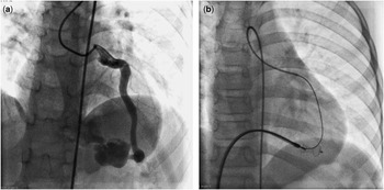
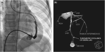
In patients with single-exit coronary artery fistula opening into the right heart structure, antegrade approach was used (Figs 1 and 3). After selective cannulation of the fistulous coronary artery with diagnostic catheter, a coronary guidewire was tracked through the fistula into the distal circulation, and then a 4–5 F multipurpose catheter was used to exchange for the 260 cm 0.035′′ Radiofocus guide wire (Terumo Corporation, Tokyo, Japan). An arteriovenous loop was established by exteriorising the guidewire by Amplatz snare through the femoral vein. Over this guidewire, a long sheath (Cook, United States of America) of appropriate size was advanced into the fistulous tract by railroad manoeuvre. The distal-most constricted site of the fistulous tract was measured during diastole in different projection, and then Cardi-O-Fix duct occluder oversized by 2 mm (Starway Medical Technology Inc., Beijing) was selected. Under fluoroscopic guidance, the device was advanced through the sheath, whereas guidewire was maintained in situ (Fig 1b). The device was deployed at the fistula exit in conventional manner as in patent ductus arteriosus device closure. Then the guidewire was pulled out from the venous end. Once the selective angiography confirmed the suitable device position and complete closure, the duct occluder was released by unscrewing it from the cable (Fig 2).
( a ): Single-exit parent coronary artery fistula (CAF) from left anterior descending coronary artery (LAD) to right ventricle (RV); ( b ): antegrade device closure of exit segment at RV by wire-maintaining technique.

( a ) Angiographic delineation of distal segment of coronary artery fistula (CAF) and device position before unscrewing the device. ( b ) Multislice spiral computed tomography (MSCT) angiography of LAD to right ventricle (RV) CAF at follow-up. LAD=left anterior descending coronary artery; LCX=left circumflex coronary artery; RCA=right coronary artery.

Retrograde (arterial) approach (n=2)
Retrograde approach was used in patients with coronary artery fistula with multiple exits. The coronary guidewire was tracked across the fistula into the distal circulation. Similar to the antegrade approach, the technique of sequential exchange of stiffer guidewire was used to track down the delivery sheath or guiding catheter to the distal-most segment of the fistula.
Surgical closure (n=2)
Of the patients with large tubular nonrestrictive fistula from the right coronary artery to the right ventricle, two were not suitable for transcatheter closure because of higher risk for procedural complications (device migration and vascular complication). Both these patients underwent surgical closure of the fistula by external plication near the parent coronary artery on the beating heart without the use of cardiopulmonary bypass.
Post-intervention management
All patients were monitored in the intensive cardiac care unit for the first 24 h and were treated with antibiotics and antiplatelet drugs. Serial electrocardiographies and cardiac enzymes were monitored until discharged from the hospital. Echocardiography was performed to look for any residual shunt, regional wall motion abnormality, and left ventricle systolic function. Patients were discharged with advice to continue aspirin and clopidogrel for 6 months, and then aspirin indefinitely.
Follow-up
Follow-up data consisted of clinical history, electrocardiography, echo evaluation for the presence of residual shunt, and wall motion abnormalities. All patients underwent conventional or multi slice computed tomography (CT) coronary angiography after a minimum of 6 months following closure.
Statistical analysis
All data are presented as percentages and means with standard deviation using SPSS 11.0 Software Package.
Results
Clinical profile
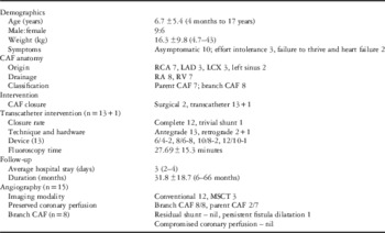
(Table 1) A total of 15 patients’ records satisfied the inclusion criteria. One patient (post-tetralogy of Fallots repair) had a very small coronary artery fistula draining into the left ventricle with insignificant shunt and was excluded from the study. There were six boys and nine girls. Coronary artery fistula intervention was attempted in 15 eligible patients by transcatheter (n=13) or surgical (n=2) closure technique. The mean age and weight were 6.7±5.4 years and 16.3±9.8 kg, respectively. Majority of the patients (n=10) in our study were asymptomatic. Of the 15 patients, 13 (87%) had continuous murmur and two patients had only systolic murmur on clinical examination. Electrocardiography was normal except nonspecific ST-T wave changes in two patients (13%). Of the patients, six had cardiomegaly on chest roentgenogram. Transthoracic echocardiography revealed gross dilatation of the respective coronary artery ostium and proximal segment in parasternal long-axis view. In two patients, (right coronary artery to right ventricle) the entire length of the tubular fistulous tract with exit segment was well delineated. However, in 13 patients, the turbulent colour flow with continuous signal and diastolic accentuation in the receiving chamber (right atrium=7, right ventricle=5) confirmed the exit point in different short- and long-axis views across the right heart structure. Left atrium and left ventricle volume overload was evident in all patients, and seven patients had mild dilatation of right atrium. Measured right ventricle systolic pressure by tricuspid regurgitation velocity on Doppler analysis was 3.16±0.4 m/s.
Clinical and procedural details of CAF patients.

CAF=coronary artery fistula; LAD=left anterior descending coronary artery; LCX=left circumflex coronary artery; MSCT=multislice spiral computed tomography; RA=right atrium; RCA=right coronary artery; RV=right ventricle
Cardiac catheterisation and coronary anatomy
The most common origin of the coronary artery fistula was the right coronary artery (n=7) followed by the left anterior descending coronary artery (n=3) and left circumflex coronary artery (n=3). In two children, the coronary artery fistula originated from the left coronary sinus of the aorta very close to the origin of the left circumflex coronary artery. All the fistulas drained into the right heart structures (right atrium=8, right ventricle=7). Of the patients, 14 had single-exit fistula, whereas one had multiple exits. In addition to this morphological analysis, the fistula was further defined on the basis of branch or parent vessel involvement. A total of eight children had fistula originating from the proximal branch of the parent coronary artery and all drained exclusively into the right atrium. Whereas in seven patients with the parent coronary artery fistula, the fistula arose from the distal segment of either the left anterior descending coronary artery or right coronary artery, and all drained into the right ventricle. One patient with the left anterior descending coronary artery fistula draining into the right ventricle had single coronary artery arising from the left coronary sinus with the absence of the contralateral right coronary artery. Preprocedure multislice spiral CT angiography scans were available in two patients.
Transcatheter closure (n=13)
(Figs 1, 2 and 3) Transcatheter closure was successfully accomplished by retrograde (n=2) or antegrade (n=11) approach. The mean size of the fistula exit was 4.89±1.63 mm. The mean fluoroscopy time was 27.69±15.3 min. Single-duct occluder was used in each patient. The most common device deployed was 8/6 mm (n=8). Post-device closure angiograms showed complete occlusion in 12 patients (93%) and trivial residual flow in one patient (7%). There were no periprocedural complications. During intensive cardiac care unit observation, no patient had ischaemic electrocardiography changes (ST-T changes, bundle branch blocks) or arrhythmias. None of the study patients had dissection or thrombosis of the parent coronary artery. The average hospital stay in the transcatheter group of patients was 3 days.
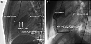
( a ) Branch CAF before device closure. ( b ) Branch CAF during follow-up angiography. CAF=coronary artery fistula; PDA=patent ductus arteriosus; RCA=right coronary artery.

Surgical closure
Of the patients, two had large non-restrictive fistula from the right coronary artery to the right ventricle and were not suitable for the device closure. The surgery was uneventful and without periprocedural complications.
Follow-up angiography
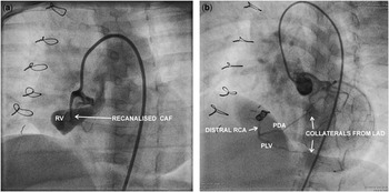
During regular clinical and echocardiography follow-up, all 15 patients were asymptomatic and none of the patients in the transcatheter group had residual flow, recanalisation, or segmental wall motion abnormality. Conventional (n=12) or multislice spiral CT coronary angiography (n=3) was performed in 15 patients at mean follow-up of 31.8±18 months. In the surgical group, an infant had continuous murmur because of recanalisation of the fistula at 6 months’ follow-up. During catheterisation, the fistula was closed by 0.038; 5/3 coil embolisation (Cook). In patients with branch (proximal) coronary artery fistula, all (n=8) had near-complete thrombotic occlusion of the fistulous tract with preserved parent coronary artery circulation. Of the seven patients (transcatheter=5 and surgical=2) with native/parent (distal) coronary artery fistula, five had complete occlusion of native coronary artery fistula extending into the nearby or distal coronary branches. However, the course and calibre of the proximal segment of the native coronary artery were preserved. Of these five patients, two had surgical closure of right coronary artery to right ventricle fistula and they had major collaterals from the left coronary artery circulation feeding the distal right coronary branches, whereas three patients with transcatheter closure had minor collaterals from the opposite coronary circulation. Only two of the seven patients with parent coronary artery fistula had preserved myocardial perfusion, and in both these patients left anterior descending coronary artery to right ventricle fistulas were closed by antegrade delivery of the device at the distal most exit into the right ventricle (Fig 2b). However, left ventricular systolic function was preserved, regardless of antegrade coronary perfusion status.
Discussion
Single origin coronary artery fistula is the most common variant (>90%), whereas bilateral coronary artery fistula s are reported in 5% cases.Reference Liberthson, Sagar and Berkoben 5 , Reference Shyam Sunder, Balakrishnan and Tharakan 10 – Reference Baim, Kline and Silverman 13 All our patients had single coronary artery fistula draining into the right atrium or right ventricle. Over 90% of the fistulas drain into the venous structures of circulation, whereas drainage into the left atrium is rare.Reference Levin, Fellows and Abrams 4 , Reference Liberthson, Sagar and Berkoben 5 , Reference Mavroudis, Backer and Rocchini 11 , Reference Holzer, Johnson and Ciotti 14 , Reference Schumacher, Roithmaier and Lorenz 15 The early clinical studies have reported that young patients are generally asymptomatic and have lower rate of complication, although unoperated patients are more likely to develop complications with increasing age, and consequent high morbidity–mortality when intervened later.Reference Fernandes, Kadivar and Hallman 3 , Reference Levin, Fellows and Abrams 4 , Reference Liberthson, Sagar and Berkoben 5 , Reference Mavroudis, Backer and Rocchini 11 Therefore, elective closure of clinically apparent – coronary artery fistula – is recommended in children, regardless of symptoms.Reference Latson 7
Transcatheter intervention
In recent years, transcatheter coronary artery fistula closure by newer devices is reported to yield a high degree of procedural success with low risk of serious complications.Reference Jama, Barsoum and Jarnason 8 , Reference Xu, Xu and Jiang 16 – Reference Qureshi and Tynan 20 The most important structural prerequisite for the successful transcatheter closure of any coronary artery fistula is the presence of single, narrow, restrictive drainage site into the cardiac chamber or vessel. As all our patients had moderate to large fistula, we attempted to close the exit site of the fistula whenever the transvenous approach was used and the duct occluder was the most preferred device. While during retrograde approach, the vascular plug has an edge over the duct occluder, as it requires smaller size sheath. In the first two patients, the duct occluder was used during the retrograde approach owing to non-availability of the plug. Our transcatheter approach differed from the conventional technique in two ways. First, we adopted the technique of sequential exchange of guidewire. We always used coronary hydrophilic guidewire to avoid inadvertent injury to the normal coronary branch while negotiating the tortuous fistula. And then it was exchanged for stiffer and supportive guidewire through the 4 or 5 F multipurpose catheter. Second, in 11 patients, the duct occluder was deployed successfully at the most distal constricted site by the antegrade approach over the arteriovenous loop with wire-maintaining technique (Fig 1b). The wire-maintaining technique has two advantages: (1) guidewire in situ maintains the antegrade access to the fistula, the most critical step in the whole procedure. This is of particular importance when we require changing the device for any reason. (2) In patients with tortuous fistula, guidewire prevents the kinking of sheath and facilitates the device advancement. This technique appears to be the preferred technique to close majority of single-exit branch coronary artery fistula (Fig 3). In addition to the facilitated delivery of the duct occluder at the desired site distant from the parent or large coronary branches, it ensures complete closure of the fistula. The retrograde approach is used in patients with coronary artery fistula with either multiple exits or drainage into the left-heart structures.
Surgery
Before the advent of the transcatheter approach, surgical closure was the conventional intervention with good short- and long-term results.Reference Mavroudis, Backer and Rocchini 11 , Reference Schumacher, Roithmaier and Lorenz 15 Cardiopulmonary bypass is used in approximately one-half of the cases, especially while using endocardial approach.Reference Mavroudis, Backer and Rocchini 11 , Reference Cheung, Au and Cheung 21 , Reference Malekahmadi and Shahmohmadi 22 Overall, the mortality rate reported in surgical series is <1%.Reference Holzer, Johnson and Ciotti 14 , Reference Xu, Xu and Jiang 16 , Reference Skimming, Gessner and Victorica 19 Recurrence or incomplete closure may occur in ~10% of cases.Reference Cheung, Au and Cheung 21 In our study, of two patients of surgical ligation, one had a significant residual fistula, which was later closed by coil embolisation. Similarly, Cheung et alReference Cheung, Au and Cheung 21 have reported 19% residual or recurrent fistula on angiography in 21 patients, with higher incidence after endocardial ligation.
Early complications
Since the first description of transcatheter closure of the coronary artery fistula in 1983, several studies have evaluated the safety and efficacy of transcatheter closure using various embolisation devices such as detachable balloon, stents, coils, double umbrella device, duct occlude, and vascular plug. Embolisation coils, the most commonly used material, may lead to permanent occlusion; however, suboptimal acute closure rate, potential risk of embolisation, and higher recanalisation rate are the major limitations.Reference Armsby, Keane and Sherwood 23 – Reference Okubo, Nykanen and Benson 26 Similarly, detachable balloons are also not suitable in children, because they require large sheaths and embolisation can occur if there is premature balloon deflation.Reference Skimming, Gessner and Victorica 19 , Reference Qureshi and Tynan 20 Another procedure-related uncommon complication is the occlusion of the fistula or coronary artery because of thrombosis, dissection, or coronary spasm.Reference Jama, Barsoum and Jarnason 8 , Reference Armsby, Keane and Sherwood 23 , Reference Collins, Mehta and Benson 25 , Reference Gowda, Forbes and Singh 27 In addition to higher acute closure rate, we did not encounter any of these procedure-related complications, possibly because of our simplified transcatheter approach, and appropriate selection and deployment of the device occluding the coronary artery fistula at the distal-most constriction site. Xu Liang has reported complete closure of the coronary artery fistula in six patients with the duct occluder deployed by antegrade approach.Reference Xu, Xu and Jiang 16
Late complications and follow-up coronary angiography
Although there are a few studies on long-term clinical and echocardiography follow-up after surgical and transcatheter closure,Reference Shyam Sunder, Balakrishnan and Tharakan 10 , Reference Mavroudis, Backer and Rocchini 11 , Reference Cheung, Au and Cheung 21 longitudinal information about the angiographic delineation of the shunt and status of native coronary circulation after transcatheter closure of coronary artery fistula in children is limited.Reference Latson 7 , Reference Jama, Barsoum and Jarnason 8 , Reference Hiraishi, Misawa and Horiguchi 9 , Reference Gowda, Forbes and Singh 27 , Reference Gowda, Latson, Kutty and Prieto 28 , Reference Horiuchi, Abe and Tanaka 29 Follow-up angiographic delineation of coronary artery fistula is imperative to look for two important late complications: first, recanalisation of previously occluded shunt, and second, coronary perfusion status of the native artery and nearby branches. Post-coronary artery fistula closure sequelae include persistent coronary dilatation, residual leak, thrombosis with or without myocardial infarction, and coronary stenosis with perfusion defects.Reference Hiraishi, Misawa and Horiguchi 9 , Reference Cheung, Au and Cheung 21 , Reference Gowda, Forbes and Singh 27 , Reference Gowda, Latson, Kutty and Prieto 28 , Reference Horiuchi, Abe and Tanaka 29 Recanalisation rate after coronary artery fistula intervention is reported as high as 22%. Of the 18 patients with follow-up angiography, four had large recanalisation requiring repeat closure in a study by Abdi Jama et al.Reference Jama, Barsoum and Jarnason 8 Another potential complication first described by Latson et alReference Latson 7 in his four patients after 4–14 years of surgical fistula closure is thrombotic occlusion of the entire parent coronary artery. In one of the most rigorous follow-up studies – 51.2% follow-up angiography – in adult patients by Cheung et al,Reference Cheung, Au and Cheung 21 all but one patient had some evidence of abnormal coronary morphology, including thread-like or completely occluded distal coronary in 10 of 21 patients and collaterals in two patients. Similarly, Hiraishi et alReference Hiraishi, Misawa and Horiguchi 9 had reported anatomical and physiological evidence of myocardial ischaemia 5–9 years after surgical closure of coronary artery fistula in two of their patients.
In our study, at 31.8±18.7 months mean follow-up, all patients were asymptomatic with adequate weight gain and had no evidence of residual or recurrent fistula. All but one patient with branch (proximal) coronary artery fistula had complete thrombotic occlusion of the fistulous tract with the patent parent coronary artery. A 14-year-old female patient with branch coronary artery fistula from right coronary artery to right atrium had persistent large cul de sac with swirling of contrast at 38 months follow-up, and she was scheduled for multislice spiral computed tomography angiography after 2 years. Among the group of parent coronary artery fistula, only two of the seven patients had shunt closure with preserved native coronary perfusion. Both these patients had closure of the distal-most exiting segment of the left anterior descending coronary artery to right ventricle fistula by antegrade technique and, except for the localised thrombotic occlusion of the fistula, the rest of the fistulous segment regressed to near-normal diameter with preserved coronary flow at mid-term follow-up. Although the minor coronary branches were compromised owing to localised thrombosis in the distal left anterior descending coronary artery segment adjoining the exit point, thrombus propagation was quite restrictive even at mid-term follow up in these two patients (Fig 2b). S Gowda et al, also observed healthy remodelling of the conduit coronary artery after transcatheter closure of medium-sized “distal” coronary artery fistula.Reference Gowda, Forbes and Singh 27 Whereas shunt closure with preserved coronary circulation established the desired outcome in these two patients, the other five patients with parent coronary artery fistula had either thrombotic or direct obliteration of the nearby or distal coronary branches evident by minor – three patients with transcatheter closure – or major – two surgical closure – collateral from the contralateral circulation. In two patients with surgical closure of large right coronary artery to right ventricle, the fistula was completely obliterated and there was absence of antegrade flow into the distal right coronary artery and branches. In fact, both these patients had major collaterals from the left coronary artery circulation filling the distal right coronary artery and branches during closure (Fig 4). Similarly, three patients with the parent fistula had minor collaterals feeding the distal circulation after transcatheter closure as well. However, in all these three patients, the targeted device closure segment was absolutely not the distal segment. In fact, one of these two patients with retrograde closure had device closure of mid segment of single-exit right coronary artery to right ventricle fistula, and probably it was the very first patient in our study. Whereas in the second patient with multiple-exit large left anterior descending coronary artery coronary artery fistula to right ventricle, the catheter could not be negotiated to the desired distal segment. The third patient with compromised distal right coronary artery circulation and minor collaterals had antegrade closure of the fistula and again, the mid segment was closed, as it was the only narrow constricted segment. The other uncommon mechanisms of revascularisation and remodelling after coronary occlusion include angiogenesis and arteriogenesis.Reference Gowda, Forbes and Singh 27 We observed that during surgical or transcatheter closure of parent coronary artery fistula, unless the distal-most exit site of the coronary artery fistula could be targeted precisely, then a more vigilant surgical closure with an additional graft to the distal vessel might possibly curtail the future risk of distal myocardial perfusion. Although patients with collateralised circulation have normal resting electrocardiography and echocardiography, long-term vigilant follow-up for myocardial perfusion is essential. Considering the risk of ongoing thrombotic occlusion of aneurysmal fistulous tract, all our patients are advised to continue antiplatelet medicine for an indefinite period.
( a ): Recanalisation of parent CAF from RCA to right ventricle (RV) after surgical closure; ( b ): transcatheter coli closure of recanalised CAF and major collateral from left coronary circulation feeding the distal branches. CAF=coronary artery fistula; LAD=left anterior descending coronary artery; PDA=posterior descending artery; PLV=posterior left ventricular branch; RCA=right coronary artery.

Owing to faster image acquisition and higher resolution, multislice spiral computed tomography confers an edge over conventional angiography in identification of anomalous origin and course of the coronaries, assessment of the complexity of the fistula, and preoperative evaluation. Multislice spiral computed tomography also delineates the drainage site into the low-pressure chamber more accurately. In one of our study patients – coronary artery fistula with single coronary artery, antegrade device closure was planned on the basis of three-dimensional images of the entire course of fistulous left anterior descending coronary artery. Although the advanced dual-source multislice spiral computed tomography with electrocardiography-gated scanning protocols have overcome the issues of cardiac CT in infants and young children with thin coronary arteries and high heart rate, the high-dose radiation to smaller body size with consequent lifetime risk for developing radiation-induced cancers constitute a major drawback. Therefore, the most important consideration is to keep the radiation dose “as low as reasonably achievable” and “as low as reasonably practicable” with the balance of interpretable image quality that needs to be diagnostic, not the optimum. As observed in our study, multislice spiral computed tomography angiography also constitutes an ideal non-invasive modality to evaluate therapeutic efficacy of coronary artery fistula closure by complete delineation of the native coronary circulation, fistulous tract, presence of thrombus, or collaterals.
Classification
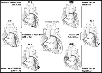
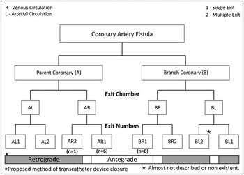
Several classifications and management guidelines of coronary artery fistula are described in literature,Reference Gowda, Forbes and Singh 27 – Reference Sakakibara, Yokoyama and Takao 30 but none of them have any implication for transcatheter device closure. Once it is decided to close the fistula, the critical step in intervention strategy is the angiographic delineation of the anatomy and morphology of the entire fistulous tract. We have proposed anatomical classification (Figs 5 and 6) based on three important variables that decide the transcatheter therapeutic approach. These include the identification of (1) fistulous structure, whether it is parent coronary – left anterior descending coronary artery/left circumflex coronary artery/right coronary artery – or one of the branches (A or B), (2) exit chamber, is either venous or arterial system circulation (R or L), and (3) single or multiple (1 or 2). Accordingly, there are eight different categories of coronary artery fistula possible. However, branch vessel fistula with multiple openings are either not reported in the literature (BL2) or detected incidentally in adult patients during angiography (BR2), and hence practically six types are clinically important in children.
Diagrammatic representation of classification of CAF. CAF=coronary artery fistula; LAD=left anterior descending coronary artery; RV=right ventricle.

Flowchart of classification of coronary artery fistula (CAF).

Type A: parent coronary artery fistula (AR1, AL1, AR2, AL2)
Where the entire parent or native coronary artery is significantly dilated and tortuous with more than one branche, perfusing the normal myocardium arises from at least the distal half of the tortuous coronary artery. This type of fistula morphology matches with the “Distal” coronary artery fistula, described in earlier classification. Salient features are: (1) they can have either single or multiple openings. (2) As the exit is either the distal coronary artery or bifurcation branches, the ventricle is the draining chamber in most cases. The common sequelae after transcatheter closure of these fistulas are either positive remodelling of the medium-sized parent/conduit coronary artery or chronic thrombotic occlusion of the large parent/conduit artery with neorevascularisation or collaterals perfusing the distal myocardium. Srinath et al have concluded from their retrospective analysis of two different series of 16 patients that this type of fistula, particularly the larger one in older patients, carries higher risk of acute or subacute thrombotic occlusion.Reference Gowda, Forbes and Singh 27 , Reference Gowda, Latson, Kutty and Prieto 28 Sparing few cases of coil closure, majority of these patients had surgical closure. Although vigilant care is mandatory during transcatheter intervention, with availability of newer Amplatzer plugs, parent coronary artery fistula can be intervened safely by retrograde or antegrade approach (AR1 – parent coronary artery fistula opening into venous circulation by single exit). Whereas surgical closure should be considered in a small infant with large, tortuous fistula presenting with heat failure.
Type B: branch coronary artery fistula (BR1, BL1)
Where the parent coronary artery is of normal calibre or only the proximal artery is dilated and one of the branch vessels is fistulous. This type of fistula morphology is also described as “proximal”. Salient features are: (1) such a fistula has only a single exit and (2) drainage site depends on the origin of the branch and myocardial perfusion territory, but commonly opens into the atria, and occasionally into the pulmonary artery.
The most common type of coronary artery fistula in paediatric patients is BR1, which is a branch coronary artery fistula with single-exit opening into the right heart structure. In our series, 60% of patients had BR1 coronary artery fistula. The overall morphology of the branch coronary artery fistula is favourable for antegrade device closure. As observed in our experience, the potential risk of thrombosis extension into the coronary artery and resultant ischaemia is almost non-existent after distal exit closure at midterm follow-up in this group. However, the current practice suggests device closure of either both the proximal origin and distal exit of the fistulous sac or only the proximal origin of the branch fistula to avoid the risk of redundant sac and secondary thrombosis.28, Reference Horiuchi, Abe and Tanaka 29 However, BL1, that is, the branch coronary artery fistula opening into the arterial circulation, is sometimes small, silent, and incidentally detected during adult catheterisation.
The next common type of coronary artery fistula is the parent coronary artery fistula. These fistulas – AR1, AL1, AR2, and AL2 – open into the right or left heart structure by single or multiple exits. The decision to intervene this complex type of coronary artery fistula is very intriguing and should be individualised. From our observation, in seven patients with parent coronary artery fistula (6 AR1 and 1 AR2), we understand that in the absence of cardiac symptoms attributed to fistula, any intervention should be different in children with small fistula. While in medium or large parent coronary artery fistula, surgical closure of fistula with distal arterial grafting should be considered, unless the fistula’s distal-most exiting segment could be a device closed without compromising the nearby normal myocardial perfusion. Fistulous anatomy in parent coronary artery fistula can be considered suitable to transcatheter closure as far as there is some constriction nearby the exit. The conventional retrograde approach is the only feasible transcatheter technique in parent coronary artery fistula, except AR1 where antegrade approach is preferred by virtue of its safety profile. Antegrade approach obviates the most difficult task of advancing the delivery system to the distal-most exit site as in retrograde technique, during which inadvertent manoeuvering may lead to acute dissection or thrombosis of the parent vessel.
Conclusion
Transcatheter device closure of the distal-most exiting segment of the branch coronary artery fistula yields complete shunt closure with preserved native coronary circulation. Surgical or transcatheter intervention of parent coronary artery fistula confirms the shunt closure and raises the concern of potential risk of compromised native coronary perfusion if the distal exiting segment is not targeted. A revised anatomical classification of coronary artery fistula can guide us about the optimal transcatheter intervention strategy.
Acknowledgement
The authors highly acknowledge the contribution by Dr Bharat Dalvi and Dr S.B. Dalal for their help, support, and critical review of the manuscript and the Classification provided.
Financial Support
This research received no specific grant from any funding agency, commercial, or not-for-profit sectors.
Conflicts of Interest
None.
Ethical Standards
Institutional ethics committee and Review Board approval was obtained for the study.