Inflammatory myofibroblastic tumours are rare and have low malignant potential, being named by Umiker and Iverson Reference Umiker and Iverson1 as “inflammatory pseudotumor”, almost 20 years after their first description in 1939. Reference Brunn2 They are characterised by the presence of proliferative myofibroblasts and mixed inflammatory cell infiltrates and can be primarily found in the soft tissues and viscera of children and adults. According to a recent review, the most common age group was paediatric (12 months–18 years, 38.2%), followed by adults (> 18 years, 34.2%) with a lower incidence among infants (< 12 months, 27.3%) and a preponderance of females (54.6% of the cases evaluated). Reference Al, Nazarullah and Shipper3 Although the intracardiac origin is extremely rare, these tumours can occur in any location within the heart, with a predilection for the right-sided chambers (56.4%) in relation to the left-sided ones (32.7%) and to both sides (10.9%). Reference Al, Nazarullah and Shipper3
The present report is of great interest due to the clinical presentation and peculiar intracardiac location of the IMT in a young male infant.
Case report
A 4-month-old male infant weighing 6.6 kg was hospitalised due to facial, cervical, and upper thoracic oedema, sweating during feedings, fontanelle bulging, convulsion, and fever. He was brought to the emergency room on some occasions with a history of intermittent fever and irritability before the hospitalisation.
On chest X-ray, there was a large increase in the cardiac area. Laboratory tests revealed elevated C-reactive protein (163 mg/l), anaemia (Hb: 8.1g/dl), total leukocytes 10.820/mm3, and platelets 419.000/mm3.
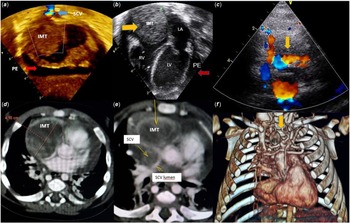
Due to the clinical presentation with syndrome of the superior caval vein and to cardiomegaly, a transthoracic echocardiogram was performed. It detected moderate pericardial effusion and a large echogenic mass measuring about 4 cm in its largest diameter, adhered to the anterosuperior free wall of the right atrium, causing obstruction to the superior caval vein flow on colour mapping and Doppler (Fig 1). The tricuspid valve and the interatrial septum were not affected by the large mass.
Transthoracic echocardiogram (Echo) showing: A- Color flow Doppler examinaton indicating turbulent jet flow in the SCV (blue arrow), large echogenic mass adhered (IMT) to the roof of the right atrium (RA) and moderate pericardial effusion (PE- red arrow) in the subcostal view; B - IMT (yellow arrow) in the four-chamber view and PE. Note that the echogenic mass spares the tricuspid valve. C - Color flow Doppler by Echo examinaton indicating turbulent jet flow besides entanglement of vessels and collaterals above the RA and in the thymus region (yellow arrow). D - Axial slices on chest angiotomography showing the huge tumor mass in the RA (4.1 cm); E- SCV almost completely occluded, with only a thin line of contrast in the lumen (SCV- RA). F - The same findings detected by Echo in C, now in the coronal reconstruction by tomography. SCV: superior caval vein; IMT: inflammatory myofibroblastic tumor ; LA : left atrium; RV : right ventricle; LV : left ventricle.

Chest angiotomography confirmed the echocardiographic findings, showing the superior caval vein almost completely occluded. A huge tumour mass was detected inside the right atrium, adhered to its superior wall, which was thickened (Fig 1). In addition, there was thymus engorgement with entangling of vessels and collaterals and moderate pericardial effusion.
Complete surgical resection of the tumour mass was performed and no signs of local infiltration were observed, but it was necessary to reconstruct the superior caval vein and the roof of the right atrium with bovine pericardium. The thymus was surgically removed.
In the immediate post-operative period, the patient had complications such as junctional tachycardia followed by a transitory atrioventricular block, and the use of a temporary pacemaker was necessary. He evolved with renal failure and metabolic disorders, requiring steroids and peritoneal dialysis. Electrocardiogram before hospital discharge demonstrated sinus rhythm and right bundle block.
The post-operative transthoracic echocardiogram showed effective resection of the right atrial mass, with increased systolic velocity at Doppler interrogation between the superior caval vein and right atrium (maximum systolic gradient of 11 mmHg) and mild tricuspid regurgitation.
The infant was discharged after prolonged hospitalisation and is being clinically followed-up with complementary exams. He does not show signs of tumour recurrence 36 months after surgery.
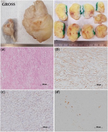
The anatomopathological evaluation of the tumour showed a well-delimited spindle cell neoplasm, with areas of necrosis and focal lymphohistiocytic infiltrates. Immunohistochemical evaluation showed positive immunoreactivity of the tumour cells for α-smooth muscle actin and desmin but negative immunoreactivity for anaplastic lymphoma kinase (ALK1), cytokeratins, and epithelial membrane antigen. CD34 immunoreactivity was seen in capillaries and the inflammatory cells were positive for CD68 and CD138 (Fig 2). The final diagnosis was an inflammatory myofibroblastic tumour.
GROSS- The upper panels show the external view (right) and cross-section surface (right) of the resected tumour: trabeculated surface and yellowish tissue with a few haemorrhagic areas. The histology panels show: a) Tumour cells are spindle-shaped and intermingled with small capillaries and inflammatory cells; b) Diffuse labelling (brown colour) for smooth muscle actin; c) Inflammatory cells characterized mainly by histiocytes (CD68 immunolabelling); d) Focal labelling of the tumour cells by desmin. a) hematoxylineosin stain; b), c) and d) immunohistochemistry for smooth-muscle actin, CD68 and desmin respectively, counter-stained with Harry’s hematoxylin. Objective magnification = 20X for all the panels.

Discussion
The differential diagnosis of inflammatory myofibroblastic tumours occurring inside the heart includes neoplasms such as rhabdomyomas, which are the most common benign primary cardiac tumours in infants and children, followed by fibromas and teratomas. Reference Becker4
Inflammatory myofibroblastic tumours have some histopathological features similar to myosarcomas, but less pleomorphism, atypia, and fewer mitotic figures. Reference Butany, Dixit, Leong, Daniel, Mezody and David5. In addition, they do not infiltrate the myocardium. It is known that the immunohistochemical expression of anaplastic lymphomakinase (ALK-1) is present in 35% of inflammatory myofibroblastic tumours although a negative result does not rule out this type of tumour. Reference Butany, Dixit, Leong, Daniel, Mezody and David5
Their aetiology remains unknown, with recent research suggesting some chromosomal aberrations at locus 2p23. Reference Li, Cerilli and Wick6
Inflammatory myofibroblastic tumours may be accompanied by systemic symptoms such as fever and anaemia, as in the case reported here, and can recur in up to 10% of cases. Reference Andersen, DiBernardo, Linardic, Camitta and Lodge7 Other systemic symptoms that may occur are polyarthritis and vasculitis, caused by the release of cytokines, especially interleukin-6. Reference Li, Cerilli and Wick6 Tests of inflammatory activity such as C-reactive protein and erythrocyte sedimentation rate show high values and should always be monitored, because they help to guide the diagnosis and possible recurrence.
The tumour location within the heart is closely related to the symptoms. In the case reported here, the infant presented syndrome of the SCV due to obstruction caused by the tumour near its entrance in the right atrium, leading to a great engorgement of the thymus and cardiomegaly. Some reports demonstrate inflammatory myofibroblastic tumours obstructing the right ventricular outflow tract, Reference Cina, Smialek, Burke, Virmani and Hutchins8 limiting the pulmonary flow and the systemic circulation. Worse prognosis is also associated with coronary and valvar involvement. In a review about cardiac tumours, Cina et al. reported around 0.06% of sudden death in patients younger than 34 years, besides poor prognosis associated with neoplasms involving the coronary arteries, cardiac valves, or ventricular outflow tracts. Reference Cina, Smialek, Burke, Virmani and Hutchins8
The primary treatment for inflammatory myofibroblastic tumours is surgical resection, as their location and size may compromise the patient’s haemodynamics and cardiac function. In the review published by Eilers et al., 73.4% of the patients required surgical resection. Heart transplantation was indicated in three cases. Reference Al, Nazarullah and Shipper3 This procedure has been suggested when the masses are considered unresectable, mainly when they are associated with vital structures, or in cases of recurrence after primary resection, or persistent heart failure after tumour excision. Reference Mull, Dahdah and Scarfone9
Some reports show that, when surgical resection is not feasible, adjunctive steroid therapy may be beneficial, based on the inflammatory/immunological aetiology of the tumour, Reference Al, Nazarullah and Shipper3 as chemotherapy is reported in the literature as an non-surgical treatment alternative Reference Mizia-Malarz, Sobol-Milejska, Buchwald and Wo¿10 .
Financial support
This research received no specific grant from any funding agency, commercial, or not-for-profit sectors.
Conflicts of interest
None.